TNF‑α and its receptors: how TNFR1 and TNFR2 connect inflammation, survival and repair
Two TNF forms, TNFR1 ubiquitin platforms, caspase-8 and MLKL, TNFR2 signaling and the limits of translating cellular mechanisms into clinical therapy.
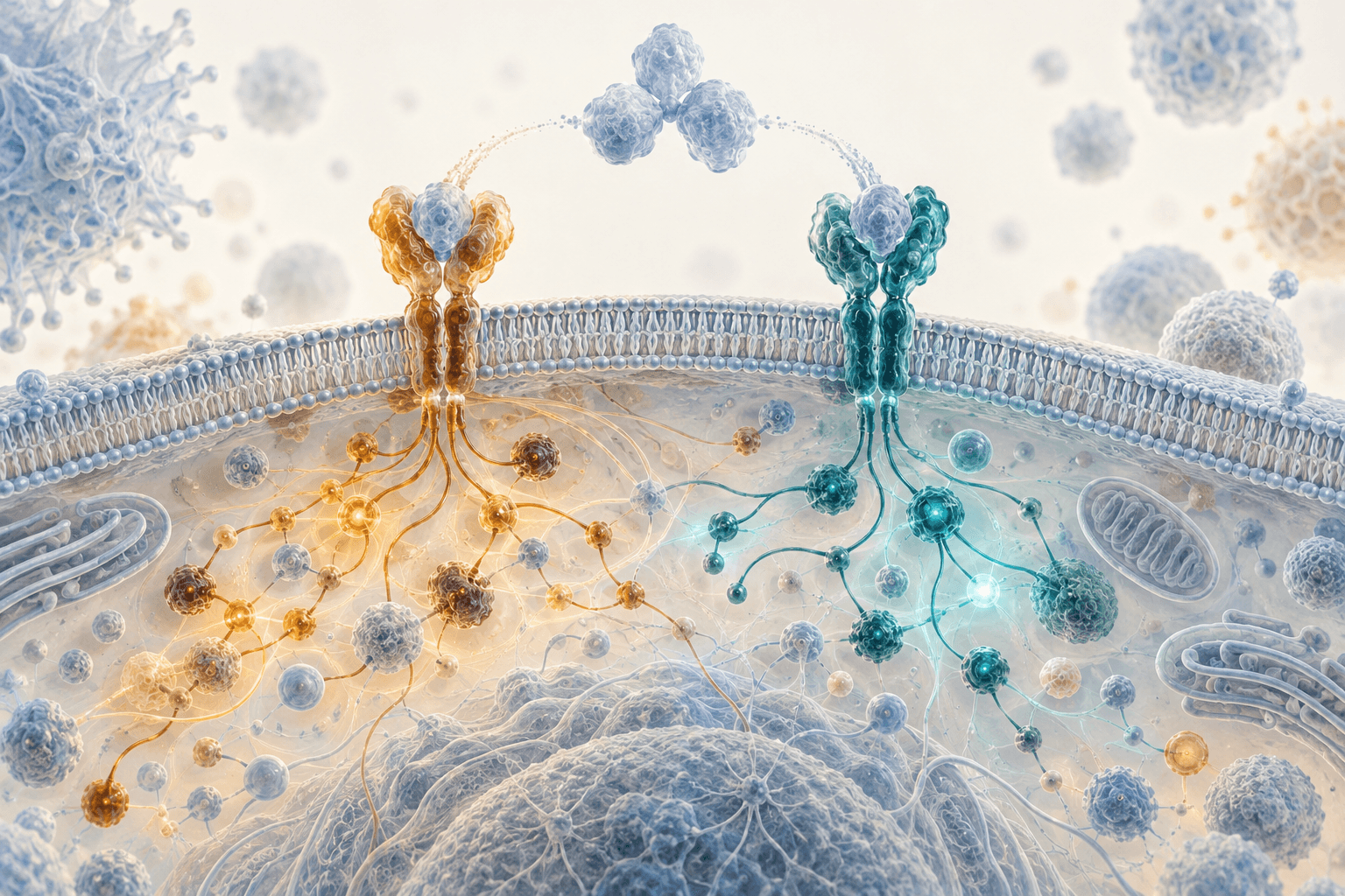
The same cytokine can support a protective inflammatory response, limit tissue damage and participate in cell death. TNF‑α illustrates why a molecule's name does not predict its action: “tumor necrosis factor” does not mean that every receptor interaction ends in necrosis. The outcome depends on ligand form, cell type, available intracellular proteins, tissue state and exposure duration. Differences between its two main receptors, TNFR1 and TNFR2, help explain this logic, but do not turn it into a simple opposition between a “bad” and a “good” signal.
Another example of context-dependent cytokine biology is classical and trans-signaling by IL‑6: changing signal delivery changes the range of responding cells. TNFR1 can initiate inflammatory transcription and survival, or apoptosis and necroptosis when control mechanisms fail. TNFR2 often supports growth, resilience and functions of particular immune and tissue cells, but these properties can be helpful during repair and undesirable in a tumor environment. Neutralizing all TNF, blocking one receptor and stimulating one receptor are therefore different interventions. Results from one cannot automatically be transferred to another.
Two forms of TNF and the importance of cell contact
TNF is synthesized as a membrane protein with its receptor-binding region facing the extracellular space. Proteolytic cleavage by ADAM17, also called TACE, produces the soluble form. Both membrane and soluble TNF can interact with receptors, but contact geometry, surface retention and formation of larger receptor clusters differ. At the cell membrane, spatial organization matters alongside molecular concentration.
The functional preference is particularly apparent for TNFR2: membrane TNF generally activates this receptor more efficiently than an ordinary soluble trimer. Grell and colleagues demonstrated this in immune-cell systems and showed that the membrane ligand could alter tumor-cell susceptibility to cytotoxic effects. Preferential TNFR2 activation by membrane TNF therefore does not mean that this signal is always anti-inflammatory. Nor should activation efficiency be replaced by an absolute claim that soluble TNF cannot bind TNFR2 at all.
TNFR1 is expressed in many cell types, whereas TNFR2 expression depends more strongly on cell state and is especially relevant in certain immune and vascular populations. Receptor expression is not equivalent to pathway activity. Signaling requires an available ligand, suitable receptor-complex organization and intracellular adapters; endocytosis, removal of receptors from the surface and negative feedback operate at the same time.
ADAM17 can also participate in shedding the extracellular portions of the receptors themselves. Soluble TNFR1 and TNFR2 bind TNF and alter its availability, but their blood concentrations do not directly measure intracellular activity of the corresponding receptor. Production, shedding and clearance, including kidney function, influence these levels. The ratio of soluble receptors therefore cannot serve as a ready-made personal scale of “destruction versus regeneration.”
TNFR1: a signaling platform comes before the cell-fate decision
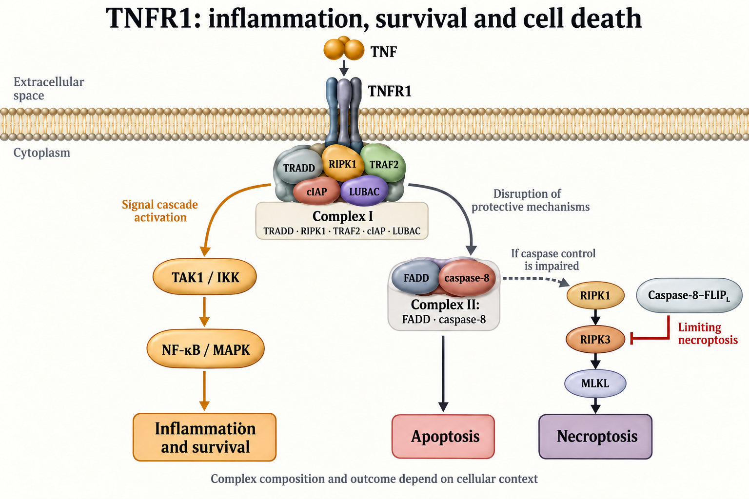
The intracellular portion of TNFR1 contains a death domain that enables protein interactions with corresponding adapters. This domain does not itself cleave proteins or function as a caspase. Its name reflects participation in families of signaling complexes capable of initiating death, but the initial TNF response often includes protective transcription. Separating these stages was a key finding of Micheau and Tschopp.
The initial complex I forms at the plasma membrane and contains the receptor, TRADD, RIPK1 and TRAF2; subsequent assembly recruits cIAP proteins and other regulators. RIPK1 has a scaffold function here that differs from its catalytic role in particular death pathways. Removing the protein and inhibiting its kinase domain are therefore not interchangeable interventions. This distinction explains why “RIPK1 appeared, so the cell will die” is an incorrect simplification.
Ubiquitin chains in complex I create binding surfaces for signaling proteins. K63-linked chains and linear M1 chains should not be treated as ordinary tags for obligatory destruction in the proteasome. They can provide a scaffold that concentrates TAK1–TAB and IKK complexes. LUBAC forms linear chains and includes catalytic HOIP with structural partners HOIL‑1 and SHARPIN. Assembly, stability and dismantling of this platform help determine whether the response remains predominantly transcriptional or becomes dangerous to the cell.
TAK1 and IKK transmit signals by phosphorylation: kinases transfer phosphate from ATP to protein substrates. Ubiquitination is different and requires sequential E1, E2 and E3 activity, with ATP expenditure during ubiquitin activation. cIAP proteins and LUBAC organize ubiquitin modifications rather than replace kinases. Distinguishing these reactions matters: the same arrow in a diagram can represent physical binding, phosphate transfer, chain assembly or proteolytic cleavage, with different consequences.
In canonical NF‑κB signaling, IKK promotes phosphorylation of IκB inhibitors, followed by their ubiquitination and proteasomal degradation, allowing transcription factors to move into the nucleus. NF‑κB regulates inflammatory mediators alongside proteins that limit death. MAPK branches also participate in transcriptional changes, cell activation and stress responses. In a particular cell, this can support survival while increasing inflammatory pressure on the surrounding tissue: cellular and tissue outcomes do not always coincide.
Checkpoints cannot be reduced to a single switch
A20, CYLD and OTULIN limit or reorganize ubiquitin signaling, but should not be grouped as enzymes that simply “remove protection and cause death.” They differ in substrates, chain types, location and additional protein interactions. A20 participates in negative feedback of inflammation and can protect cells in particular conditions. The outcome depends on the complex being modified and the compensatory mechanisms that remain available.
Protective transcription is only part of the control system. Phosphorylation of individual components, cIAP availability, integrity of the ubiquitin scaffold and caspase‑8 activity limit progression toward death before gene expression changes. A cell can therefore die after disruption of early checkpoints even when a conventional diagram presents NF‑κB as the only main barrier. Conversely, finding inflammatory transcripts does not automatically establish necroptosis.
Apoptosis and necroptosis: different complexes and enzymatic tasks
Cytoplasmic complexes containing FADD and caspase‑8 can form after changes to the initial membrane platform. In the classical model they are called complex II. Subtypes IIa and IIb and the term “ripoptosome” describe different compositions and assembly conditions, and cannot be made unconditional synonyms. To understand an experiment, it is more useful to establish whether TRADD and RIPK1 participate, whether RIPK1 kinase activity is required and which opposing proteins are present.
Caspase‑8 is a cysteine protease that cleaves particular protein substrates after aspartate residues. Activation on a suitable platform can initiate executioner caspases and apoptosis. In some cell types, BID cleavage connects this process to mitochondrial amplification: tBID promotes BAX/BAK activation and permeabilization of the mitochondrial outer membrane. Cytochrome c release supports apoptosome assembly and caspase‑9 activation, but dependence on this amplification is cell-context specific.
FLIP_L resembles caspase‑8 structurally, but its own caspase-like domain does not perform ordinary protease activity. This does not mean that the FLIP_L–caspase‑8 heterodimer must be entirely inactive. Oberst and colleagues showed that a proteolytically active caspase‑8–FLIP_L complex can limit RIPK3-dependent necrotic death without initiating full apoptosis. The same caspase can thus execute one form of death and prevent another.
Necroptosis involves activation of RIPK3 and its substrate MLKL. In TNF-dependent conditions, RIPK1 can participate in assembly of a signaling platform with RIPK3; the importance of this step depends on the system and available alternative activators. Phosphorylated MLKL changes conformation, oligomerizes and interacts with membranes, contributing to loss of their integrity. MLKL is not an executioner caspase, so necroptosis cannot be described as another form of caspase-dependent apoptosis.
Membrane disruption favors release of intracellular components and amplification of inflammation, but “apoptosis is quiet, necroptosis is always inflammatory” also requires context. Timely clearance of dead cells, surrounding tissue state and the number of damaged cells matter. Measuring total cell death without characterizing its mechanism does not establish a particular branch. A scientific conclusion requires several concordant features rather than one universal marker.
TNFR2: coordinating survival and immune function
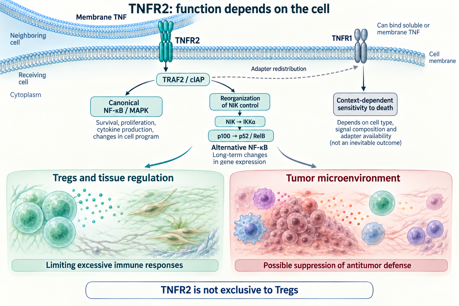
TNFR2 has no death domain and uses a different set of initial interactions. It recruits TRAF2 and associated cIAP proteins and can activate canonical NF‑κB, MAPK and contextually connected growth and survival pathways. These events do not guarantee tissue repair. Growth of a useful regulatory cell and growth of a tumor cell can use partly overlapping mechanisms despite opposite consequences for the organism.
An important TNFR2 feature is its effect on availability of the TRAF2–TRAF3–cIAP complex, which helps keep NIK levels low at rest. Reorganization of this system can allow NIK accumulation and IKKα activation. Subsequent limited proteasomal processing of p100 produces p52, which participates with RelB in noncanonical NF‑κB signaling. Processing p100 differs from complete destruction of IκB: proteolysis creates a functional transcription-factor form.
Canonical and noncanonical branches differ in organization and kinetics, but an identical schedule in minutes or hours cannot be assigned to every cell. Speed depends on initial protein levels, stimulus intensity and duration, receptor expression and parallel signals. Saying that the noncanonical pathway usually requires NIK accumulation and protein-complex reorganization is more accurate than giving a universal timing table without an experimental model.
When TNFR2 increases sensitivity to TNFR1
The receptors share some intracellular resources. Under certain conditions TNFR2 redistributes, or contributes to reduced availability of, TRAF2/cIAP, weakening TNFR1 protective mechanisms. The cell can then become more sensitive to death after TNF stimulation. This does not mean that TNFR2 has acquired a death domain: the effect arises through cross-regulation of the signaling environment.
This connection matters when discussing selective drugs. Preserving TNFR2 during TNFR1 blockade is different from stimulating TNFR2 while TNFR1 remains active. Effects depend on expression of both receptors, ligand form and the cell's capacity to maintain protective complexes. The useful question is therefore which cell receives which signal in a particular tissue and disease phase, rather than which receptor is more beneficial.
Regulatory T cells: tissue protection with a contextual cost
TNFR2 is often associated with activated, functionally prominent regulatory T cells, but is not their exclusive marker. It also occurs in other immune populations, including activated conventional T cells. Describing a Treg requires attention to origin, transcriptional program, FOXP3 stability and the functional capacity to suppress a particular immune response. TNFR2 staining alone is insufficient.
IL‑2 and its STAT5 signaling pathway remain important for Treg maintenance. TNFR2 costimulation can complement this system, but should not be depicted as directly and obligatorily initiating the same receptor-associated JAK–STAT mechanism. Similarly, changing FOXP3 expression does not always establish stable epigenetic remodeling of every regulatory region. A claim of stability should rest on specific measurements and observation duration, rather than a short-term marker increase.
Mensink and colleagues studied human blood Tregs using transcriptomics, proteomics, functional migration assessment and comparison with tissue signatures. TNFR2 costimulation supported tissue-effector Treg features, while comparison with CD28 costimulation revealed distinct differentiation programs. This provides a strong basis for a mechanistic hypothesis and development of cellular approaches. It is not a therapeutic trial demonstrating organ repair or clinical efficacy in patients.
In autoimmune or injured tissue, regulatory function can limit excessive immunity. In a tumor microenvironment the same function can impede antitumor defense. The result also depends on tumor composition, effector cells and direct receptor expression by tumor cells. Stimulating TNFR2 for one clinical purpose can therefore conflict with blocking it for another without creating a contradiction in the underlying biology.
Nervous tissue: remyelination in mice is not treatment of MS
Arnett and colleagues linked TNF and TNFR2 to oligodendrocyte precursor proliferation and remyelination in a mouse cuprizone model. This helps establish that TNF can participate in myelin repair. However, toxic myelin injury differs from a multifactorial human immune disease. Causes of injury, temporal sequence and involvement of peripheral immunity differ.
These experimental findings cannot be retold as proof that increasing TNFR2 necessarily restores myelin in a patient. That conclusion requires clinical trials with a defined population, drug form, sufficient exposure and outcomes reflecting function and disease course. A cellular-marker change or increased precursor numbers is an important intermediate observation, but does not replace a clinical outcome.
The lenercept trial illustrated the danger of directly transferring a hypothesis. A double-blind randomized trial enrolled 168 predominantly relapsing-remitting MS patients; intravenous lenercept at 10, 50 or 100 mg every four weeks or placebo was given for up to 48 weeks. MRI measures did not differ significantly, but exacerbations were more frequent and occurred earlier with lenercept. EDSS was not significantly affected; antidrug antibodies and low detectability of trough concentrations limited exposure interpretation.
Lenercept was soluble TNFR1–Fc binding TNF. It neutralized the ligand rather than selectively blocking membrane TNFR1 while guaranteeing preservation of every other TNF interaction. Its negative result is therefore not a direct trial of every modern receptor-selective approach. It nevertheless demonstrates that TNF involvement in inflammation does not make suppressing the entire system automatically beneficial in MS.
Clinical efficacy depends on the disease and chosen outcome
Positive anti-TNF results in rheumatoid arthritis do not require the assumption that TNF is always harmful. ATTRACT randomized 428 patients with active arthritis on stable methotrexate to placebo or four infliximab regimens: 3 or 10 mg/kg followed by administration every four or eight weeks. At 30 weeks, ACR20 was achieved by 53%, 50%, 58% and 52% in the corresponding groups versus 20% with placebo plus methotrexate. ACR50 was achieved by 29%, 27%, 26% and 31% versus 5%.
ACR20 is a defined composite improvement criterion, not complete disappearance of disease in every responder. The trial evaluated adding infliximab to methotrexate in a selected population, without isolating TNFR1 and TNFR2 contributions. Thirty weeks of observation also does not replace assessment of rare complications during prolonged use. Clinical benefit from a particular regimen and mechanistic appeal of a receptor hypothesis represent different evidence levels.
Etanercept contains the extracellular portion of TNFR2 joined to Fc and binds the ligand. It cannot be called an agonist of cellular TNFR2 simply because its construction uses that receptor fragment. Likewise, TNF-binding antibodies and receptor-binding antibodies have different immediate targets. Discussing a drug requires identifying what it binds, neutralizes or clusters, rather than inferring its action from the name of a protein fragment.
Heart failure: reducing a marker does not guarantee benefit
The double-blind randomized pilot ATTACH trial studied 150 patients with moderate-to-severe chronic heart failure, NYHA functional class III–IV and ejection fraction no greater than 35%. Participants received infliximab 5 or 10 mg/kg or placebo at weeks 0, 2 and 6, with follow-up through 28 weeks. The primary clinical-status assessment at 14 weeks did not show improvement from infliximab. Reducing inflammatory measures did not translate into corresponding clinical benefit.
With 10 mg/kg, the risk of all-cause death or hospitalization for heart failure through 28 weeks was higher: hazard ratio 2.84, 95% confidence interval 1.01–7.97, nominal p=0.043. This concerns a specific dose and selected population, with a wide confidence interval and pilot-trial limitations. It does not prove that every anti-TNF treatment is equally dangerous for every patient. It does refute the universal assumption that a lower inflammatory marker must mean better organ function.
Safety reflects TNF's physiological role
TNF participates in normal immune defense and coordination of cell interactions, so blocking it changes more than pathological inflammation. Safety assessment includes infectious complications, concurrent immunosuppressive therapy and comorbidities. A single risk multiplier covering every drug, dose, infection and population would be incorrect. A short trial showing no group difference in serious infections does not exclude risks revealed during longer and broader use.
Prescribing a clinical drug depends on its own indications and restrictions, rather than a general receptor diagram. Clinical choices rely on diagnosis, disease history, assessment and monitoring; a signaling article helps explain their basis. Replacing prescribed therapy independently with a supplement or an untested “selective” strategy transfers a mechanistic hypothesis to a decision level it does not substantiate.
Diet and lifestyle are not proven receptor switches
Metabolic state, dietary composition and chronic inflammatory stimuli can influence the immune environment. This does not establish that an individual dietary molecule selectively switches off TNFR1 and switches on TNFR2 in humans. In cell studies, polyphenols, fatty acids or microbiota metabolites can change gene expression, redox balance and signaling proteins. Laboratory concentration, exposure duration and chemical form may differ substantially from exposure following ordinary food or supplements.
Butyrate, for example, does more than potentially change individual inflammatory markers: it is a metabolite involved in receptor-associated and epigenetic regulation. Specialized pro-resolving mediators derived from omega‑3 fatty acids likewise have numerous interactions. None of these observations alone proves selective clinical control of the two TNF receptors. Claims of guaranteed necroptosis blockade or a universal several-fold TNFR2 increase require direct evidence in a defined system.
Vitamin D as a nutrient, its circulating metabolites and active calcitriol cannot be treated as one identical laboratory exposure. Likewise, resveratrol or EGCG effects in cultured cells cannot be turned into dosing protocols for autoimmune disease. Practical conclusions require evidence on bioavailability, metabolites, clinical outcomes and adverse effects. A mechanistic article does not establish high-dose supplement prescriptions to modify TNFR1/TNFR2.
Reading future receptor-selectivity studies
An informative study should separate target, cell context and outcome. For a TNFR1-directed drug, it matters whether it blocks ligand binding, clustering or a particular intracellular response. For TNFR2 stimulation, it matters which cells actually receive the signal and whether TNFR1 remains active. Increased Treg numbers or a changed inflammatory protein can confirm pharmacodynamics without proving patient benefit.
Human cells outside the body, animal models and clinical trials must also be distinguished. Human cultures lack many features of drug distribution and tissue interaction; animal models do not fully reproduce human disease. Clinical trials require attention to baseline severity, background drugs, duration, primary outcome and adverse events. Negative results are particularly valuable because they expose limits of plausible biological explanations.
The TNF system exemplifies contextual biology: cell preservation, inflammation reduction and organ-function recovery do not always occur together. TNFR1 organizes both protective and lethal complexes, while TNFR2 supports functions whose usefulness depends on tissue and purpose. Understanding enzymatic reactions, compartments and cross-regulation enables more precise discussion of therapy. It does not remove the need to test the clinical result of each specific intervention.
FAQ
Does TNFR1 always cause cell death?
No. Initial complex I can support NF-κB, inflammatory transcription and survival. Progression to apoptosis or necroptosis depends on checkpoints and cellular context.
Does TNFR2 always support repair?
No. Regulatory functions can limit excessive immunity, but in tumors they can impede antitumor defense. The receptor also occurs outside Tregs.
Does etanercept stimulate TNFR2?
Its construction contains an extracellular TNFR2–Fc fragment that binds the ligand. This does not make the drug an agonist of cellular TNFR2.
Can supplements switch the receptors?
Selective clinical control of TNFR1/TNFR2 with supplements has not been established. Cellular effects do not define a dosing protocol for treating autoimmune disease.
Sources
- The transmembrane form of tumor necrosis factor is the prime activating ligand of the 80 kDa tumor necrosis factor receptor
- Induction of TNF receptor I-mediated apoptosis via two sequential signaling complexes
- Recruitment of the linear ubiquitin chain assembly complex stabilizes the TNF-R1 signaling complex and is required for TNF-mediated gene induction
- Catalytic activity of the caspase-8-FLIP(L) complex inhibits RIPK3-dependent necrosis
- Mixed lineage kinase domain-like protein mediates necrosis signaling downstream of RIP3 kinase
- Mixed lineage kinase domain-like protein MLKL causes necrotic membrane disruption upon phosphorylation by RIP3
- Tregs from human blood differentiate into nonlymphoid tissue-resident effector cells upon TNFR2 costimulation
- TNF alpha promotes proliferation of oligodendrocyte progenitors and remyelination
- TNF neutralization in MS: results of a randomized, placebo-controlled multicenter study
- Infliximab versus placebo in rheumatoid arthritis patients receiving concomitant methotrexate: a randomised phase III trial
- Randomized, double-blind, placebo-controlled, pilot trial of infliximab in patients with moderate-to-severe heart failure: results of the ATTACH trial
- Noncanonical NF-kappaB activation requires coordinated assembly of a regulatory complex of the adaptors cIAP1, cIAP2, TRAF2 and TRAF3 and the kinase NIK
- Apoptotic crosstalk of TNF receptors: TNF-R2-induces depletion of TRAF2 and IAP proteins and accelerates TNF-R1-dependent activation of caspase-8
- The tumor necrosis factor receptor 2 signal transducers TRAF2 and c-IAP1 are components of the tumor necrosis factor receptor 1 signaling complex