
Uridine and synaptic membrane synthesis: mechanisms and clinical limits
How UMP connects to CTP, the Kennedy pathway, choline and DHA; what synapse models and nutritional-mixture trials show, and where evidence ends and unsupported protocols begin.
How UMP connects to CTP, the Kennedy pathway, choline and DHA; what synapse models and nutritional-mixture trials show, and where evidence ends and unsupported protocols begin.
Neuroplasticity requires more than electrical activity and gene expression: a growing projection needs new membrane, and a synapse needs a coordinated system of lipids, receptors and proteins. Uridine attracted researchers because its metabolism contributes to the production of CTP, a nucleotide required for phosphatidylcholine and phosphatidylethanolamine synthesis. This is a persuasive biochemical connection, but several distinct levels of evidence separate replenishing a nucleotide pool from improving human memory.
Uridine 5′-monophosphate, or UMP, is a nucleotide: uridine with a phosphate attached to the 5′ carbon of ribose. Free uridine is a nucleoside without that phosphate group; UTP contains three phosphates. These distinctions matter for absorption, intracellular reactions and receptor signalling. Oral UMP cannot be treated as a ready-made extracellular P2Y receptor agonist, and a positive result for a mixture containing UMP, DHA and vitamins cannot be attributed to uridine alone.
The presynaptic terminal contains neurotransmitter-filled vesicles. Their membranes undergo cycles of fusion with the plasma membrane and subsequent recovery, so lipid metabolism supports both growth and maintenance of existing contacts. The SNARE complex participates directly in membrane fusion, whereas synapsin-1 helps organise vesicle pools and synaptophysin is associated with synaptic vesicle membranes. These proteins cannot be grouped together as a single “exocytosis enzyme”.
Across the synaptic cleft lie receptors and the postsynaptic density. In an excitatory contact, AMPA receptors, including complexes containing GluA1/GluA2, and NMDA receptors, which may contain GluN2B, are organised together with scaffold proteins. PSD-95 belongs to the MAGUK family and primarily provides structural organisation; the family name does not mean that PSD-95 acts here as an active guanylate kinase. Connections through additional adaptors, including a network involving Shank, link receptor complexes to the spine's actin cytoskeleton. A simple PSD-95—Shank—actin line in a diagram represents an organisational network, not necessarily direct binding between every adjacent pair.
Phosphatidylcholine (PC), phosphatidylethanolamine (PE), phosphatidylserine (PS), sphingomyelin and cholesterol make different contributions to membrane properties. Composition varies with cellular compartment, synapse type and isolation method; universal percentages for all synaptic membranes are misleading. DHA can enter the acyl chains of several phospholipid classes, often at the sn-2 position. A DHA-containing lipid and an ordered cholesterol–sphingomyelin domain are nevertheless different aspects of bilayer organisation: the presence of DHA does not automatically make an area a “lipid raft”.
New membrane area alone does not determine where a projection grows or which contact becomes functional. Growth signals, adhesion, protein delivery, cytoskeletal remodelling and network activity are also needed. Increases in PC, PSD-95 or spine number are therefore interesting, but each measurement answers a different question. None replaces an electrophysiological assessment of transmission or a clinical assessment of memory.
Following UMP intake, much of its journey involves dephosphorylation and absorption as uridine. Intestinal phosphatases and nucleotidases, nucleoside transport and subsequent metabolism in the liver and other tissues contribute. Plasma concentration depends on more than the dose: chemical form, product composition, food and utilisation rate all matter. Increased blood uridine does not measure neuronal UTP concentration or reveal how many new synapses have formed.
Two major families transport nucleosides. ENT1/ENT2, encoded by SLC29A1/SLC29A2, provide equilibrative transport down a gradient; direction can change with concentrations. CNT1/2/3 belong to SLC28 and can use a cation gradient. CNT1 preferentially transports pyrimidine nucleosides, CNT2 purines but also uridine, and CNT3 has broader selectivity. Experiments expressing recombinant human transporters in Xenopus oocytes demonstrated a Na⁺:nucleoside ratio of 1:1 for CNT1/2 and 2:1 for CNT3; CNT3 can also operate with H⁺ at 1:1. These are properties of the studied proteins, not an established map of their contribution to the human blood–brain barrier.
The blood–brain barrier consists of a layer of endothelial cells with tight junctions. A transcellular route first crosses the blood-facing membrane, then endothelial cytoplasm and a second, tissue-facing membrane; the nerve cell's own membrane remains to be crossed. This route cannot be reduced to a single “blood–neuron” boundary, with identical transporters assigned to both sides without evidence. Brain entry of uridine and changes in brain nucleotides have been demonstrated in experimental models, but they do not establish precise human pharmacokinetics for every supplement.
In the salvage pathway, uridine–cytidine kinases UCK1/UCK2 phosphorylate uridine. CMPK1 and nucleoside diphosphate kinases of the NME family then support sequential UDP and UTP formation. UMP, UDP and UTP denote phosphate number here, not different bases. Kinase reactions consume energy-bearing nucleotides and are metabolically regulated; exogenous uridine does not become membrane without an energy cost.
Uridine + ATP → UMP + ADP — UCK1/2, EC 2.7.1.48.
UMP + ATP ⇌ UDP + ADP — CMPK1, EC 2.7.4.14.
UDP + ATP ⇌ UTP + ADP — NME/NDPK, EC 2.7.4.6.
The other possibility is de novo synthesis of the pyrimidine ring. The CAD complex carries out early reactions, mitochondrial DHODH converts dihydroorotate to orotate, and UMPS participates in UMP formation. Enzymes of this pathway have been detected in adult human brain tissue; cell experiments also connect pyrimidine metabolism with differentiation. It is therefore incorrect to claim that the mature brain almost completely lacks de novo synthesis and can obtain CTP only from circulating uridine. The relative importance of salvage and de novo synthesis varies with cell type, state and substrate availability. Cytidine can also be phosphorylated to CMP and subsequently produce CDP/CTP.
CTP synthetases CTPS1/CTPS2 convert UTP to CTP using the amide nitrogen of glutamine and ATP-derived energy. This is not the addition of another phosphate: the chemistry of the pyrimidine base changes. The overall reaction is:
UTP + glutamine + ATP + H₂O → CTP + glutamate + ADP + Pi.
Pi denotes inorganic phosphate. The glutaminase domain releases nitrogen, and the synthetase domain uses it to aminate activated UTP. CTP participates in feedback; GTP can regulate glutamine-dependent activity. Human CTPS can form filaments whose function depends on the isoform and enzyme state. A scheme in which “an inactive dimer becomes an active tetramer only because of GTP”, and a single Ki for all conditions, are consequently too crude.
A gerbil experiment measured changes in brain uridine, UTP, CTP and CDP-choline following oral UMP. This supports a relationship between precursor availability and the metabolic pool in that model. Plasma or cerebrospinal-fluid uridine, however, cannot be substituted directly for the intracellular concentration in a UCK kinetic equation: that comparison mixes compartments. Similarly, increased intracellular UTP does not mean that the same UTP reached the extracellular space and activated a receptor.
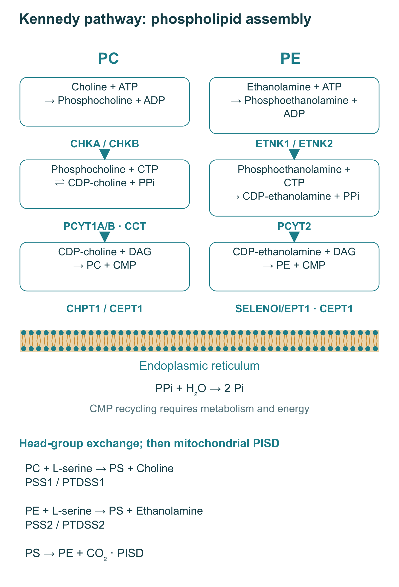
After entering a cell, choline is phosphorylated by choline kinases CHKA/CHKB. CTP:phosphocholine cytidylyltransferase, or CCT, transfers the cytidylyl group from CTP to phosphocholine. The resulting CDP-choline is an activated donor of the future PC polar head group. Membrane phosphotransferases then join it to diacylglycerol, or DAG, in the endoplasmic reticulum and related compartments.
Choline + ATP → phosphocholine + ADP — CHKA/CHKB.
Phosphocholine + CTP ⇌ CDP-choline + PPi — CCT, PCYT1A/PCYT1B, EC 2.7.7.15.
PPi + H₂O → 2 Pi — inorganic pyrophosphatase; a separate reaction that helps drive flux.
CDP-choline + DAG → PC + CMP — CHPT1 or CEPT1.
CTP does not become part of the final phospholipid: its cytidine moiety leaves as CMP. CMP can re-enter nucleotide metabolism, but that requires enzyme reactions and energy; it should not be depicted as a free, self-regenerating “catalyst”. The modern name for choline phosphotransferase is CHPT1. The older abbreviation CPT1 can be confused with carnitine palmitoyltransferase and is therefore not used in the diagrams.
The high-affinity transporter CHT1/SLC5A7 is particularly important in cholinergic terminals and acetylcholine synthesis. Other transport routes, including CTL1/SLC44A1, also contribute to phospholipid metabolism. One CHT1 arrow cannot describe choline uptake into every brain cell. Furthermore, PC undergoes transport and acyl-chain remodelling after synthesis; DHA-containing DAG is one possible substrate, not a mandatory molecule for every newly synthesised PC.
The parallel CDP-ethanolamine branch explains why changes in pyrimidine pools can also affect PE. ETNK1/ETNK2 produce phosphoethanolamine, PCYT2 uses CTP to synthesise CDP-ethanolamine, and SELENOI/EPT1 or CEPT1 completes assembly with DAG. The principle of head-group activation resembles the CDP-choline pathway, but enzyme regulation and product distribution are not identical.
Ethanolamine + ATP → phosphoethanolamine + ADP.
Phosphoethanolamine + CTP → CDP-ethanolamine + PPi.
CDP-ethanolamine + DAG → PE + CMP.
Mammalian PS is produced by polar head-group exchange. The branch using PC is written PC + L-serine → PS + choline, involving PSS1/PTDSS1; the PE branch is PE + L-serine → PS + ethanolamine, involving PSS2/PTDSS2. This scheme shows major substrate preferences, not absolute specificity in every experimental system. PS can also be decarboxylated to PE in mitochondria: PS → PE + CO₂, catalysed by PISD. These reactions connect lipid classes without making PC the universal immediate precursor of every membrane.
CCT is an important regulated step of the CDP-choline pathway. Activity depends both on substrate availability and membrane interaction: an amphipathic segment can bind the bilayer, changing autoinhibition of the catalytic site. Lipid composition, charge and packing defects influence this transition. Structural studies explain why membrane binding changes catalytic activity and apparent CTP affinity; they do not imply the same manyfold acceleration in every neuron after UMP intake.
| CCTα region | Function | Interpretation |
|---|---|---|
| N-terminal region | Regulation of localisation, including nuclear distribution | CCTα cannot be described solely as a cytosolic protein |
| Catalytic region | CDP-choline formation from phosphocholine and CTP | Rate depends on substrates and regulatory conformation |
| Membrane-binding domain M | Amphipathic bilayer interaction and relief of autoinhibition | Membrane properties matter; free DHA and lipid-bound DHA are different |
| C-terminal phosphorylated region | Modulation of membrane affinity and regulation | Phosphorylation is not a universal “completely inactive” switch |
Understanding saturation is more useful than universal amino-acid positions and fixed Km values. In the simplest enzyme case, v = Vmax × [S] / (Km + [S]), where v is rate, Vmax the limiting rate under the specified conditions, [S] the available substrate concentration, and Km the concentration at which rate equals half of Vmax. This is an explanatory model, not a personal dose calculation: CCT has several substrates and membrane-dependent regulation. As [S] rises, rate approaches a limit rather than increasing exponentially. A rate-limiting step also does not mean that uridine is always the only limitation.
The three components participate in different parts of metabolism: uridine can replenish pyrimidine pools, choline supplies the PC polar head group, and DHA contributes to lipid acyl chains. This prompted the idea of jointly increasing precursor availability. The “Wurtman triad” names a research concept and a series of experiments, not an established clinical treatment standard. The familiar claim that “all other enzymes are already saturated except these three” does not describe a universal state of brain metabolism.
In a four-week gerbil experiment using a choline-containing diet, UMP and DHA, brain PC increased by approximately 45% with the combination. Separate interventions in that study produced PC changes of 13–22%; other phosphatides and individual proteins also changed. These percentages refer to a particular species, diet, exposure and method of normalising tissue measurements. They cannot be transferred to humans as the expected percentage of membrane recovery, and not every change can be assigned to synthesis of new functional synapses.
A separate study measured apical dendritic spines in adult gerbil CA1. With DHA at 300 mg/kg/day and UMP at 0.5% of a choline-containing diet for four weeks, spine density was 36% higher than control; DHA without UMP produced a 19% increase, while UMP alone did not significantly increase this outcome. These are animal-experiment doses and conditions, not a human dosing schedule. The small number of animals, selection of a particular hippocampal region and distinction between a spine and a confirmed synapse limit generalisation. In related experiments EPA and DHA altered lipid and protein measures whereas arachidonic acid did not reproduce that effect; this does not mean arachidonic acid is “harmful” or unnecessary for the brain.
DHA is written 22:6 n−3, or 22:6 Δ4,7,10,13,16,19: a 22-carbon chain with six cis double bonds. Its conformational flexibility influences lipid packing, protein interactions and bilayer properties, but the effect depends on the complete lipid environment. Reduced bending energy in a model membrane does not automatically imply enhanced AMPA trafficking or long-term potentiation in a living brain. Those conclusions require separate functional measurements.
Synapsin-1, synaptophysin, PSD-95 and glutamate-receptor subunits provide information about different parts of a contact. A change in protein content in a homogenate can reflect synthesis, degradation, redistribution or tissue composition. Agreement between several measures strengthens a structural hypothesis but does not turn an immunoblot into a direct count of functioning contacts. This distinction matters in practice: a promise of “new synapses” needs stronger evidence than an increase in one marker.
UTP has a second role: outside the cell it can activate certain P2Y receptors. For this to happen, an intracellular pool must be connected to nucleotide release; extracellular enzymes further constrain its concentration. Release mechanisms and local concentrations depend on the cell and conditions. A UMP supplement should not be depicted as a stream of UTP directly to neuronal receptors, nor should UTP transport through every proposed channel be presented as established.
P2Y₂ is activated by ATP and UTP, while P2Y₄ shows substantial species differences. In recombinant systems, ATP acted as an agonist at rat P2Y₄ but an antagonist at human P2Y₄; findings also depend on the experimental system and measurement timing. A universal claim that “UTP is 10–50 times stronger than ATP at P2Y₄” does not replace specifying species and conditions. For this diagram, identifying the UTP signal and receptor specificity is more useful.
In the Gq/11-coupled branch, PLCβ catalyses PIP₂ + H₂O → IP₃ + DAG. IP₃ acts on endoplasmic-reticulum IP₃ receptors and can release Ca²⁺ into the cytosol. DAG and Ca²⁺ contribute together to activation of conventional PKC isoforms; other isoforms are regulated differently. This is a receptor-signalling sequence, not evidence that oral UMP induces the entire cascade in the human hippocampus.
Pooler's experiments used PC12 cells differentiated with nerve growth factor, NGF. Uridine increased neurite growth and branching; UTP reproduced part of the effect, and apyrase and agents acting on P2 receptors suppressed it. The observation supports extracellular nucleotide signalling alongside a metabolic mechanism. However, suramin, PPADS and Reactive Blue 2 lack sufficient selectivity to establish the precise receptor subtype on their own. Hemicholinium-3 likewise cannot be treated as an unconditionally selective switch for one Kennedy reaction: it is known to affect choline transport, and interpretation depends on concentration and system.
Arthur's separate experiments investigated P2Y₂ and TrkA using ATPγS, PC12 cells, sensory neurons and genetic approaches. Enhanced differentiation was associated with interaction between P2Y₂ and NGF/TrkA in the presence of NGF. These findings do not support a general scenario in which “uridine activates TrkA and TrkB without neurotrophins”. A more accurate scheme shows conditional enhancement of NGF signalling rather than replacement of NGF or BDNF by a supplement.
Downstream of TrkA lie Ras—Raf—MEK—ERK and PI3K—Akt branches associated with cell growth and survival. Regulation of early genes, including c-Fos and Egr1, mTOR, GSK3β, the microtubule proteins MAP1B/tau and the actin growth cone belongs to the wider biology of plasticity. Listing these molecules, however, does not establish that every step was measured in a UMP experiment or causes a clinical effect. The diagram distinguishes links demonstrated in particular studies from the general growth context; its specification does not invent phosphorylated-residue numbers.
In Wang's study, aged rats received a diet containing 2.5% UMP for six weeks. Striatal dopamine release was measured by microdialysis; the increase concerned the response to potassium stimulation. This differs from basal transmitter levels, receptor activity and the condition of a person after psychostimulant use. Even unchanged basal release in a model does not guarantee the absence of excitation, interactions or other adverse effects in people.
A more developed vesicle membrane system might affect transmitter storage and release, but microdialysis does not directly measure VMAT2 abundance or quantal content. Thus “UMP → more VMAT2 → normalised D₁/D₂” remains a hypothesis, and the claim of a 15–25% increase in receptor density is not supported by the study cited in the original article. A universal percentage of dopamine-receptor loss per decade is also unsuitable for explaining an individual's fatigue or motivation.
Another study produced a partial unilateral striatal lesion with 6-OHDA in rats. UMP and DHA were started before the neurotoxin; their combination reduced amphetamine-induced rotations and altered dopaminergic measures on the lesioned side. This design includes a preventive component and does not fully model progressive Parkinson's disease. Increased tyrosine hydroxylase (TH) activity or protein does not establish preservation of substantia nigra neuron numbers. These findings cannot be extended to a rotenone model, complex I recovery or clinical treatment of parkinsonism.
These experiments are practically valuable for generating testable questions about membrane metabolism and signalling. They do not justify prescribing UMP for “dopamine depletion”, withdrawing medication or self-directed recovery after stimulants. Such complaints require attention to medication burden, sleep, mood, substance use and neurological status; a biochemical hypothesis cannot replace diagnosis.
Post-mortem studies by Terry and Scheff associated declining synaptic measures with cognitive impairment and found changes at early disease stages. This explains interest in maintaining synapses but does not establish the primary cause of disease. Amyloid and tau pathology, inflammation, vascular factors, energy metabolism and loss of connections interact; correlation with synapse number does not exclude other mechanisms. Correlation coefficients from one small series cannot be treated as a universal measure of causality.
Mild cognitive impairment (MCI) is a clinical syndrome with several causes, not a synonym for prodromal Alzheimer's disease. LipiDiDiet selected people using prodromal-disease criteria combining cognitive signs with supporting evidence. Disease progression may involve early contact dysfunction, spine changes, subsequent atrophy and neuron loss, but this sequence has no single established duration for every patient. The scheme does not assume that everyone with MCI has a CTP deficiency or that a supplement can reverse cell death.
The clinical programme investigated Souvenaid with Fortasyn Connect, a multicomponent nutritional mixture. Uridine is only one ingredient. The table gives the composition of the 125 mL research serving consumed once daily; it characterises the trial intervention rather than providing individual prescriptions. Amounts of different chemical forms, the mass of a UMP salt, choline mass and the mass of its source must not be substituted for one another.
| Component | Amount per serving | Biochemical context |
|---|---|---|
| UMP | 625 mg | Pyrimidine-precursor source; not a direct UTP agonist |
| DHA | 1200 mg | Acyl chains of membrane lipids |
| EPA | 300 mg | Lipid metabolism and precursors of signalling mediators; not an established cause of the mixture's effect |
| Choline | 400 mg | Phosphocholine and acetylcholine metabolism |
| Phospholipids | 106 mg | Components of the nutritional mixture |
| Folic acid | 400 μg | One-carbon metabolism |
| Vitamin B₁₂ | 3 μg | B₁₂-dependent methionine synthase |
| Vitamin B₆ | 1 mg | Including transsulfuration enzymes |
| Vitamin E | 40 mg α-TE | Antioxidant metabolism; α-TE means α-tocopherol equivalents |
| Vitamin C | 80 mg | Water-soluble antioxidant system |
| Selenium | 60 μg | Selenoproteins, including antioxidant-defence enzymes |
Each explanation in the final column describes a possible nutrient role. Testing the complete mixture does not determine which ingredient is necessary for an effect, nor establish that replacing choline with citicoline or α-GPC reproduces it. In patients with impaired nutrition, providing energy and protein remains a separate clinical issue: a product designed for a particular research question does not replace a full nutritional assessment.
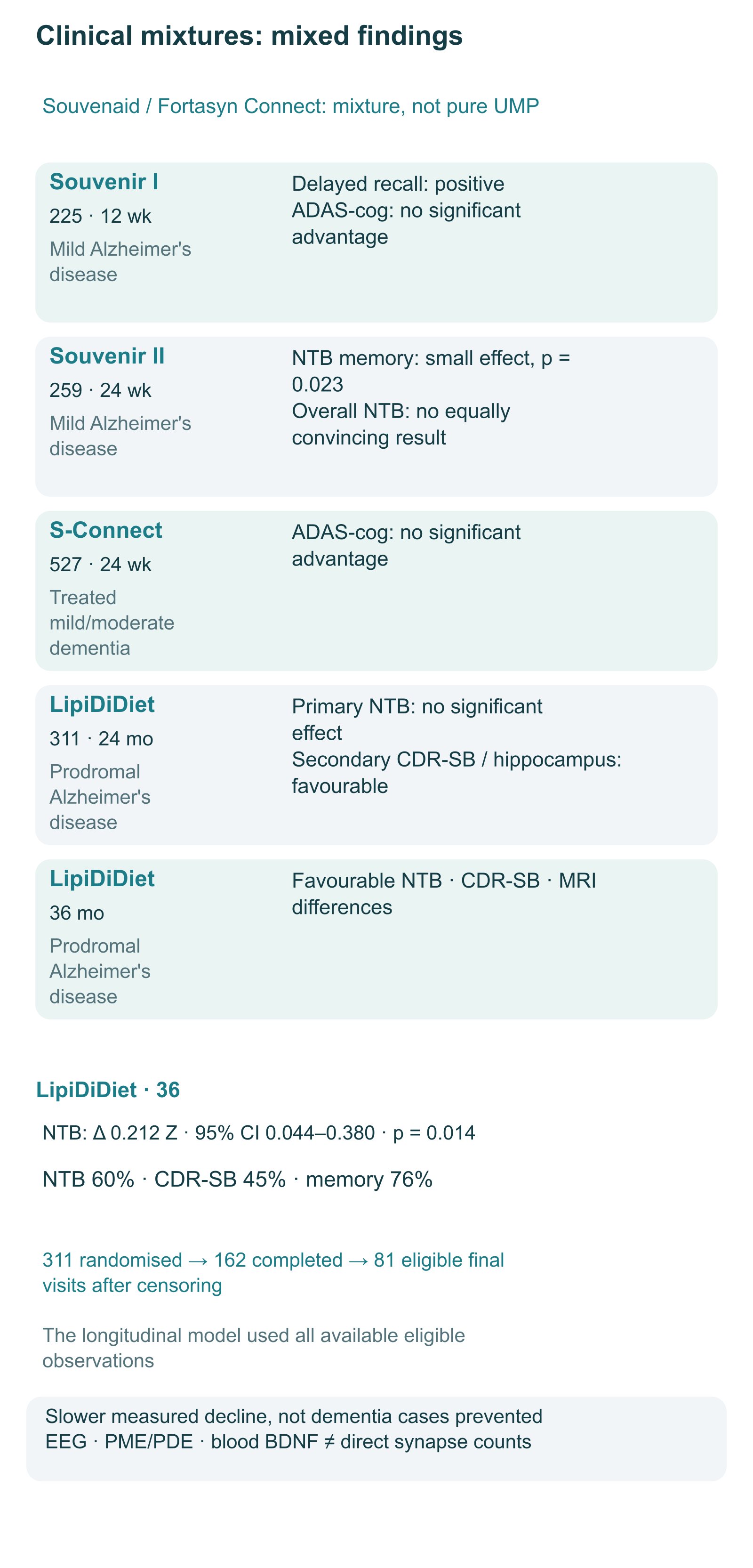
Souvenir I included 225 patients with mild Alzheimer's disease who were not receiving disease-specific medication. After 12 weeks, delayed verbal recall showed a positive result, but the other co-primary endpoint, modified ADAS-cog, showed no advantage. This is a mixed result, not proof of general cognitive restoration.
Souvenir II involved 259 patients over 24 weeks. The NTB memory-domain difference was statistically significant but small; the overall composite NTB did not provide equally convincing evidence. Differences in EEG connectivity, including the delta band, were secondary functional observations. EEG does not directly demonstrate an increase in synapse number, so “proven slowing of synaptic-network breakdown” overstates the findings.
S-Connect investigated 527 patients with mild or moderate dementia already receiving standard medication for 24 weeks. No advantage was found on the primary ADAS-cog endpoint. This negative result limits use of the mixture in that population but does not establish that failure was caused solely by neuron death or a lack of living “targets”. The authors specifically discussed the inability to distinguish insufficient effect at a later stage from lack of additional effect on top of medication.
| Trial | Population and duration | Main finding | Limit of inference |
|---|---|---|---|
| Souvenir I | 225; mild Alzheimer's disease; 12 weeks | Positive delayed recall; no advantage on co-primary ADAS-cog | Primary cognitive outcomes were not uniformly positive |
| Souvenir II | 259; mild Alzheimer's disease; 24 weeks | NTB memory domain: p = 0.023; small effect | EEG and protein hypotheses are not direct evidence of synaptogenesis |
| S-Connect | 527; treated mild/moderate dementia; 24 weeks | Primary ADAS-cog: no significant advantage | An add-on effect was not confirmed in this group |
| LipiDiDiet, 24 months | 311 randomised; prodromal Alzheimer's disease | Primary five-item NTB: no significant effect; selected secondary measures favourable | A selected secondary endpoint cannot replace a negative primary result |
| LipiDiDiet, 36 months | Extension of the same cohort | Favourable differences in five-item NTB, memory, CDR-SB and MRI | Extension, attrition and censoring require caution |
In the 24-month LipiDiDiet analysis, the primary NTB result did not reach statistical significance. Secondary analyses found differences in CDR-SB and hippocampal atrophy, but the rate of dementia diagnosis showed no significant advantage. These results support further investigation rather than establishing dementia prevention.
In the 36-month extension, the mixed model estimated a between-group five-item NTB difference of 0.212 Z units, 95% CI 0.044–0.380, p = 0.014. Its relative reduction in deterioration was 60%; the corresponding figures were 45% for CDR-SB and 76% for memory. Relative differences in rates of change in hippocampal, whole-brain and ventricular volumes were 33%, 22% and 20%, respectively. These estimate changes in selected measures, not percentages of prevented dementia cases or restored neurons.
Of 311 randomised participants, 162 completed 36 months, and final-visit data from 81 met efficacy-analysis conditions after censoring. The longitudinal model used available observations and was not limited to those 81 participants, but attrition and exclusion of data after starting open-label product or medication matter when assessing reliability. Extending the same cohort is not an independent new trial. Differences between disease stages and follow-up durations do not establish a precise universal “therapeutic window”.
A small patient study also examined the metabolic mechanism itself: after four weeks of the mixture, magnetic resonance spectroscopy showed changes in the phosphomonoester-to-phosphodiester ratio, PME/PDE, and choline-containing signals. These are surrogate measures of lipid synthesis and breakdown, not direct measurements of membrane composition or contact number. Thirty-four people were enrolled and 33 analysed; the study informs biological activity of the mixture but does not replace a clinical efficacy trial.
Later research with other nucleotide mixtures likewise cannot be treated as testing pure UMP. For example, a three-group study in physically independent older adults compared a yeast product, a monophosphate-nucleotide mixture and placebo, assessing cognition, physical measures and BDNF. Even with positive changes, that design does not isolate uridine's contribution from other ingredients. Blood BDNF does not directly measure the formation of new neurons or synapses in the human brain, despite “neurogenesis” appearing in the paper's title.
UMP, free uridine and triacetyluridine (TAU, uridine triacetate) differ chemically and pharmacokinetically. TAU's three acetyl groups increase lipophilicity; their removal by esterases produces uridine. Higher exposure does not mean greater cognitive efficacy or justify replacing one form with another without calculation.
| Form and route | What is known | What is not established for cognitive use |
|---|---|---|
| Oral UMP | Used in nutritional mixtures; its route involves dephosphorylation and uridine metabolism | Universal absolute bioavailability of 80%, a single Tmax of 60–90 minutes, and precise brain exposure for every product |
| Sublingual UMP | Marketed in supplements, but the chemical name does not guarantee mucosal absorption | Superiority to oral use, Tmax of 15–25 minutes, and equivalence of a dose reduced two- to threefold |
| Oral free uridine | Investigated pharmacokinetically; high doses can cause diarrhoea | A universal half-life and optimal memory-enhancing dose |
| Oral TAU | In the studied TAU-containing product, uridine Cmax and AUC exceeded those after equimolar free uridine | Interchangeability with UMP and established benefit for a healthy brain |
In a small healthy-volunteer study, a TAU-containing product produced peak uridine at 1–2 hours and approximately fourfold higher Cmax and AUC than equimolar uridine. These parameters belong to a particular formulation and research dose. Cmax is the maximum concentration, AUC the integrated exposure during observation, and Tmax the time of the peak; half-life describes a different part of the curve. Tmax must not be confused with duration of action.
Clinical experience with citicoline further illustrates species specificity: in studied humans, intake increased plasma choline and uridine while cytidine remained very low. This does not mean every choline form necessarily increases uridine: α-GPC and phosphatidylcholine have different chemical structures. Measuring circulating metabolites also does not establish an increase in synapses after citicoline.
There is no established universal UMP regimen for intellectual workload, chronic fatigue, asthenia or “recovery after stimulants”. The popular protocol ranges of 250–300, 500–700 or 600–1000 mg do not become therapeutic doses because they fit a biochemical scheme. The only specific daily UMP exposure in the cognitive trials discussed here characterises a complete multicomponent product. It does not determine the efficacy of a stand-alone capsule, another combination or sublingual use.
Nor is there an established general rule of “a course lasting at least 6–12 months, with effects only after 8–12 weeks”. Studies used different durations, from weeks to years, and different endpoints. Membrane turnover cannot calculate an individual's time to clinical response. For clinicians, the value is discussing a particular studied intervention in a suitable patient, rather than constructing treatment from presumed lipid turnover speed.
Besides the CDP-choline pathway, PC can be formed by methylating PE. The hepatic PEMT pathway is particularly important: PE + 3 SAM → PC + 3 SAH. SAM is S-adenosylmethionine, a methyl-group donor; SAH is S-adenosylhomocysteine, the transfer product. Three successive methylations change the ethanolamine head group into a choline head group rather than adding fatty acids. The hepatic role of this pathway cannot automatically be declared the main route of neuronal PC synthesis.
In the methionine cycle, methionine becomes SAM; donation of a methyl group produces SAH, whose metabolism is linked to homocysteine. B₁₂-dependent methionine synthase MTR uses 5-methyltetrahydrofolate to remethylate homocysteine to methionine. A betaine-dependent BHMT branch also exists in the liver and kidney. Another fate of homocysteine is transsulfuration through CBS and CTH, using B₆ as pyridoxal phosphate, with cystathionine and then cysteine formation. B₆ should not be drawn as a direct cofactor of folate/B₁₂-dependent remethylation.
SAH can inhibit methyltransferase reactions, and its metabolism is linked to homocysteine concentration. Nevertheless, a single homocysteine level of 10–12 μmol/L is not an established threshold that “switches off” phospholipid synthesis. Experimental effects of homocysteine on neurons, receptors and endothelium likewise do not supply such a clinical switch. Correcting an identified B₁₂ or folate deficiency matters independently; it does not establish obligatory synergy between high-dose vitamins and UMP.
Uridine is a pyrimidine, so its base does not follow the purine route to uric acid. Uridine phosphorylase produces uracil and ribose 1-phosphate; DPYD, DPYS and UPB1 then successively transform uracil, dihydrouracil and N-carbamoyl-β-alanine. The final overall step is N-carbamoyl-β-alanine + H₂O → β-alanine + CO₂ + NH₃. At physiological pH, nitrogen is predominantly present as ammonium; its subsequent disposal and the fate of β-alanine are different processes. Thymine produces a different carbon product, β-aminoisobutyrate, so all pyrimidines cannot be assigned one end metabolite.
The absence of direct uric-acid production does not prove supplement safety for everyone with gout. Such a conclusion requires clinical data on the product, comorbidities and medication. “Nontoxic” also does not mean that high metabolite concentrations, impaired hepatic disposal or inherited catabolic defects are irrelevant.
In a mouse study, prolonged uridine administration at 400 mg/kg/day for 16 weeks was associated with liver fat accumulation and impaired glucose tolerance. Insulin signalling, FABP1 and O-GlcNAc modification of FOXO1 were considered; this is not an established universal mechanism through hepatocyte P2Y receptors. The conditions differ from human nutritional-mixture trials, and direct conversion of mg/kg does not establish a human risk threshold. At the same time, one cannot claim adverse effects occur only above 1–2 g/kg or that years of any 250–1000 mg regimen are unquestionably safe.
Tumour-cell dependence on nucleotide metabolism deserves attention but does not by itself establish an absolute uridine contraindication for every cancer history. Fluoropyrimidines provide a more specific situation: uridine triacetate is used as a medicinal antidote for overdose or early severe toxicity from 5-fluorouracil/capecitabine. Timing is critical for the emergency indication, generally within 96 hours after administration ends. This is medical-team treatment and cannot be transferred to ordinary supplemental UMP.
The same ability to oppose toxic metabolites means that unsupervised uridine-product use during anticancer treatment should be discussed with an oncologist. Neither “membrane support” nor the distinction between pyrimidines and purines eliminates drug interactions. For pregnancy, breastfeeding, paediatric cognitive use and severe organ failure, there are insufficient data here to propose a general regimen. Good tolerability of a particular mixture in a trial is not a complete safety profile of pure UMP or TAU in all those groups.
Start with the clinical question rather than choosing a “membrane stack”. Memory complaints call for clarification of onset and progression, everyday functioning, medication with anticholinergic or sedative effects, sleep, mood and diet. Where needed, patients undergo neurological and neuropsychological assessment; suspected prodromal Alzheimer's disease needs the appropriate diagnostic context. Fatigue, forgetfulness and MCI are not interchangeable diagnoses.
Next, compare the exact product with the studied intervention. Chemical form, UMP amount, salt or active-component mass, choline and fatty-acid content, added vitamins, overall nutrition and medication all matter. A complete mixture, a collection of individual supplements and a higher-bioavailability TAU product cannot be considered equivalent. Additional ingredients sometimes duplicate existing products, and metabolic “enhancement” does not justify dose increases without evidence.
| Practical question | What this article provides | What it does not justify |
|---|---|---|
| Why does uridine matter? | Connections between salvage, CTP and lipid metabolism | Diagnosing “brain uridine deficiency” from complaints |
| Who resembles trial participants? | Specific Alzheimer's disease groups and particular mixtures | Treating every MCI, fatigue or stimulant after-effect |
| How should benefit be assessed? | Clinical tests and functioning in an appropriate context | Counting new synapses from subjective feelings or blood BDNF |
| Which laboratory tests are needed? | Clinically indicated investigations, including nutritional problems | A universal UMP-titration panel |
| When is medication caution necessary? | Particular attention to anticancer treatment and product composition | Self-administering UMP as an antidote |
Homocysteine, the omega-3 index and hs-CRP can answer independent clinical questions but are not a validated system for monitoring uridine-associated “synaptogenesis”. Targets of homocysteine <7–8 μmol/L, omega-3 index >8–11% and hs-CRP <1 mg/L cannot be presented as a mandatory UMP protocol. Laboratory decisions follow the person's condition and diagnostic task; normalising one marker does not guarantee a cognitive effect.
When clinician and patient discuss a studied nutritional mixture, expectations should concern measured outcomes and account for effect size and uncertainty. Nutrition, correction of genuine deficiency, sleep, physical activity and vascular risk factors remain independent tasks. A mechanistically plausible supplement does not replace these measures or treatment of an established disease.
Uridine metabolism helps explain the material requirements of plasticity: nucleotide availability connects to lipid synthesis, and extracellular nucleotides can participate in signalling. The most direct membrane and spine data come from experimental systems; human trials mainly concern particular multicomponent interventions and yield mixed findings. This distinction makes biochemical knowledge useful for explaining mechanisms, reading research correctly and discussing clinical possibilities without promising proven brain restoration.
Related topics: choline, the liver and neurotransmitter metabolism, EPA and DHA: metabolism, selection and safety.
No. Direct lipid, protein and dendritic-spine observations mainly concern cell and animal models. Human trials mostly investigated multicomponent mixtures; cognitive tests, EEG and spectroscopy do not directly count new contacts.
Biochemically these components contribute to different parts of lipid metabolism, but clinical equivalence of a homemade combination is not established. The studied product also contained EPA, phospholipids, vitamins and selenium. Replacing the choline form or substituting TAU for UMP changes the intervention.
They are different chemical forms. UMP contains phosphate, free uridine does not, and TAU has three acetyl groups whose removal produces uridine. Pharmacokinetic differences do not establish interchangeability or cognitive superiority.
No universal evidence-based dose has been established for these purposes. UMP 625 mg characterises a serving of one nutritional mixture; animal doses are not human regimens. Complaints following stimulants require assessment of sleep, medication, substance use and neuropsychiatric status.
In the 36-month analysis it is the relative reduction in deterioration of the composite NTB; the corresponding CDR-SB estimate was 45%. It is not the proportion of prevented dementia cases. The 24-month primary result was negative, and the extension involved attrition and censoring.
Correction of an established deficiency has independent indications. It does not prove obligatory high-dose synergy with UMP; B₆ participates in transsulfuration rather than directly in the folate/B₁₂-dependent MTR reaction. Universal laboratory targets for UMP titration are not established.
Self-directed use should be discussed with an oncologist, especially during fluoropyrimidine treatment. Medicinal uridine triacetate provides emergency treatment for particular toxicity; ordinary UMP is not a replacement. A cancer history alone does not support one absolute contraindication for every case.
These measures can have separate clinical importance but do not measure uridine-associated synaptogenesis or form a validated UMP-monitoring panel. Testing follows the diagnostic question and the patient’s condition.