Taurine in the CNS: Astrocytes, VRAC, GlyR, and tRNA — NutriFit
Taurine in the Brain: Astrocyte Osmoregulation, Receptors, and Mitochondria
How taurine moves between astrocytes and neurons, contributes to cell-volume regulation, and modifies mitochondrial tRNA. Where the biochemistry is established and where clinical promises still outpace the evidence.
Taurine is useful to discuss at the intersection of two processes that are usually considered separately. When an astrocyte swells, it releases some taurine along with other osmotically active substances to restore its volume. Outside the cell, the same molecule can interact with neuronal receptors. This is an intriguing link between water balance and inhibitory signalling, but it does not mean that taking taurine will stop brain swelling or an epileptic seizure.
Taurine is 2-aminoethanesulfonic acid. It is not incorporated into ordinary proteins, but it is present in many tissues as a free molecule. In the brain, its intracellular concentration is much higher than its concentration in interstitial fluid; this reserve is maintained by synthesis, the TauT transporter, and cellular retention. We will follow the molecule from sulfur-containing amino acids to the astrocyte, receptor, and mitochondrial tRNA, then consider which of these mechanisms have actually been studied in people.
A molecule cells can accumulate
Taurine has the formula C₂H₇NO₃S and a molar mass of about 125.15 g/mol. Unlike α-amino acids, its amino group is attached to the second carbon from the acidic group, and it has a sulfonate instead of a carboxylate. At physiological pH, the zwitterion H₃N⁺–CH₂–CH₂–SO₃⁻ predominates: the amino group is positively charged and the sulfonate is negatively charged. This polar molecule dissolves readily in water and crosses lipid membranes poorly by simple diffusion. Its movement is therefore governed by transporters and channels, not just by the concentration on either side of a membrane.
This combination of properties matters for osmoregulation. Taurine can contribute to the number of dissolved particles in the cytosol without replacing ions involved in enzyme function or disrupting protein structure as sharply as some other solutes. But being a “compatible osmolyte” does not mean the supply is unlimited: its amount depends on intake, synthesis, reuptake, and renal reabsorption.
How taurine is made and delivered
The main pathway starts with sulfur-containing amino acids. Through the methylation cycle, methionine gives rise to homocysteine; the transsulfuration pathway, involving cystathionine β-synthase (CBS) and cystathionine γ-lyase (CSE), leads to cysteine. Cysteine dioxygenase (CDO) adds oxygen to cysteine sulfur to form cysteine sulfinic acid. Cysteine sulfinic acid decarboxylase (CSAD), which requires pyridoxal 5′-phosphate—the active form of vitamin B₆—then removes CO₂ to produce hypotaurine. Oxidation of hypotaurine completes taurine synthesis:
There is also an additional branch: coenzyme A breakdown produces pantetheine and then cysteamine; cysteamine dioxygenase (ADO) oxidizes it to hypotaurine. The contribution of these pathways differs across tissues and species. CSAD activity is lower in humans than in many animal models used in experiments, but this does not by itself establish the exact shares of dietary and endogenous taurine or prove that taurine is unconditionally essential for every adult. Intake and metabolism are especially important to assess in preterm infants and in particular nutritional states.
Taurine is found mainly in animal-derived foods, including fish and seafood; plant foods generally provide little. However, the presence of taurine in the diet does not show how much reaches the brain. TauT, encoded by SLC6A6, takes up taurine together with Na⁺ and Cl⁻; the commonly described stoichiometry is 2 Na⁺ : 1 Cl⁻ : 1 taurine. Na⁺/K⁺-ATPase activity maintains the sodium gradient that makes accumulation possible. Hypotaurine and β-alanine compete for the transporter. During hypertonic stress, the NFAT5 transcription factor promotes expression of osmolyte-accumulation genes, including SLC6A6; regulation of TauT positioning in the membrane also changes uptake rate.
The blood–brain barrier makes systemic and local pools non-equivalent. TauT is found in brain capillary endothelium and neural tissue cells, but transport is limited and regulated on both sides of the barrier. Therefore, a rise in plasma taurine after taking a supplement cannot be equated with the same rise in an astrocyte or a specific neuronal ensemble.
There is an important biochemical fork in this chain. Cysteine sulfinic acid does not have to become taurine: it can enter other pathways of sulfur metabolism. CDO activity alone therefore does not determine final taurine output; substrate availability, PLP-dependent CSAD, and tissue state matter. Hypotaurine is not only an intermediate: its oxidation can accompany reactions with oxidants. But it would be inaccurate to turn this observation into a blanket claim that dietary taurine directly “traps all free radicals” in the brain.
Where the reserve is located
Concentrations depend on age, species, brain region, and measurement method. Experimental studies describe higher content in some regions of the cerebellum and olfactory system, and different distributions in the cortex, striatum, hippocampus, brainstem, and spinal cord. Comparing micromoles per gram of wet tissue with millimoles per litre of intracellular water requires knowing the cellular composition; it does not yield a simple table of universal human “normal” values.
Compartment
What is measured
How to interpret the result
Astrocytes and neurons
Intracellular free taurine, often in the millimolar range in experimental systems
A large reserve for osmotic responses; the value varies by cell, age, and state
Interstitial fluid
A much smaller, usually micromolar pool
Even a small release from the cellular reserve can change the local concentration
Plasma and BBB endothelium
Substrate available to TauT
The blood level is not a direct measure of the concentration in the brain
The regional pattern is interesting beyond average levels. In the cerebellar cortex, taurine is found in Purkinje cells and granule cells; in the olfactory system, in mitral cells and interneurons. The striatum, hippocampus, sensory cortex, brainstem, and spinal cord differ in glial proportions, neuronal maturity, and inhibitory-circuit composition. Tabulated ranges for each region should not be transferred into a clinical reference guide: comparative measurements from tissue samples across species and methods cannot be treated as a single standard for human blood, MR spectroscopy, or cerebrospinal fluid.
TauT is not the only regulator of the cellular pool. If the surrounding environment becomes hypertonic, NFAT5 helps increase the accumulation of organic osmolytes to prevent cellular dehydration; during swelling, the priority shifts to their release. Phosphorylation and movement of the transporter between the membrane and intracellular compartments can alter uptake rate, although the specific effects of PKC and PKA depend on the cell system studied. Even commonly cited TauT affinity values cannot automatically support the conclusion that the transporter is “always saturated” at usual blood concentrations: transport rate also depends on the ion gradient, transporter density, direction of transport, and taurine availability on each side of the BBB.
The difference between intracellular and extracellular environments explains why taurine can rapidly leave a swelling cell down its gradient through an available pathway. It also explains a limitation of dietary intervention: a rise in blood taurine must still cross the barrier and be taken up by the relevant cell.
What happens when an astrocyte swells
Astrocytes wrap their endfeet around brain capillaries. The water channel AQP4 is concentrated in these membranes, so osmotic changes are quickly reflected in cell volume. Within the rigid cranial cavity, the combined volumes of tissue, blood, and cerebrospinal fluid are limited; substantial tissue swelling can dangerously raise intracranial pressure. This clinical danger is real, but findings from an isolated astrocyte do not prove that taurine supplementation prevents herniation or preserves perfusion in a patient.
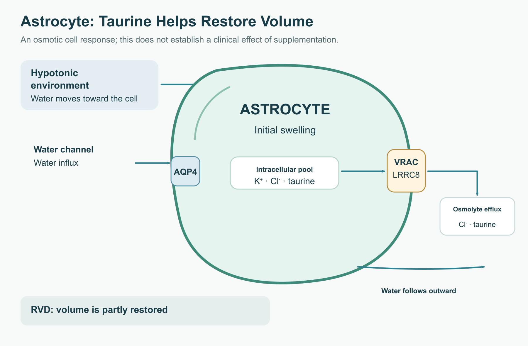
When the extracellular environment becomes less concentrated, water enters the astrocyte. The cell responds with regulatory volume decrease (RVD): it releases K⁺ and Cl⁻ as well as organic osmolytes, including taurine. The loss of dissolved particles reduces the osmotic pull of water into the cytosol; if the cell's adaptive capacity is intact, some water leaves and cell volume decreases. This does not “switch off” edema: the outcome depends on the cause, duration, and severity of the disturbance and on the state of ion pumps and neighbouring cells.
Astrocyte endfeet containing AQP4 lie next to capillaries, but water moves down an osmotic gradient; AQP4 does not “pump” it. A fall in plasma sodium is one example of a hypotonic disturbance in the source literature. Sodium concentration alone, however, does not determine the rate or severity of cerebral edema: osmolality, duration, cellular adaptation, and other dissolved substances matter. During chronic hyponatremia, cells gradually lose osmolytes; restoring extracellular osmolality too quickly risks a different mechanism of injury. This transition shows why RVD is not a simple instruction to “add taurine.”
One route for taurine release is linked to the volume-regulated anion channel VRAC. Its obligatory subunit LRRC8A forms heteromers with other proteins in the LRRC8 family; LRRC8D is particularly important for the permeability of a range of organic compounds. Channel architecture and substrate selectivity depend on the heteromer composition, and opening is linked not only to membrane stretch but also to changes in intracellular ionic strength and cell signalling. Different LRRC8C, LRRC8D, and LRRC8E subunits alter heteromer permeability; highlighting LRRC8D's role for organic compounds does not mean that every channel in every astrocyte has the same composition. Electrophysiological characterization of VRAC as an anion current and observation of efflux of a molecule that is net-neutral as a zwitterion describe different properties of a permeable pore. Potassium channels, including the studied BK/IK and KCNQ channels, and chloride-efflux pathways also contribute to volume loss. Ionic currents maintain electroneutrality, while a reduction in the total number of particles creates the driving force for water movement.
In experiments, taurine loss from a swollen cell can occur faster than TauT-mediated reuptake. Once conditions recover, the cell must replenish its osmolyte pool.
Ischemia is more complex than hypotonic stress. ATP depletion weakens Na⁺/K⁺-ATPase and calcium pumps; Na⁺ and other ions accumulate in the cell, followed by water. Swollen astrocytic processes can narrow extracellular space and affect microcirculation. In vasogenic edema, by contrast, the barrier is disrupted and fluid with plasma components leaves the vessels. These mechanisms often coexist in stroke or trauma. The post-ischemic microcirculatory no-reflow phenomenon has several contributors: cell swelling, endothelial injury, pericyte changes, and microthrombi. Astrocyte involvement does not prove that an adequate taurine reserve will preserve capillary patency or stop the penumbra from progressing to infarction. Brain trauma and subarachnoid hemorrhage also involve vascular, inflammatory, and mechanical factors. Preserving extracellular space may facilitate diffusion, but the clinical importance of this one pathway must be tested against patient outcomes.
Experimental evidence that taurine participates in RVD cannot replace sodium management, reperfusion, intracranial pressure control, or other intensive care measures.
From osmolyte to signal between cells
Release from an astrocyte creates a local opportunity for taurine to act on neuronal receptors. Taurine resembles glycine and GABA in the combination of a positively charged amino group and a negatively charged acidic end. This structural similarity does not make it a full substitute for either transmitter: the response depends on the concentration at the receptor, channel subunit composition, tissue age, and the neuron's chloride gradient.
Comparing pharmacophores helps explain the research hypothesis: glycine is written as H₃N⁺–CH₂–COO⁻, taurine as H₃N⁺–CH₂–CH₂–SO₃⁻, and GABA as H₃N⁺–(CH₂)₃–COO⁻. The distance and orientation of charged groups matter, but a similar two-dimensional drawing does not establish equal agonist efficacy or the concentration reached at a receptor in vivo.
Glycine receptors (GlyR) are pentameric channels that primarily conduct Cl⁻. They are especially important for inhibitory transmission in the spinal cord and brainstem; extrasynaptic pools also occur in other regions. Taurine can activate GlyR, while strychnine blocks the corresponding receptor component in experimental systems. The GlyR family includes several α subunits and a β subunit. Adult and developing tissues differ in their subunit composition, and receptor distribution also varies—from synapses of spinal motor neurons and interneurons to extrasynaptic receptors in certain forebrain regions. A concentration–response curve measured in a culture or at a recombinant receptor therefore cannot be treated as a norm for all neurons. Pharmacological separation also matters: strychnine identifies a GlyR component of a response, whereas bicuculline blockade points to certain GABA_A components; the lack of an effect from one antagonist does not mean that receptors do not interact in a living network.
Gephyrin anchors synaptic receptors in a complex with the cytoskeleton. But the electrical effect cannot be determined by the single phrase “Cl⁻ enters, so the membrane hyperpolarizes”: it depends on the Cl⁻ equilibrium potential. In a mature network, opening the channel often hyperpolarizes the cell or shunts excitatory current; the chloride gradient may differ in developing or damaged tissue.
Ionotropic GABA_A receptors also conduct Cl⁻. Classical GABA_A receptors assemble from several subunit types; combinations of α, β, γ, or δ subunits alter their location, kinetics, and pharmacology. Extrasynaptic α4/α6-containing variants with a δ subunit have been described as supporting a prolonged tonic current. This differs from the brief channel opening of phasic synaptic inhibition. The thalamus, dentate gyrus, and cerebellar granule cells have been studied in different reports; they should not be treated as one uniform receptor pool.
Taurine activates some GABA_A variants in experiments, including extrasynaptic thalamic receptors that contain the δ subunit. These receptors support a prolonged tonic current, unlike the brief synaptic response. Direct binding of taurine as an agonist at a high local concentration and proposed allosteric enhancement of GABA action are different hypotheses. Release from glia could create brief local concentrations above the interstitial-fluid average, but measurements of the microenvironment are needed to compare them with a response curve for a specific receptor. An experimental estimate of affinity should not be turned into a required “therapeutic concentration” after an oral dose.
Results from specific subunits cannot establish one universal affinity, obligatory allosteric potentiation of all GABA_A receptors, or the same effect throughout the brain. Evidence for interaction with metabotropic GABA_B receptors is less clear; the canonical GABA_B pathway itself involves Gi/o, reduced cAMP, opening of GIRK channels, and suppression of presynaptic Ca²⁺ entry. Direct activation of this pathway by taurine should be considered an experimental hypothesis, not an established clinical effect.
Why discuss this in relation to epilepsy? In an excitable network, repeated depolarization and cell synchrony can produce a paroxysmal depolarization shift followed by a series of action potentials. Opening Cl⁻ channels lowers membrane input resistance: for the same excitatory current, the voltage change is smaller, as expressed by ΔV = I × Rᵢₙ. When conditions allow, this makes it harder to reach the Naᵥ-channel threshold and propagate a discharge. GABA_B receptors are fundamentally different from GlyR and GABA_A: rather than forming a chloride pore, they initiate a G-protein cascade. Their postsynaptic branch can open GIRK channels and produce slow hyperpolarization through K⁺; the presynaptic branch reduces calcium entry and transmitter release. These well-described effects of GABA_B activation do not establish that taurine ordinarily reaches the receptor at an effective concentration. For GlyR and GABA_A, primary shunting can also reduce EPSP amplitude without necessarily driving the membrane toward the illustrative value of −75 mV shown in the original electrophysiology diagram.
Possible changes in glutamate release, GABA turnover, and GAD65/67 activity are studied separately; without additional measurements, they cannot be combined into a guaranteed “antiseizure package” for a patient.
Seizure models using chemical convulsants and kindling can indeed test seizure latency and severity after taurine administration. However, animal doses, routes of administration, and network properties are not equivalent to taking a supplement in humans. Small, older clinical observations are heterogeneous and do not confirm sustained efficacy as monotherapy or as an add-on to modern treatment for refractory epilepsy. Reports of a temporary effect cannot confidently be explained solely by reduced TauT without direct transporter measurements. These mechanisms are not a basis for changing antiseizure treatment.
Mitochondria: two distinct stories about taurine
The first concerns calcium overload. During ischemia and strong neuronal excitation, Ca²⁺ enters through NMDA receptors and voltage-gated channels, including L-type channels. Mitochondria take up some Ca²⁺ through the MCU uniporter. If energy is also scarce and oxidative stress rises, loss of membrane potential and a shift toward high inner-membrane permeability amplify injury. Calcium activates calpains, which cleave cytoskeletal proteins such as spectrin. In severe injury, cytochrome c release is linked to the cell-death programme.
In a resting cell, free cytosolic Ca²⁺ is maintained far below its extracellular level. Even brief opening of NMDA receptors or Caᵥ channels can therefore sharply change the intracellular signal. During energy failure, Na⁺/K⁺-ATPase is less able to maintain the sodium gradient, and NCX may run in reverse, adding Ca²⁺ to the cytosol. PMCA requires ATP to remove Ca²⁺. Mitochondria temporarily take on part of the load through MCU, but the cost of this buffering rises as the energy reserve is depleted. The combination of Ca²⁺, reactive oxygen species, and a fall in ΔΨₘ creates conditions for the membrane to shift to high permeability; collapse of the ion gradient impairs ATP synthesis.
Calpains are calcium-dependent proteases, not a distinct “taurine programme.” Their activation during injury alters spectrin, microtubule proteins, and other structures in dendrites and axons. At the same time, the balance of anti-apoptotic and pro-apoptotic Bcl-2-family proteins affects mitochondrial outer-membrane integrity and cytochrome c release. These branches converge in the injured cell, but they are measured separately in experiments; improvement in one marker does not prove that neuronal death has been prevented in humans.
In cell and animal models, taurine has been associated with a smaller rise in cytosolic Ca²⁺, preservation of mitochondrial potential, and less calpain activation. The sodium–calcium exchanger NCX, calcium pumps PMCA, and Bcl-2- and Bax-family proteins are discussed. For NCX, it is important to distinguish its forward mode, which extrudes Ca²⁺ from the cell, from reverse mode under a changed sodium gradient: the usual exchange moves three Na⁺ ions for one Ca²⁺ ion. These experiments do not show that taurine directly “closes” NMDA channels, always switches NCX mode, or demonstrably blocks the mPTP in humans. The molecular composition of the mPTP itself remains under discussion; cyclophilin D is a known regulator of pore sensitivity, not grounds for describing one definitive structure.
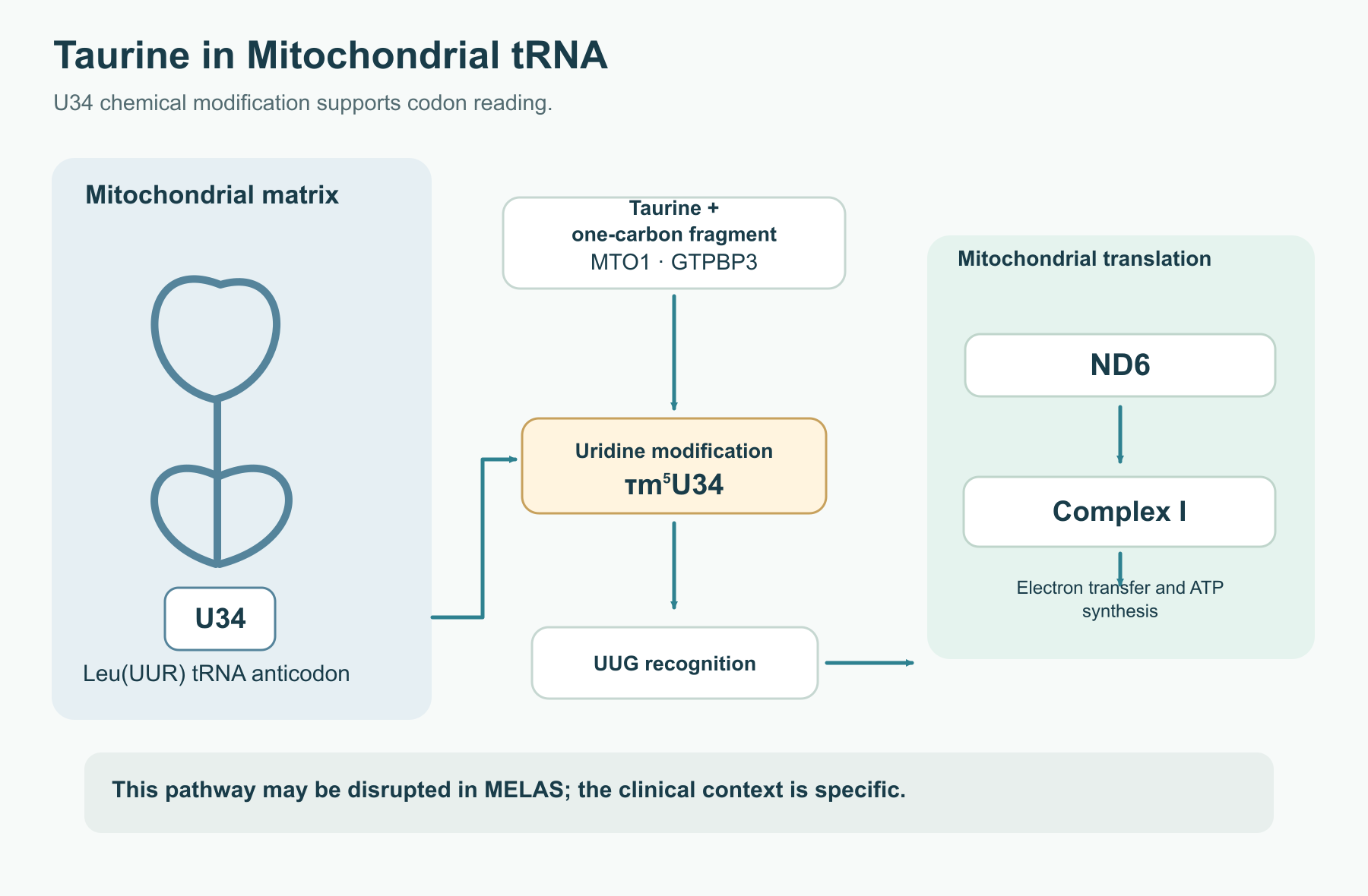
The second story is much more specific biochemically: taurine participates in modifying uridine in the anticodon of certain mitochondrial tRNAs. At position U34, tRNA^Leu(UUR) is modified to 5-taurinomethyluridine (τm⁵U); 5-taurinomethyl-2-thiouridine (τm⁵s²U) has been described in tRNA^Lys. The proteins GTPBP3 and MTO1 participate in the process, and the one-carbon fragment is linked to folate metabolism. It is important not to conflate two kinds of chemistry here. Taurine remains free in the cytosol as an osmolyte; in tRNA, it becomes part of a covalently modified nucleoside. “Wobble” refers to the third codon position recognized by anticodon U34. Changing the chemical group at U34 can improve recognition of particular codons, affecting the speed and accuracy of translation of polypeptides encoded by the mitochondrial genome. Codon UUG is particularly discussed for tRNA^Leu; other taurine-dependent tRNAs have a different context. It would be inaccurate to claim that one unmodified nucleoside completely stops synthesis of all Complex I in every cell.
The modification helps recognize the relevant codons and supports translation of proteins made by mitochondria themselves. The mitochondrial genome encodes 13 polypeptides: seven for Complex I (ND1–ND6 and ND4L), cytochrome b for Complex III, COX1–3 for Complex IV, and ATP6/ATP8 for Complex V.
The m.3243A>G mutation in the mitochondrial gene for tRNA^Leu is often associated with MELAS and impaired wobble-uridine modification. Studies link it to less efficient reading of UUG, including during synthesis of the ND6 subunit, and to Complex I dysfunction. The degree of impairment cannot be reduced to one fixed percentage of ATP loss: the fraction of mutant mitochondrial DNA copies, tissue, and age all matter. The mutation affects tRNA structure; it does not simply “lock out” one taurine molecule. The fraction of mutant mitochondrial genomes in a cell—heteroplasmy—helps explain why tissue manifestations of MELAS vary so much. Complex I receives seven subunits from the mitochondrial genome, while other respiratory-chain components are partly encoded in the nucleus. The U34 → UUG reading → ND6 → Complex I link is therefore informative as a specific pathway, not as the sole cause of every stroke-like episode.
An open-label, multicentre phase III study tested high-dose taurine to prevent stroke-like episodes in MELAS. This is a specific clinical setting with its own protocol, not evidence of universal neuroprotection or of the effectiveness of ordinary supplements for every brain disorder.
Where the model ends and clinical evidence begins
Area
What has been shown
What cannot yet be concluded
Epilepsy
Receptor and network effects; antiseizure results in some animal models; limited and heterogeneous observations in people
That taurine replaces antiseizure medicines or reliably lowers seizure frequency with current treatment
Ischemia and trauma
Cellular osmoregulation, calcium and mitochondrial markers; neuroprotection in several experimental models
That oral intake prevents edema, reduces a stroke lesion, or restores cerebral blood flow in people
Diabetic encephalopathy
Experimental links with oxidative stress, inflammation, and altered metabolism
That taurine treats cognitive impairment in diabetes
Tinnitus
Reduced symptoms in an animal model; mechanistic interest in inhibition along auditory pathways
That a similar effect has been established in people
MELAS
An open-label clinical study in a specific mitochondrial syndrome
That its results apply to normal ageing or other CNS disorders
Diabetic encephalopathy is discussed in relation to chronic hyperglycaemia, vascular changes, inflammation, and altered oxidative metabolism. In experimental tissue, taurine may affect some of these measures, but “encephalopathy” covers different cognitive and neurological phenotypes. Without a defined outcome, control group, and clinical sample, it is not possible to assess whether the intervention changes memory or everyday functioning in patients.
In ischemia, inflammation, endothelial injury, and reperfusion stress occur alongside cytotoxic edema. Animal studies have examined lesion size, blood–brain barrier integrity, activity of antioxidant pathways including Nrf2/HO-1, and neuronal death. These measures explain the research interest but do not justify prescribing taurine instead of established care for acute stroke. The same boundary applies to trauma and hyponatremia: cellular osmotic behaviour is one part of pathophysiology, while clinical intervention must take the whole body and the rate of sodium correction into account.
Similar caution is needed when discussing “adjunctive” epilepsy treatment. Valproate, levetiracetam and brivaracetam, and lamotrigine act through different targets; regimens are selected according to seizure type, tolerability, and interactions. Theoretical complementarity between GlyR/GABA_A and SV2A or sodium channels is not clinical evidence of synergy. Cell experiments suggesting protection against valproate toxicity also do not prove prevention of medication complications in people. Taurine should not be described as a way to avoid increasing the dose of an antiseizure medicine.
Tinnitus particularly illustrates the risk of translating a model too directly. After cochlear injury, auditory pathways may change their signal gain: spontaneous activity and reduced inhibition are studied in the dorsal cochlear nucleus, inferior colliculus, and auditory cortex. In the study by Brozoski and colleagues, taurine supplementation reduced tinnitus-like signs in animals. But subjective tinnitus in people is heterogeneous; peripheral injury, central adaptation, and sound perception are intertwined. This model alone does not establish a human dose or “restoration of GlyR in the auditory cortex.”
Intake, doses, and caution
Supplement composition and delivery route matter: free taurine, a food matrix, and a combination product do not necessarily produce the same concentration curve. Intestinal uptake and tubular reabsorption use transporters; at high intake, the fraction of taurine excreted in urine may change. Hepatic CYP450 is not a main route of taurine metabolism, but that does not mean pharmacodynamic interactions are absent. Individual bioavailability and Tₘₐₓ values reported in source studies should not be treated as properties of every product form without considering the study conditions.
Taurine is absorbed in the intestine, appears in blood, and is excreted mainly by the kidneys with regulated tubular reabsorption. A plasma peak after a single dose is not the same as the duration of changes in the brain: transport across the BBB is saturable and depends on tissue state. Findings on time to peak, bioavailability, or half-life from individual studies should not be turned into universal parameters for every formulation and patient. Nor should one assume that barrier damage always improves delivery to the place where taurine is needed.
The source literature describes experimental doses and a specific therapeutic regimen for MELAS, but there are no evidence-based standard dosing regimens for “edema prevention,” epilepsy, post-traumatic conditions, cognitive “homeostasis,” or tinnitus. The table below therefore separates contexts; it does not offer prescriptions.
Context
Appropriate practical interpretation
Usual diet
Taurine intake depends on diet; low levels in plant foods alone do not prove a neurological deficiency
Dietary supplements
Studies have used different doses and durations; a laboratory receptor effect does not determine an effective oral dose
Epilepsy, stroke, TBI, hyponatremia
Self-treatment and replacing prescribed therapy are not appropriate; no clinical regimen is established from VRAC or GlyR mechanisms
MELAS
The high-dose regimen belongs to specialist care and does not transfer to other conditions
Magnesium participates in voltage-dependent blockade of the NMDA receptor, and pyridoxal 5′-phosphate is required by CSAD and GAD enzymes. These are real biochemical links, but they do not prove that combining magnesium, vitamin B₆, and taurine enhances seizure treatment or prevents ischemic injury. L-theanine is studied separately; it would be unjustified to claim that it reliably blocks AMPA/kainate receptors in people or has clinical “synergy” with taurine. Excess vitamin B₆ can cause neuropathy, making the automatic addition of high PLP doses to taurine especially inappropriate.
Historical estimates of safe intake and high-dose studies in MELAS address different questions. Neither automatically establishes an “ideal profile” for lifelong self-directed use or treatment of edema. Contraindications and limits should account for age, pregnancy, kidney function, and coexisting conditions; evidence is limited for some groups. Combinations with drinks containing caffeine and other ingredients cannot be assessed by considering taurine alone.
Taurine has been generally well tolerated under the conditions studied, but observations in a limited number of participants over limited periods do not establish lifelong safety at every dose. Kidney function, coexisting illnesses, and medicines require individual assessment. There is no established rule that taurine necessarily potentiates valproate, carbamazepine, levetiracetam, lamotrigine, or benzodiazepines. With lithium, changes in fluid and electrolyte balance matter for monitoring drug concentration; with hypotension or antihypertensive treatment, blood pressure and symptoms should be considered. Compatibility with all medicines cannot be claimed merely because taurine is not a typical CYP450 substrate.
Taurine is an unusual example of a molecule linking cell volume, receptor physiology, and a precise chemical modification of mitochondrial tRNA. The most definite facts concern its molecular structure, TauT, its role in osmoregulation, and taurine-dependent tRNA nucleosides. Effects on seizure networks, edema, or auditory pathways remain important areas of research. For most neurological conditions, the clinical question is still open: a mechanism explains what to investigate, but it does not replace testing benefits and safety in patients.
How this review was prepared
This is a narrative review of taurine in the central nervous system, based on a literature collection assembled earlier. Editorial work was completed on 4 October 2026. We did not conduct a systematic search under a predefined protocol, a meta-analysis, or a formal risk-of-bias assessment for each study; the collection does not claim to cover the literature exhaustively.
The explanations of osmoregulation, receptor effects, and mitochondrial metabolism are kept separate from clinical conclusions. Species, age, tissue, route of administration, and measurement method matter when interpreting quantitative results: animal measures cannot be transferred to humans as diagnostic reference values. A cellular protective effect or a biologically plausible mechanism alone does not establish that a supplement benefits a neurological disease. Assessing use requires evidence for the specific formulation, population, and clinical outcome.\n
FAQ
Why does taurine leave a swollen astrocyte?
In response to osmotic changes, the cell releases ions and organic osmolytes, including taurine. Reducing the number of dissolved particles helps water leave and cell volume recover.
Does taurine help with epilepsy?
Receptor effects and animal-model findings are of research interest. There is no established modern clinical regimen of taurine for epilepsy, and it should not replace prescribed medicines.
Can taurine be used for tinnitus?
A result in an animal tinnitus model cannot be transferred to people. Efficacy and a dosing regimen for this purpose have not been established.
How is taurine related to MELAS?
Taurine participates in mitochondrial tRNA modification, which may be disrupted in MELAS. A high-dose regimen has been studied in a specialist open-label clinical trial and cannot be extrapolated to other diseases.
Does a high blood taurine level mean a high level in the brain?
No. Passage across the blood–brain barrier and cellular uptake depend on regulated, limited TauT transport.