L-theanine: alpha rhythms, glutamate, stress, and caffeine
An in-depth examination of L-theanine chemistry, transport, synaptic models, and human trials: what is known about attention, anxiety, sleep, dosing, and safety, and which mechanisms cannot yet be translated into clinical practice.
L-theanine is often described as an aid to “calm concentration”: a person supposedly reacts less strongly to stress while retaining mental clarity. There is an experimental basis for this description, but it combines different phenomena—subjective tension, task performance, cortical electrical activity, and sleep. A change in one does not automatically predict changes in the others. Understanding theanine’s place in practice requires following the entire path from chemical structure and transport to the results of specific trials.
Theanine is indeed interesting as a small molecule resembling ordinary amino acids without being incorporated into proteins during ribosomal synthesis. This property connects it with amino acid transporters and research on glutamatergic transmission, while its presence in tea connects it with familiar caffeine exposure. Tea, isolated L-theanine, and a combination of the two substances nevertheless constitute different interventions. This article examines each of these levels, including situations in which a physiologically plausible explanation still runs ahead of clinical evidence.
Chemical structure: resemblance to glutamate does not mean identical effects
What the molecule contains
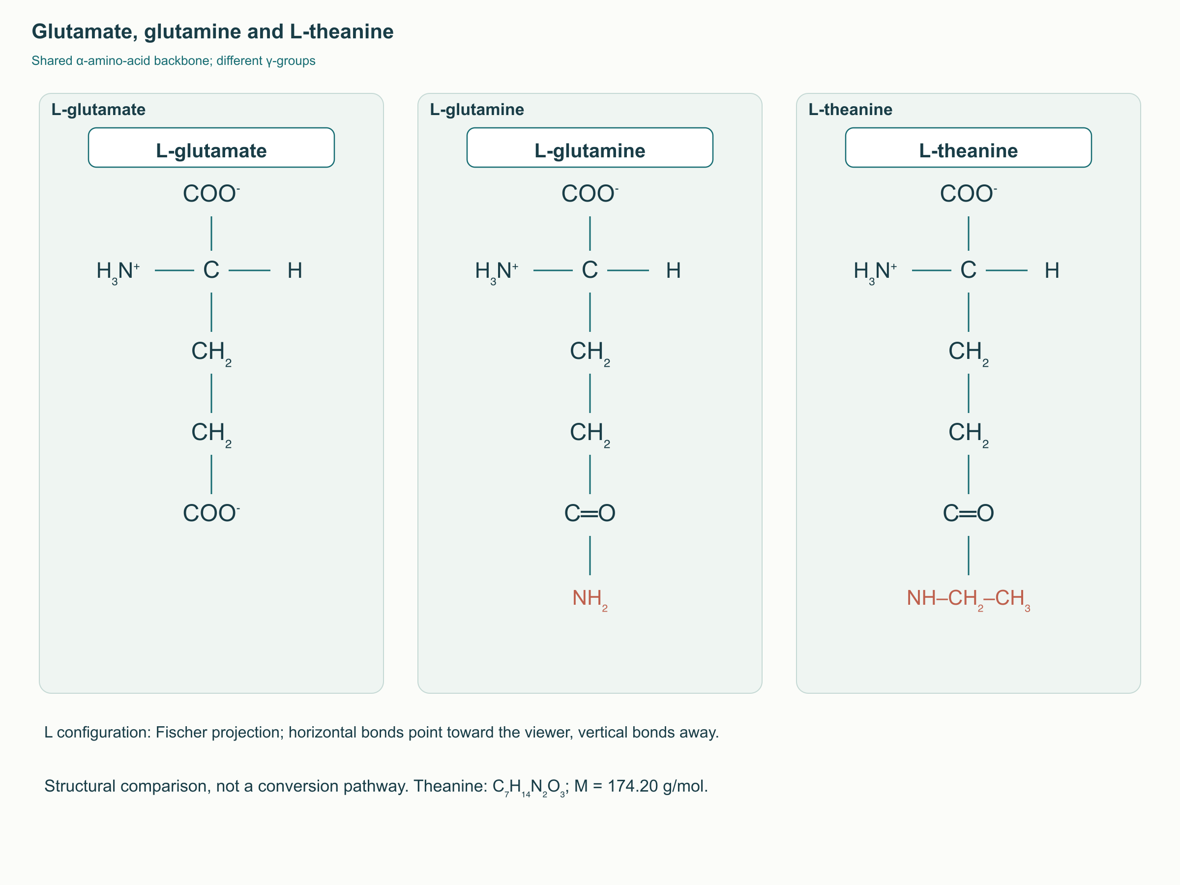
L-theanine is γ-glutamylethylamide, also called N-ethyl-L-glutamine, with the formula C₇H₁₄N₂O₃ and a molar mass of 174.20 g/mol. Comparing it with glutamate and glutamine is useful: all three retain an amino acid backbone, but the far end of the side chain differs. In glutamate it is a carboxyl group, in glutamine an amide group, and in theanine an ethyl group is attached to the amide nitrogen. This small change affects side-chain charge and protein recognition, but does not by itself indicate whether the substance will activate or inhibit a receptor.
The main dietary source is Camellia sinensis leaves; tea beverages contain theanine alongside caffeine and numerous polyphenols. The amount in a cup depends on the variety, processing, quantity of leaf, and brewing, so the mass of dry leaves cannot be directly converted into a supplement dose. Attention studies generally used fixed amounts of purified substance, whereas tea is a variable mixture. The tens of milligrams per cup reported in the literature provide a reference for a particular beverage rather than a universal property of every tea.
Spatial configuration and solubility
The letter L indicates the molecule’s spatial configuration, not its purity or the product’s “strength.” The spatial arrangement of groups does matter to transport proteins and enzymes, so results obtained with the L-form should not automatically be extended to the D-form or an isomer mixture. However, there is no precise human rule establishing a tenfold difference in brain penetration between these forms. In practice, the composition of the studied and purchased product needs to be known, rather than assigning identical pharmacokinetics to anything labelled “theanine.”
At physiological pH, theanine’s amino group is predominantly protonated and its free carboxyl group deprotonated. This produces a zwitterion: the molecule carries both positive and negative charges although its net charge is close to zero. The ethyl fragment does not turn this structure into a fat-soluble substance that freely crosses every membrane. Discussion of central effects should therefore begin with transporters rather than the claim that theanine “readily dissolves in brain lipids.”
Protein binding depends not only on molecular size but also on the arrangement of hydrogen-bond donors and acceptors, local charge, and the conformational flexibility of the side chain. Structural similarity can permit transporter recognition while making the substance a weak competitor of the natural substrate. It does not prove that theanine becomes GABA or occupies all glutamate receptors at an ordinary dose. Distinguishing chemical similarity from functional outcomes is particularly important when interpreting cell experiments using high concentrations.
Absorption, transport across barriers, and metabolism
From the intestinal lumen to plasma
After oral administration, theanine appears in plasma relatively quickly, but its specific route through a human enterocyte is less well studied than its concentration–time profile. Cell experiments identified a contribution from system L—neutral amino acid transporters sensitive to competition with leucine and the experimental inhibitor BCH. Direct transport measurements confirmed LAT1 and LAT2 involvement in particular model systems. This supports a carrier-mediated mechanism but does not establish a universal sequence of “B⁰AT1 on entry, LAT2 on exit” in humans.
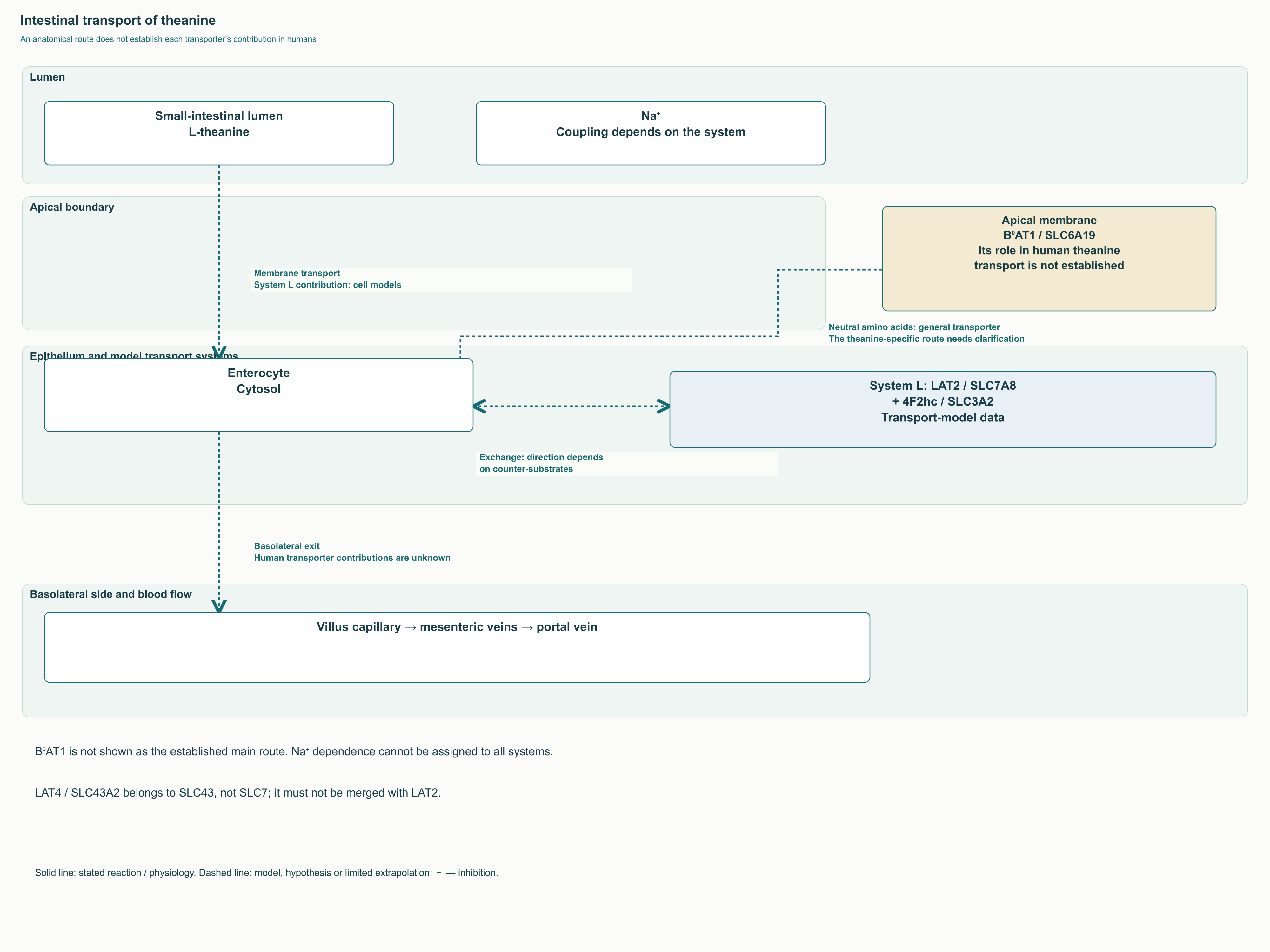
In intestinal physiology, B⁰AT1/SLC6A19 does transport neutral amino acids at the apical surface, while LAT2/SLC7A8, together with the heavy chain 4F2hc/SLC3A2, participates in amino acid exchange across the membrane. LAT4/SLC43A2 belongs to a different family and should not be identified as an SLC7 member. However, the presence of these proteins in an enterocyte does not establish each one’s contribution specifically to theanine transport. The intestinal diagram therefore distinguishes established anatomical compartments, findings from model experiments, and transporters whose involvement in human pharmacokinetics remains to be clarified.
The most useful human study compared the same 100 mg dose in a capsule and a tea beverage in 12 healthy participants using a crossover design. Peak plasma concentration occurred at approximately 0.8 hours; Cmax was 24.3 ± 5.7 μmol/L after the capsule and 26.5 ± 5.2 μmol/L after tea. Additional observations at 50 and 200 mg involved only three people, so they cannot establish broad linearity across arbitrary doses. Similarity between two oral forms also does not prove 100% absolute bioavailability: that conclusion requires an appropriate intravenous reference.
| Parameter | What can be used in interpretation | What the parameter does not show |
|---|---|---|
| Cmax | Maximum measured plasma concentration; approximately 24–27 μmol/L after 100 mg in the cited trial | Synaptic concentration and receptor occupancy |
| Tmax | Time to peak; approximately 0.8 hours in the capsule–tea comparison | The optimal dosing time for every individual |
| t½ | In small studies, plasma concentration declined with a half-life of about an hour | Duration of brain effects or sleep duration |
| AUC | Integrated systemic exposure during observation | Proof of clinical effectiveness |
| Vd and protein binding | Require a specific human study and method | Cannot be assigned from similarity to other amino acids |
In another human study, Van der Pijl and colleagues investigated 15 healthy men aged 18–28, using 25–100 mg in water and tea preparations. A one-compartment model described rapid absorption and plasma elimination with a half-life of approximately 65 minutes. This result helps plan measurement times but applies to the studied range and simple food matrices. It establishes neither a brain half-life of 3–4 hours nor the size of a delay following a protein-containing meal.
The blood–brain barrier and substrate competition
The blood–brain barrier is not a single impermeable wall but a system of endothelial cells with tight junctions, transporters, and interacting cells in the vascular environment. For an amino acid analogue, the routes across both endothelial membranes and the availability of exchange substrates matter. In rat experiments, theanine was detected in the brain, while transport findings are consistent with systems carrying neutral amino acids. This explains the possibility of central effects but does not provide a precise distribution map or time to peak in the human brain.
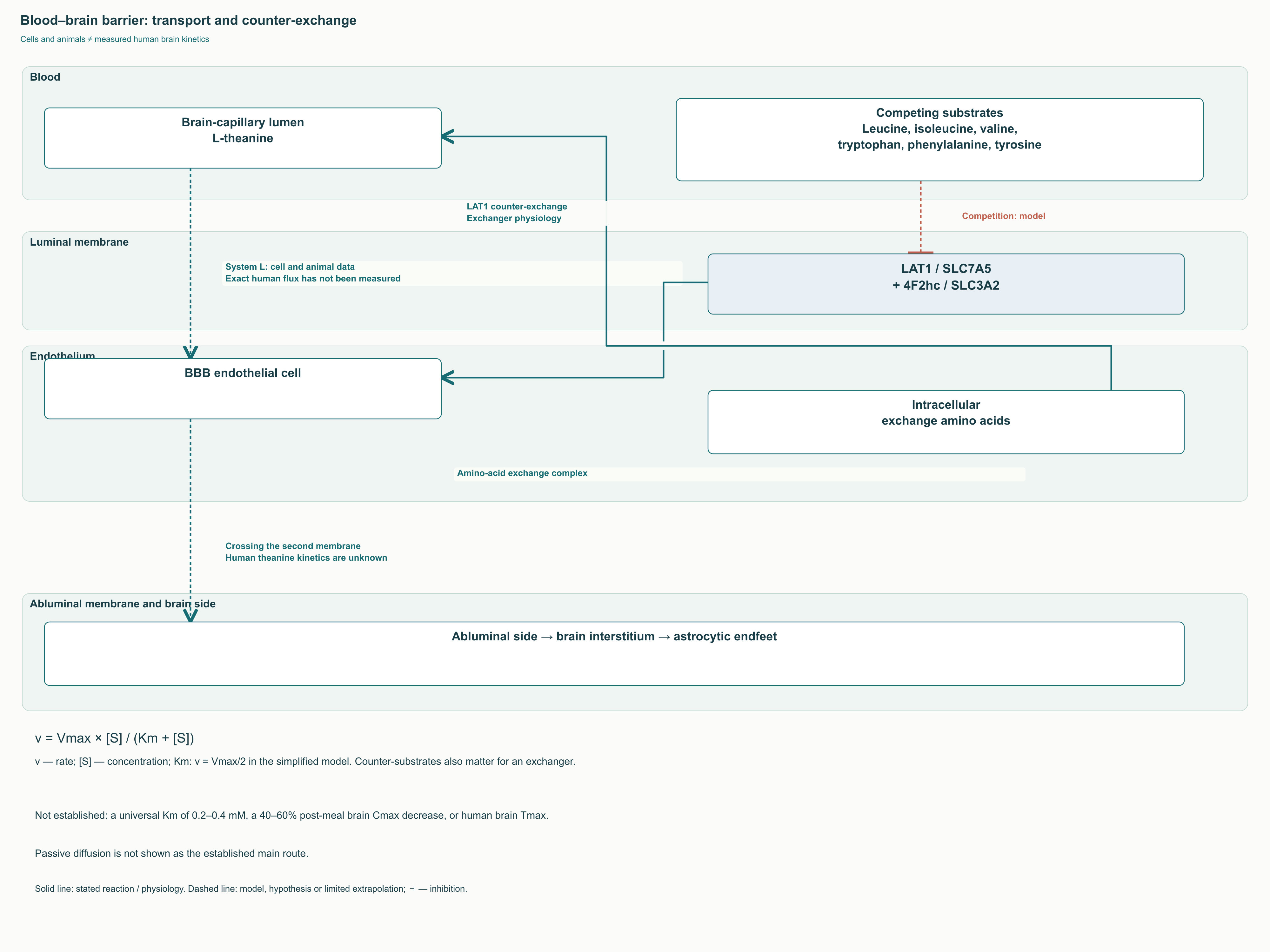
LAT1/SLC7A5 forms a complex with 4F2hc/SLC3A2 and operates as an exchanger: movement of one substrate is coupled to the opposite movement of another. This differs from a simple pump that accumulates a substance in one direction regardless of the composition on both sides of the membrane. Leucine and other large neutral amino acids can compete for a shared transport system, but the effect’s size depends on concentrations and context. Demonstrated competition in a cell model is not equivalent to a proven reduction in cognitive effects after a protein-rich breakfast.
Saturation kinetics are often explained using v = Vmax × [S] / (Km + [S]), where v is transport rate, [S] substrate concentration, Vmax maximum rate, and Km the concentration at half Vmax under the model’s conditions. For an exchanger this is only a simplified illustration: counter-substrates, several competing amino acids, and both sides of the membrane influence flux. Km therefore cannot be interpreted as a universal “dose needed by the brain.” Values of 0.2–0.4 mM and recommendations to take theanine on an empty stomach to guarantee maximum brain exposure lack sufficient human support here.
Hydrolysis and the subsequent fate of the products
The stoichiometry of theanine breakdown can be written clearly as L-theanine + H₂O → L-glutamate + ethylamine. In a human pharmacokinetic study, theanine and ethylamine levels in blood and urine increased after administration, supporting metabolic biotransformation. However, this reaction does not establish which enzyme and organ contribute to it in a particular person. The term “theaninase” refers to specific experimental enzyme systems and should not imply a separate established human drug-metabolism pathway.
The resulting glutamate enters general amino acid metabolism: it can become glutamine, participate in transamination, and connect with α-ketoglutarate and energy metabolism. Ethylamine has the formula CH₃CH₂NH₂, but its subsequent oxidation and the relative contributions of different pathways cannot be quantified from a diagram alone. In particular, it is incorrect to assume that every glutamate molecule produced automatically becomes an excitatory brain neurotransmitter. Barriers, cellular transporters, and local control of synthesis and release separate systemic metabolism from the synaptic pool.
The glutamate synapse: receptors, transport, and the limits of a causal model
NMDA, AMPA, and kainate receptors
Glutamate triggers rapid excitatory responses through ionotropic receptors, but these receptors are not interchangeable. AMPA receptors usually generate a fast depolarizing current; their calcium permeability depends, among other factors, on subunit composition and GluA2 editing. NMDA receptors require glutamate and a co-agonist—glycine or D-serine—and their current also depends on membrane potential and magnesium block. This combination of chemical and electrical conditions enables their involvement in plasticity while also making them important in pathological calcium overload.
In a classic 2002 study, theanine altered radioligand binding to AMPA and kainate receptors and the glycine site of the NMDA receptor in rat cortical preparations. This was a binding experiment, not a direct recording of current through a human receptor after supplement ingestion. IC50 is the concentration that reduces the measured signal by 50% in that experiment; it does not automatically equal Ki, a clinical dose, or brain concentration. Arbitrary tables giving “NMDA Ki of 150–200 μM” therefore do not reliably explain human effects.
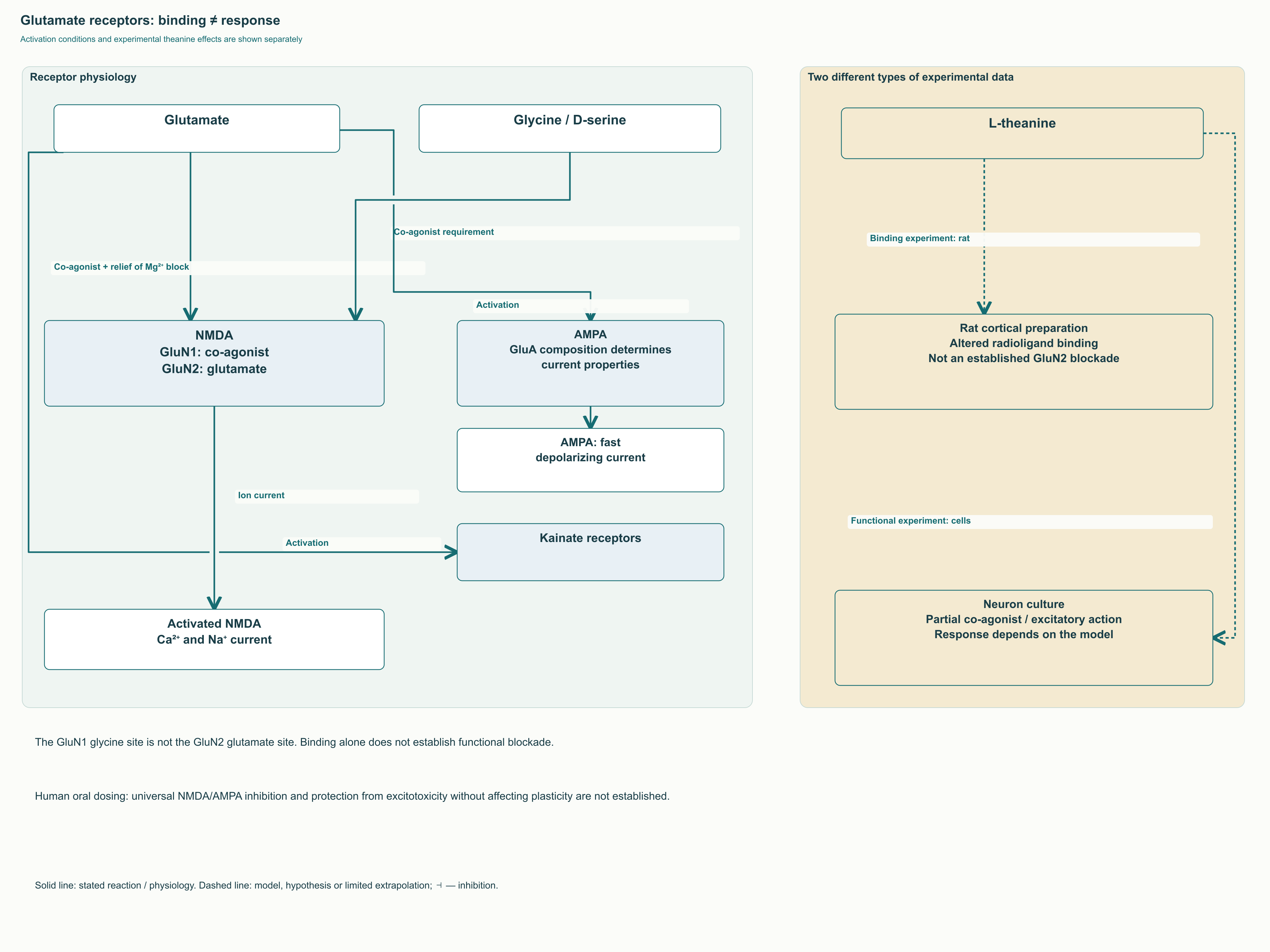
Later experiments complicate the picture: in cultured cortical neurons, theanine increased intracellular Ca²⁺, and the response was suppressed by the NMDA antagonists AP-5 and MK-801. Another study in hippocampal neuron cultures described partial co-agonist activity at NMDA receptors. These results do not justify depicting theanine as an unequivocal blocker of the GluN2 glutamate site. Nor do they establish dangerous excitotoxicity at ordinary human doses: model, concentration, and exposure duration each matter independently.
Excitotoxicity means injury from excessive, prolonged excitatory activity, for example during ischemia, when energy metabolism and ionic gradients are disrupted. Normal glutamatergic transmission, by contrast, is essential for perception, learning, and memory formation. An important limitation follows: reduced injury in a model does not confirm “brain protection” in a healthy person, and improved attention does not prove excitotoxicity blockade. A clinical promise must rest on an outcome actually measured rather than an appealing receptor name.
The glutamate–glutamine cycle and the role of astrocytes
After release, glutamate must be removed from extracellular space or the signal loses temporal precision. EAAT transporters play an important role, including astrocytic EAAT1/SLC1A3 and EAAT2/SLC1A2. In the astrocyte, glutamine synthetase converts glutamate into glutamine; glutamine can then leave the cell and enter a neuron. Neuronal glutaminase returns it to the glutamate pool, which connects with energy metabolism and neurotransmitter packaging into vesicles.
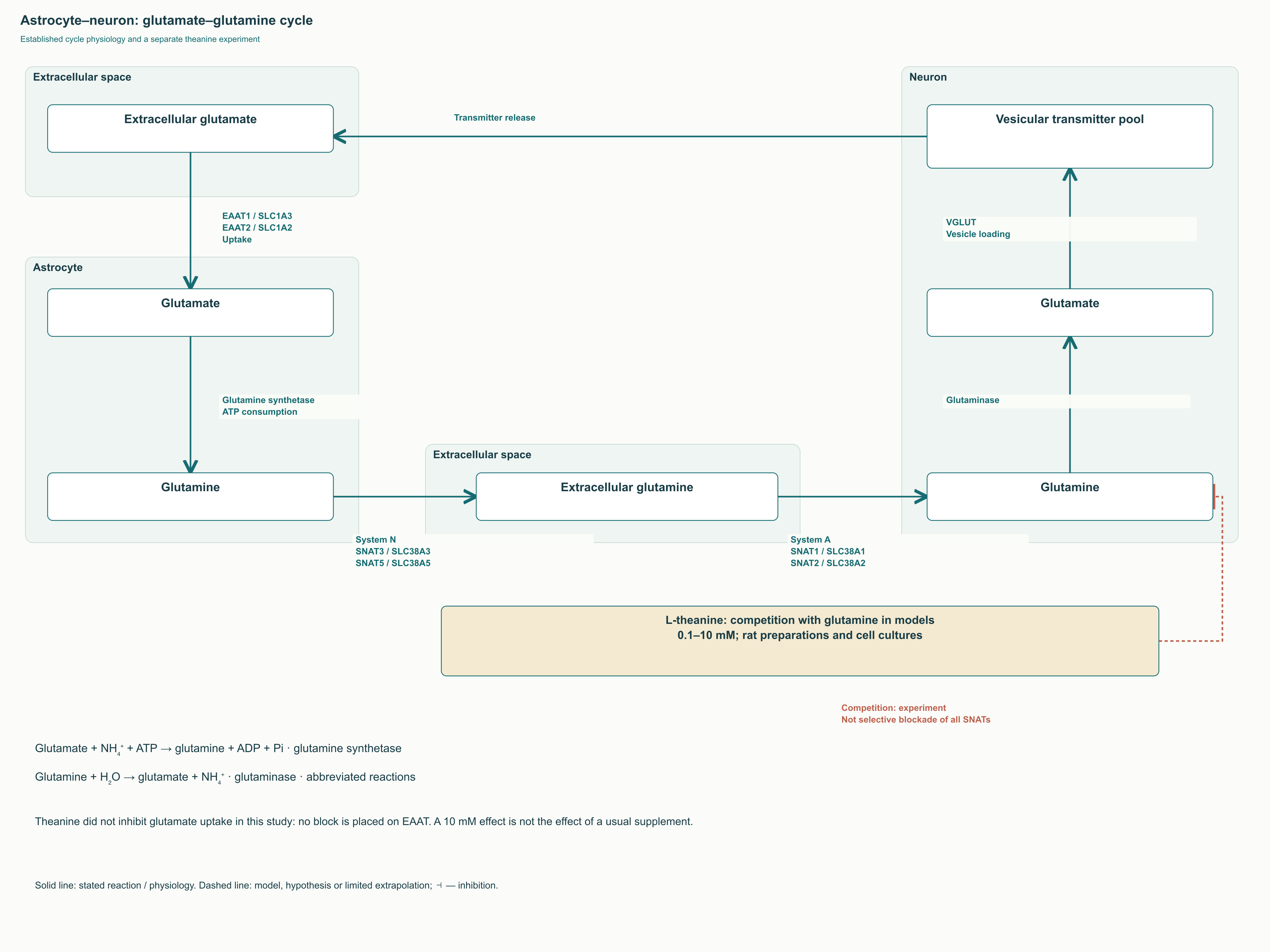
Glutamine export from astrocytes is associated with system N, including SNAT3/SLC38A3 and SNAT5/SLC38A5, and neuronal uptake with system A, including SNAT1/SLC38A1 and SNAT2/SLC38A2. However, this general physiological map does not mean that theanine selectively and equally inhibits every named protein. Experiments with synaptosomes, neurons, and astrocytes investigated competition with glutamine at 0.1–10 mM; prolonged exposure to 10 mM reduced extracellular glutamate release. The authors did not find comparable suppression of glutamate uptake itself.
Millimolar concentrations in that experiment differ substantially from the tens of micromoles per liter in plasma after 100 mg in humans. Even matching concentrations would not guarantee direct extrapolation because the cellular environment, substrate exchange, and local exposure differ. Furthermore, changes in Slc38a1 expression were described in a P19 cell model, concerning regulation over time rather than immediate competitive inhibition. For a specialist, these are two distinct causal chains: an effect on transporter function and a change in transporter abundance.
GABA, monoamines, and neurotrophic cascades
Why “increases GABA” needs qualification
GABA is synthesized from glutamate by glutamate decarboxylases GAD65 and GAD67 with pyridoxal phosphate: L-glutamate → GABA + CO₂. This reaction is part of the normal activity of inhibitory neurons and does not mean that a structural glutamate analogue serves as the same substrate. Measuring an increase in tissue GABA after theanine does not identify a single cause either: synthesis, degradation, transport, or distribution between cellular pools may change. Without appropriate experiments, the effect cannot be attributed to direct inhibition of GABA transaminase.
Changes in concentrations of several neurotransmitters and neurotrophin expression have been described after theanine exposure in developing rat brains. Such findings help generate mechanistic hypotheses but do not equal a proven increase in inhibitory current in an adult human. GABA findings cannot automatically be extended to particular extrasynaptic δ-containing GABA-A receptors or a specific degree of “tonic inhibition.” Still less do they establish that theanine acts like a benzodiazepine with fewer adverse effects.
Dopamine and serotonin: regional specificity matters more than a slogan
Dopamine is involved not only in motivation but also in tuning working memory, selecting actions, and processing signal salience. Serotonin acts through numerous receptors that can alter network activity in different ways. Phrases such as “optimizes dopamine” and “balances serotonin” therefore conceal several questions instead of explaining them: which region, which animal, which dose, and which measurement method? Even a neurotransmitter concentration in a tissue homogenate does not directly indicate the intensity of synaptic transmission.
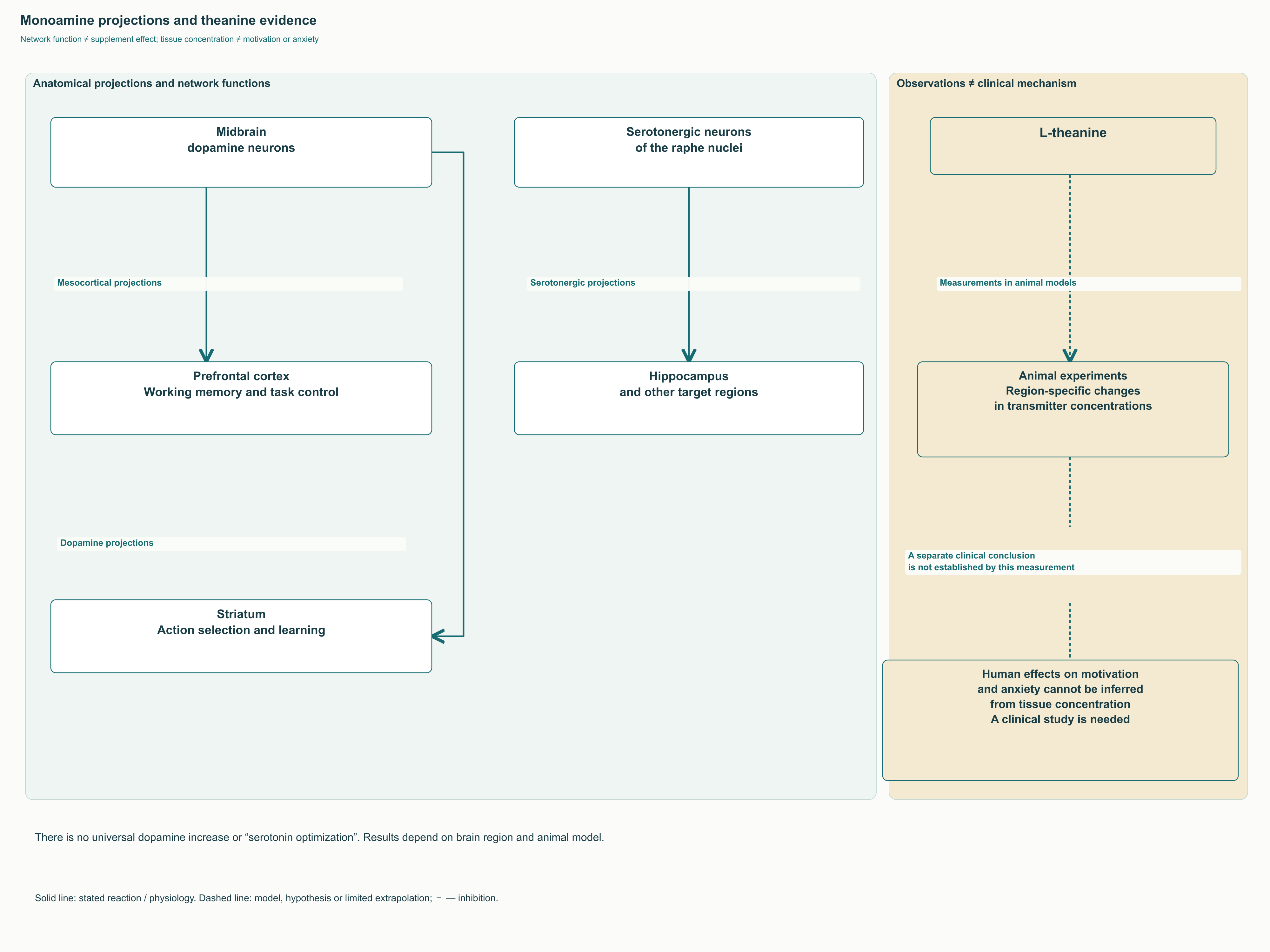
Animal experiments have indeed detected regional monoamine changes after theanine, but a universal scheme of enhanced mesocorticolimbic transmission in humans has not been established. Projections from the midbrain to the striatum and prefrontal cortex, along with serotonergic projections from the raphe nuclei, remain an important anatomical basis for discussion. The diagram separates them from hypothetical supplement effects so that normal anatomy is not converted into evidence for prescribing.
BDNF, neurogenesis, and Nrf2: different experimental questions
BDNF is a neurotrophic factor that acts through TrkB in regulating cell survival, synaptic properties, and plasticity. Mouse experiments reported increased hippocampal BDNF after three weeks of theanine exposure; studies in developing rats examined markers of cell division and neuronal maturation. In one such experiment, increased BDNF depended on measurement timing and was not maintained equally at every developmental age. These results cannot become a promise of new functional neurons in an adult supplement user.
Nrf2 and Keap1 belong to the cellular response system for oxidative and electrophilic stress. When appropriately activated, Nrf2 changes the expression of protective genes, including NQO1 and HMOX1; its influence on SLC7A11 and GPX4 also connects this system with resistance to ferroptosis. A 2025 study examined theanine in sleep-deprived mice and an HT22 cell model, assessing protein markers and animal behavior. This maintains scientific interest in the cascade but does not prove that a standard human dose activates it sufficiently to prevent neurodegeneration.
Alpha rhythms: what EEG measures and why task context matters
A rhythm is a network property, not a property of one molecule
Alpha activity generally refers to oscillations at approximately 8–12 Hz, although the band boundaries depend on methodology and individual peak frequency. It is prominent over posterior head regions during quiet wakefulness with closed eyes and changes with visual activity. However, EEG records the aggregate synchronized electrical activity of large neuronal populations, not the “amount of relaxation.” Amplitude depends on electrode position, reference, the person’s state, and signal processing, so a single microvolt range is not a certificate of theanine effectiveness.
Cortical and thalamic networks participate in rhythm generation, including excitatory relay neurons, inhibitory neurons, and cortical feedback projections. The thalamic reticular nucleus uses GABA, whereas most corticothalamic and thalamocortical projections use glutamate. T-type calcium channels matter for some firing modes, but not all alpha activity can be reduced to their automatic switching at 10 Hz. Primary recordings from the cat lateral geniculate nucleus, for example, revealed distinctive interactions between high-threshold firing and inhibitory interneurons.
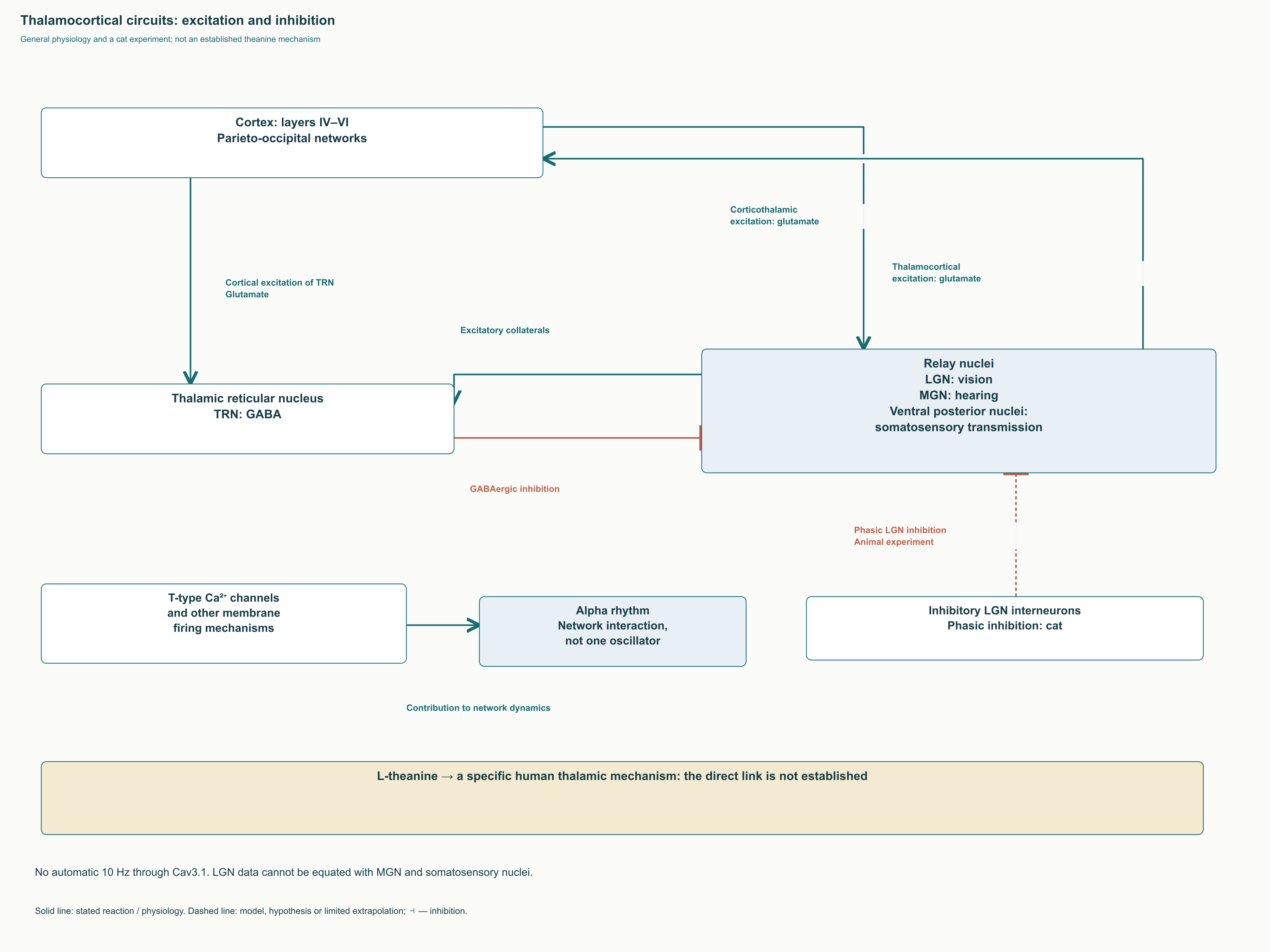
The lateral geniculate nucleus belongs to the visual pathway, the medial geniculate nucleus to the auditory pathway, and the ventral posterior nuclei to somatosensory transmission. Their involvement in sensory circuits does not prove identical responses to theanine in every nucleus. No established human chain links “oral dose → increased δ-GABA-A activity in the reticular nucleus → alpha peak → better memory.” A plausible network hypothesis remains useful when each transition makes clear what was measured and what merely connects results from different models.
Quiet rest and directed attention produce different responses
A small Nobre study used 50 mg of theanine and recorded eyes-closed EEG 45–105 minutes after administration. The groups initially included 16 theanine participants and 19 controls; the authors described increased alpha activity relative to control. This is an electrophysiological finding in a particular state, not a direct measurement of clinical anxiety or productivity. The small, brief publication also cannot establish how reproducible the effect is across people with different baseline states.
During a task, the direction of change may differ. The Gomez-Ramirez study used high-density recordings with 168 electrodes and analyzed preparation for switching attention between modalities. Theanine was associated with reduced background alpha activity but an enhanced, distinct anticipatory task-related effect in the right parieto-occipital region. This finding directly shows why “more alpha is better” cannot replace analysis of the signal’s spatial and temporal organization.
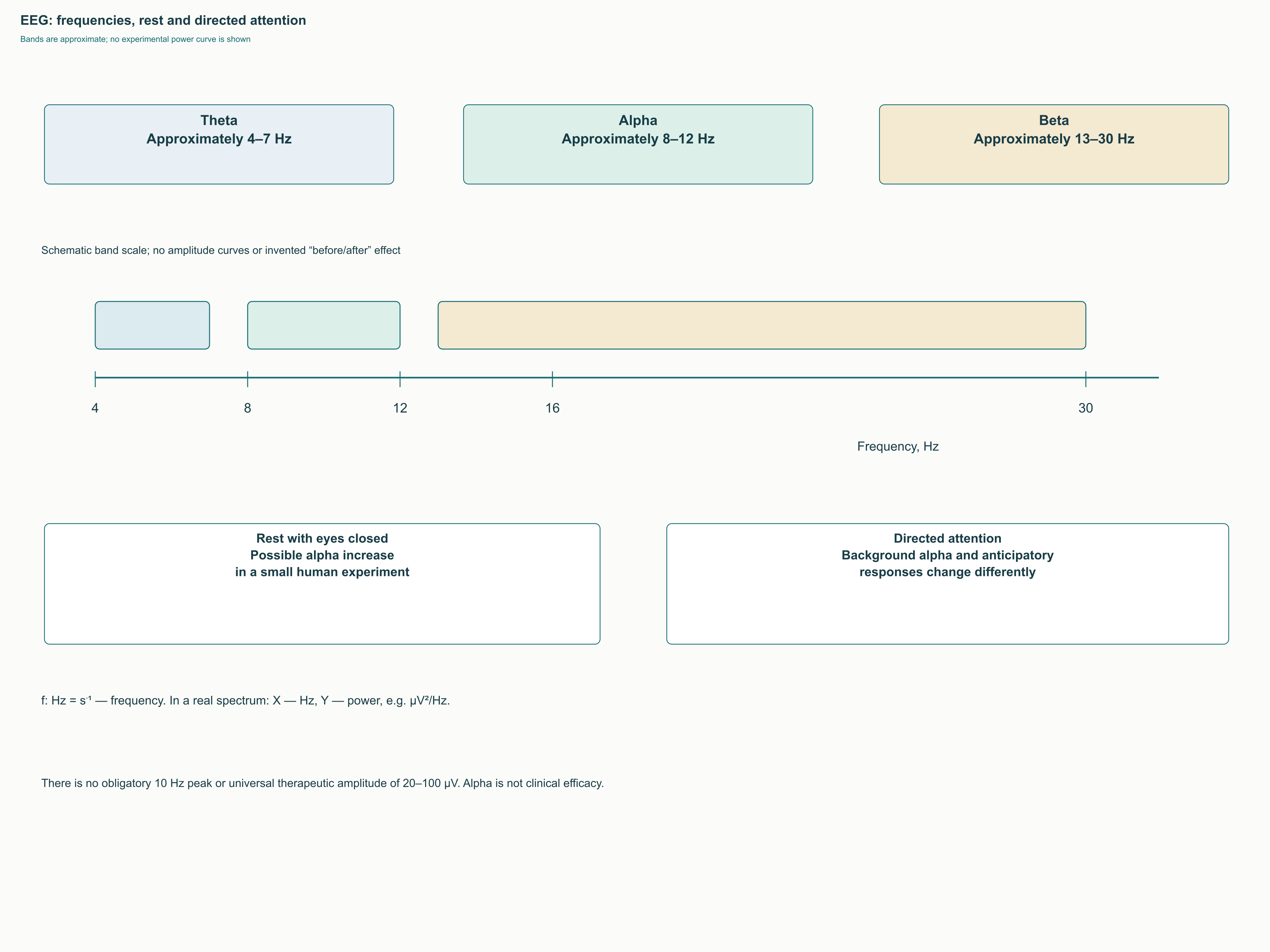
Spectral power describes the contribution of oscillations at a particular frequency to the recorded signal; it differs from peak frequency, phase, and connectivity between regions. The theta band, approximately 4–7 Hz, and beta band, approximately 13–30 Hz, likewise have no single psychological meaning. Without original numerical data, a spectrum should not resemble an experimental “before and after” curve. The illustration therefore shows frequency bands and the difference between rest and task performance, rather than an invented universal peak increase after a capsule.
In the Kelly experiment, 100 mg of theanine and 50 mg of caffeine were assessed separately and together in a visuospatial attention task. The combination increased the proportion of detected targets and signal discriminability d′ relative to placebo, while caffeine alone increased d′ and theanine alone did not show a comparable effect. d′ accounts for signal–noise discrimination and differs from simply counting correct button presses. This study connects electrophysiology with behavior but does not confirm a universal alpha mechanism for every improvement in attention.
Inhibitory control of attention
Under the inhibitory gating concept, increased alpha activity in a particular region may help restrict processing of irrelevant information. At the same time, reduced alpha in another region may accompany its engagement in the current task. This is a spatial means of allocating resources, not a uniform shutdown of the cortex. For theanine, this interpretation makes sense only in relation to the specific EEG experiment design and participants’ behavior.
In practice, an EEG measure cannot substitute for learning outcomes, accuracy in professional work, or subjective well-being. Even a well-measured effect can be small, task-dependent, and fail to translate into everyday benefit. Conversely, the absence of a general alpha increase does not rule out every change in attention. When choosing an outcome, specialists should define in advance which improvement matters to the person rather than selecting a target to fit an attractive physiological image.
Stress responses, heart-rate variability, and vascular effects
Autonomic responses to a challenge
Psychological challenges change sympathetic and parasympathetic regulation, heart rate, and vascular tone. However, cardiovascular responses are not determined by anxiety alone: breathing, posture, sleep, fitness, and the task itself influence them. In the Kimura experiment, 12 young men received 200 mg of theanine or a control before an arithmetic challenge in a crossover design. Reduced heart-rate and salivary immunoglobulin A responses were observed, but this finding does not provide a complete profile of all autonomic measures.
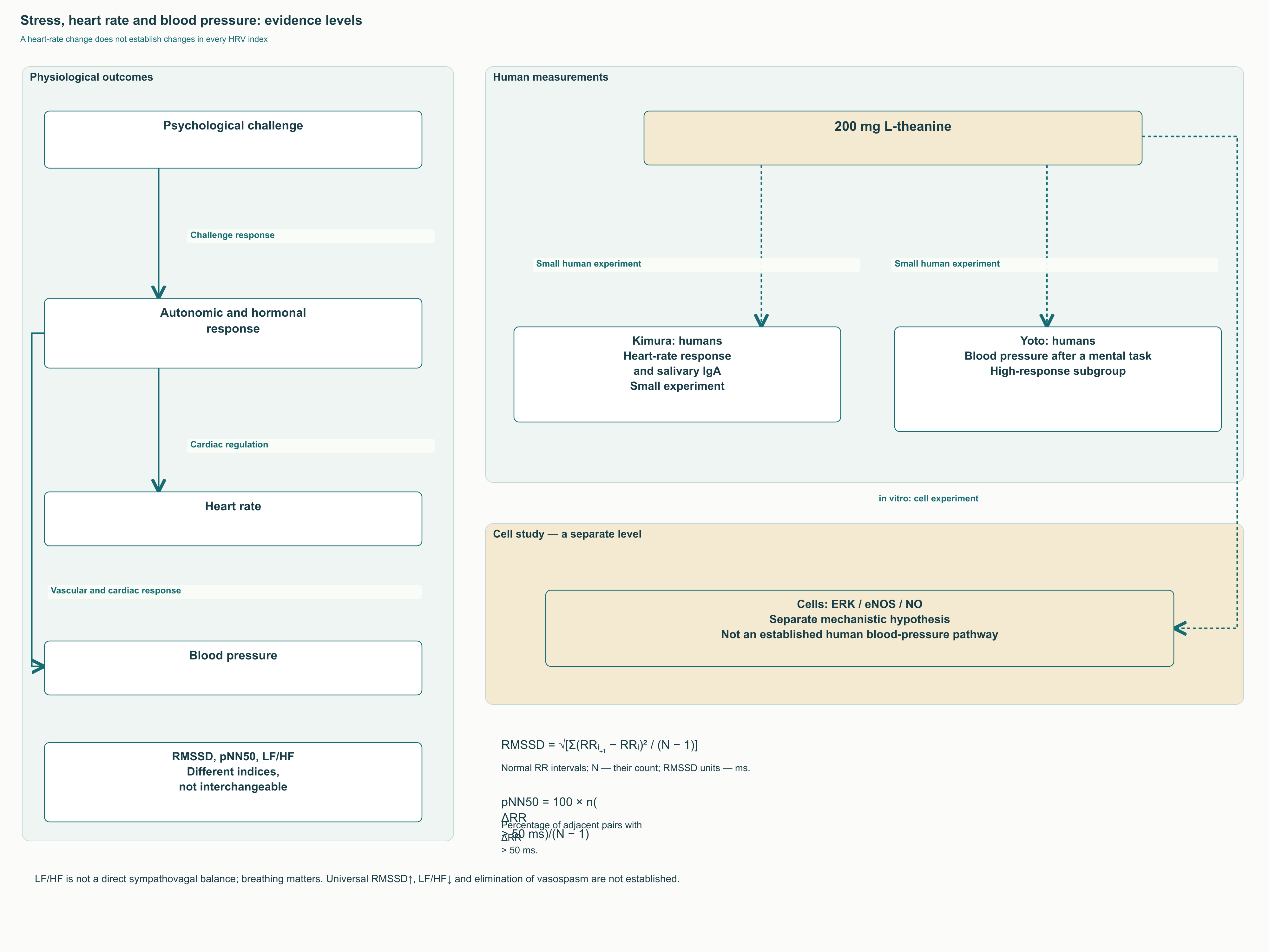
RMSSD is calculated as √[Σ(RRᵢ₊₁ − RRᵢ)² / (N − 1)], where RR represents intervals between successive normal beats and N the number of intervals. pNN50 is the percentage of adjacent pairs differing by more than 50 ms. These measures characterize particular features of variability, often associated with vagal regulation, but depend on recording quality and conditions. They do not directly measure GABA concentration and cannot be attributed to theanine simply because heart rate changed after a task.
The LF/HF ratio requires still greater caution: the low-frequency component is not pure sympathetic activity, while the high-frequency component depends on breathing. A decrease in LF/HF therefore does not prove restoration of a universal “autonomic balance.” Professional interpretation requires recording duration, frequency bands, respiratory conditions, and a prespecified outcome. Comparing arbitrary watch readings, a brief laboratory recording, and a 24-hour Holter recording as one supplement effect is methodologically incorrect.
Blood pressure and the endothelial hypothesis
The Yoto study analyzed 14 participants who received 200 mg of theanine, 100 mg of caffeine, or placebo on separate occasions. A smaller blood-pressure increase after mental tasks was found primarily in a subgroup with a high placebo response, identified after the experiment. No convincing comparable effect was demonstrated during a physical cold challenge. This is therefore a small experiment involving stress responses and subgroup analysis, not proof of hypertension treatment or a sustained reduction in blood pressure.
A separate explanation concerns endothelial nitric oxide synthase, eNOS: the NO produced can influence vascular tone through smooth-muscle signaling pathways. In a cell study, theanine increased NO production, while interventions targeting ERK/eNOS altered the response; vascular responses were examined in an ex ovo system. Such a design helps establish causal links in a model but does not reveal the size of blood-pressure reductions in humans. On a clinical diagram, ERK → eNOS → NO must be distinguished from the actually measured blood-pressure response to a psychological task.
Theanine and caffeine: interaction without a universal “ideal ratio”
What caffeine provides and what theanine does not cancel
At ordinary dietary doses, caffeine’s central effects are primarily associated with antagonism of adenosine A₁ and A₂A receptors. Adenosine signaling participates in sleep–wake regulation, so blocking it can temporarily reduce feelings of sleepiness. Phosphodiesterase inhibition and massive intracellular calcium release require a different concentration context and should not become the main explanation for the effect of a cup of coffee. Theanine is not a chemical antidote to caffeine and does not automatically resolve the need for sleep.
Caffeine sensitivity depends on habitual intake, metabolic rate, timing, and baseline state. ADORA2A variants may be associated with anxiety responses but do not predetermine their severity in every person. Later fatigue after stimulation does not prove “neurotransmitter depletion”: persistent sleep loss, fluctuations in arousal, and withdrawal from habitual caffeine can contribute. Adding theanine does not justify exceeding a tolerable caffeine dose or using stimulation instead of recovery.
Which doses were actually compared
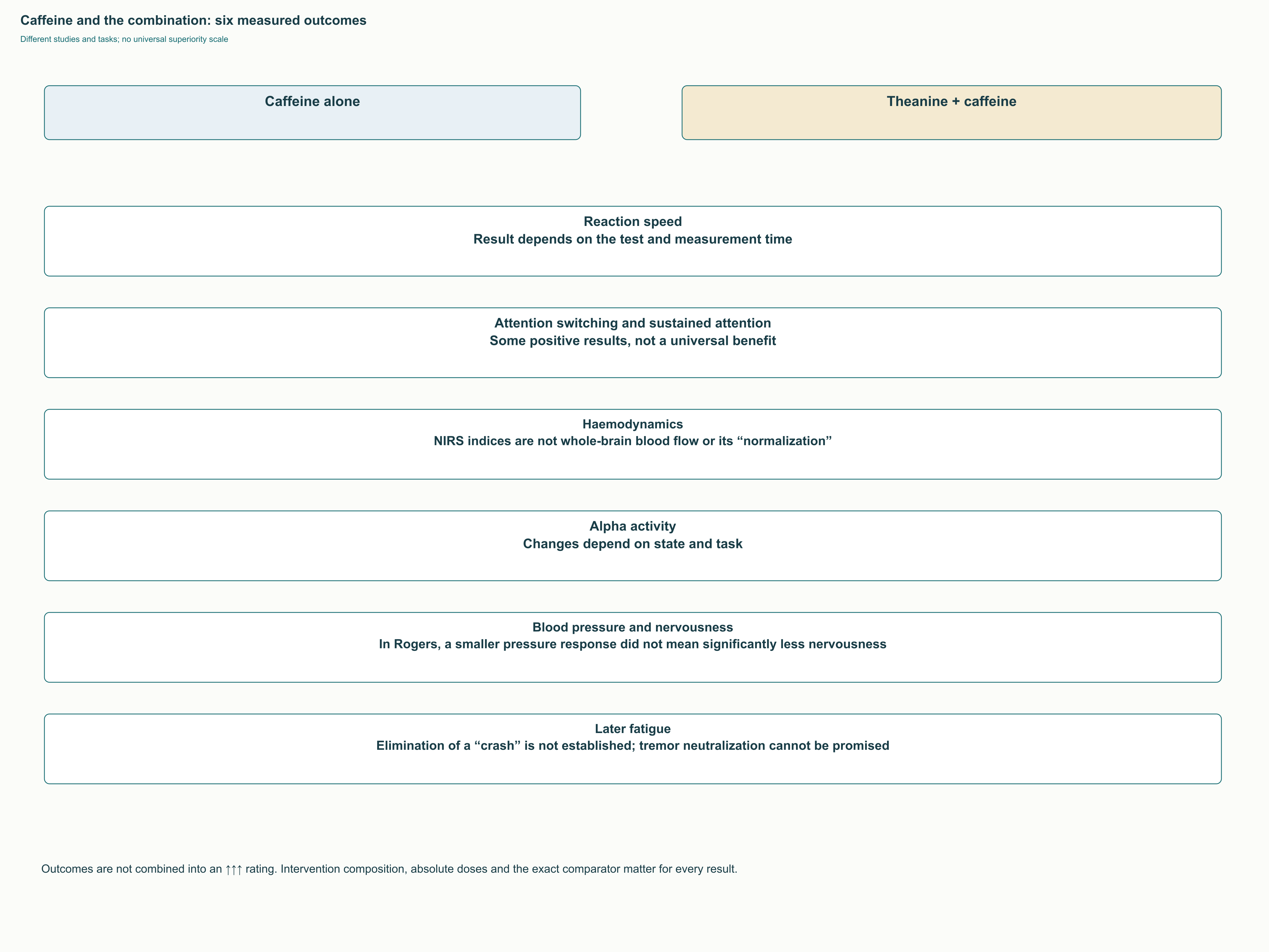
The Owen crossover trial enrolled 27 healthy adults and used 100 mg of theanine with 50 mg of caffeine, caffeine alone, and placebo. The combination improved selected attention-switching and distractor-resistance measures at specific times 60–90 minutes later. This design is useful because it includes a caffeine comparison, but it does not prove identical results for every cognitive function. Still less did it investigate 200 mg plus 100 mg as the optimal recommendation for everyone.
Haskell and colleagues studied 24 participants in a crossover design with 250 mg of theanine, 150 mg of caffeine, their combination, and placebo. The Giesbrecht study compared a beverage containing 97 mg of theanine and 40 mg of caffeine against placebo in 44 young adults, with measurements at 20 and 70 minutes. In the latter trial, task-switching accuracy and subjective alertness improved, but not all tests, including visual search, choice reaction, and mental rotation. Moreover, the absence of a caffeine-only group prevents determining how much theanine itself added to caffeine’s effects.
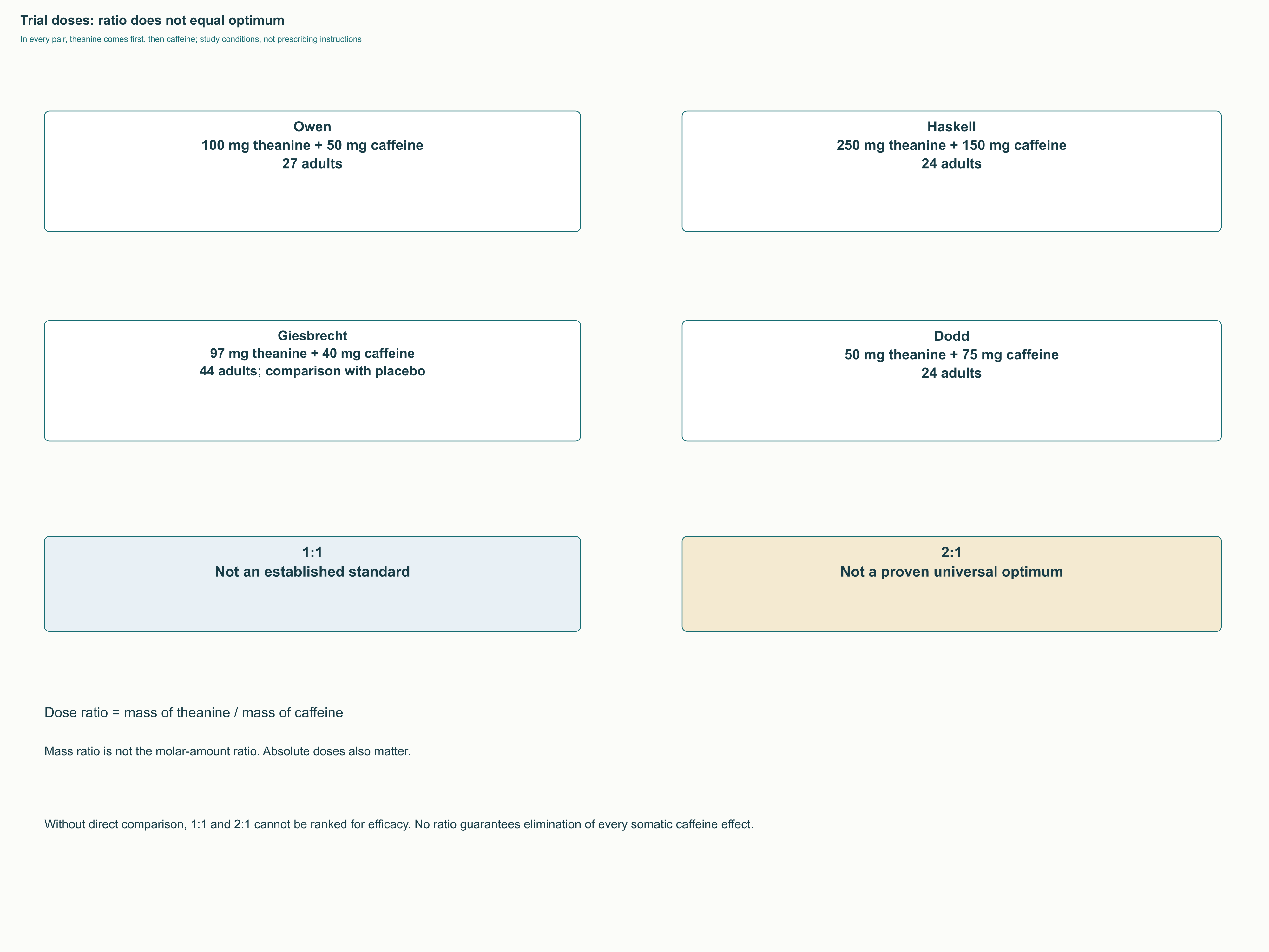
A 2:1 ratio is often convenient for describing a product, but it is not a physiological constant. Published trials used 100:50, 250:150, 97:40, and 50:75 mg, with differences in formulation, design, and tasks. These studies cannot be compared as a direct race between 1:1 and 2:1: effects may depend on absolute doses rather than the fraction alone. A claim that 200:100 mg completely eliminates somatic responses requires a direct trial of that outcome.
Cognitive tests measure different aspects of performance: task switching measures the cost of changing a rule, RVIP detects target sequences in a continuous stream, and Stroop and Flanker tests assess interference from conflicting stimuli. More correct responses without accounting for false alarms may reflect a strategy change rather than better inhibitory control. Results from one task therefore cannot be transferred to another or used as proof of specific cortical gating. A claim of d2-test normalization in adults with ADHD at 200:100 mg lacks sufficient support in the primary studies considered.
Early evidence of counteracting caffeine stimulation included EEG experiments in rats using intravenous administration, where responses depended on theanine dose. This is a separate model that cannot be converted into an oral mass-ratio instruction for humans. Equal molar amounts and equal masses are also different quantities because the substances have different molar masses. Such an experiment supports investigation of the interaction but does not prove relief of human anxiety or subsequent fatigue.
Blood flow, pressure, and subjective state
In the Dodd study, 24 participants—equal numbers of habitual and nonhabitual caffeine consumers—received 75 mg of caffeine, 50 mg of theanine, their combination, or placebo. Oxygenated and deoxygenated hemoglobin measures were examined using near-infrared spectroscopy, alongside attention and mood. Theanine altered part of the hemodynamic response to caffeine, but some beneficial behavioral effects of caffeine were also eliminated. Attenuation of a particular vascular measure therefore does not imply a universal improvement in brain function.
Near-infrared spectroscopy reflects local hemoglobin changes and has limitations in assessing signal depth and source. It does not provide a simple number indicating that “blood supply to the whole brain has normalized,” and vascular responses are not equivalent to task performance quality. The diagram must separately show the measured hemodynamic variable and behavioral outcome. Their divergence is precisely what makes such studies useful for interpretation, rather than an inconvenient exception to omit.
In the Rogers trial, 48 healthy adults received 250 mg of caffeine, 200 mg of theanine, both substances, or a control. Theanine attenuated the caffeine-related blood-pressure increase but did not significantly reduce jitteriness or alter alertness or other aspects of mood. This substantially limits the popular promise to “remove shaking and anxiety from coffee.” When a stimulant causes unpleasant reactions, the first practical step remains reconsidering the stimulant itself rather than trusting a second capsule’s protective effect.
Anxiety, ADHD, and sleep: human trials answer different questions
A stress response is not treatment of an anxiety disorder
A brief laboratory task allows responses to a challenge to be measured but does not reproduce every manifestation of an anxiety disorder. The Kimura and Yoto experiments studied arithmetic and sensory tasks, not demonstrated recovery from anxiety. Heart rate, blood pressure, salivary markers, and questionnaire responses can change independently. Invented percentages for cortisol and heart-rate increases in a “Trier test” therefore cannot be used to illustrate these studies.
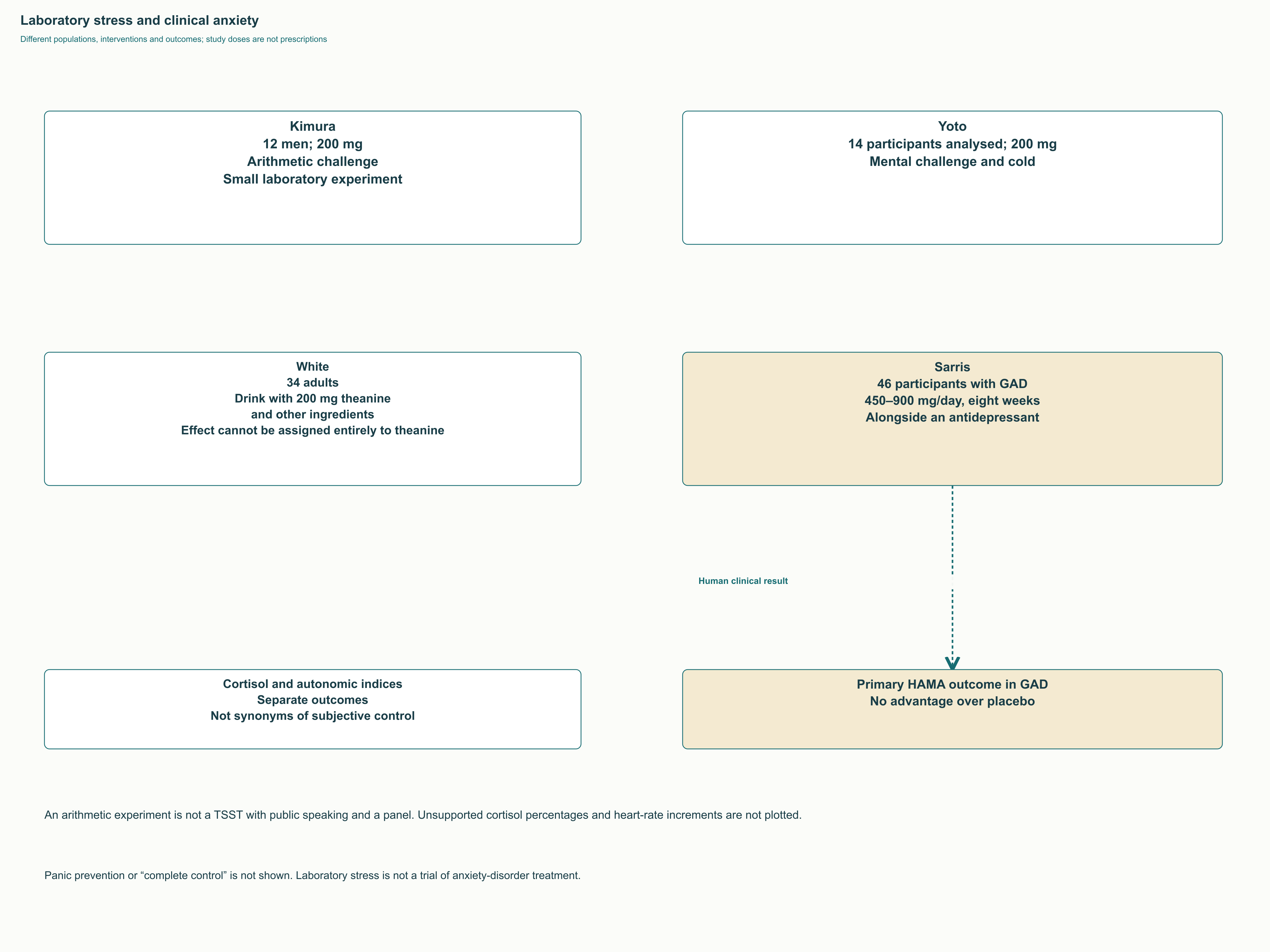
The hypothalamic–pituitary–adrenal axis links central appraisal of a challenge with cortisol secretion, but time of day and preceding state influence its measurement. In the White trial, 34 adults received a beverage with 200 mg of theanine and additional ingredients; subjective responses to a multitasking challenge decreased after one hour and cortisol responses after three hours. Because the beverage also contained phosphatidylserine, α-GPC, and chamomile, the entire result cannot be attributed to theanine alone. Changes in alpha activity did not correlate with changes in subjective stress or cortisol, further limiting a simple causal chain.
In the Unno study, 20 students were assigned to theanine or placebo groups in a single-blind design during pharmacy practice. They received 200 mg twice daily, beginning one week before practice and continuing through its first ten days. Theanine participants reported less stress, and morning salivary α-amylase was lower than in the placebo group. This everyday context complements laboratory experiments, but the small sample and incomplete blinding limit confidence in generalizing the result to other groups.
In the Hidese study, 30 adults without a major psychiatric disorder received 200 mg daily for four weeks and placebo in a crossover design. Some measures improved from the start of the theanine period, but within-group change is not equivalent to superiority over placebo. Between conditions, overall depressive-symptom and PSQI scores did not reach the accepted significance threshold: p = 0.084 and p = 0.073, respectively. Differences in individual sleep components and subgroup analyses warrant further investigation but do not turn a small trial into confirmation of broad antidepressant or anxiolytic effects.
Sarris and colleagues examined a different clinical question: 46 participants with generalized anxiety disorder received 450–900 mg of theanine daily or placebo alongside stable antidepressant treatment. Over eight weeks of the double-blind phase, no advantage emerged on the HAMA anxiety scale, p = 0.73, or the total ISI insomnia severity index, p = 0.35. Some secondary sleep-satisfaction assessments appeared more favorable, but the primary results remained negative. Increasing the dose therefore did not confirm reliable anxiety treatment in this population.
Differences between these studies help set an appropriate clinical goal. Subjective tension in a healthy person, a diagnosed disorder, and sleep disturbance during treatment require different outcomes and observation periods. If symptoms persist, interfere with work, or accompany a marked decline in mood, a supplement does not replace investigating their causes. Specialists gain more from considering a specific negative finding than from grouping every condition under “stress.”
What was shown in children with ADHD
The Lyon study enrolled 98 boys aged 8–12 with established ADHD. For six weeks they received 400 mg of theanine daily—200 mg at breakfast and after school—or placebo. Sleep was assessed using five nights of actigraphy before and after intervention; some measures of sleep percentage and sleep efficiency improved. Sleep onset latency did not change, and reduced wakefulness after sleep onset was only a statistically nonsignificant trend.
Actigraphy estimates sleep mainly from movement and does not identify N1, N2, N3, and REM stages with the precision of polysomnography. This trial therefore does not confirm longer deep sleep or more organized dreaming. Nor does it establish therapeutic effects on core ADHD symptoms or automatically extend to girls, adults, or every age group. The study dose describes a research intervention rather than an independent recommendation for a child.
For a child with sleep problems, schedule, coexisting conditions, and the duration of action of prescribed stimulants matter. Changing medication or adding several substances at once makes it harder to understand what helped and what caused an adverse reaction. Inclusion of some children taking stimulants does not prove that theanine neutralizes tachycardia, tics, or anxiety from any medication. Those separate outcomes require direct study.
Sleep architecture and measurable effects
Nighttime sleep consists of repeated cycles involving wakefulness, N1, N2, N3, and REM; their proportions change with age and circumstances. Polysomnography uses EEG, eye movements, and muscle tone to identify stages, whereas a questionnaire reflects perceived sleep. Sleep onset latency is the time taken to fall asleep, WASO total wakefulness after sleep onset, and sleep efficiency the proportion spent asleep within the corresponding time-in-bed interval. None of these should be replaced with the general phrase “brain recovery.”
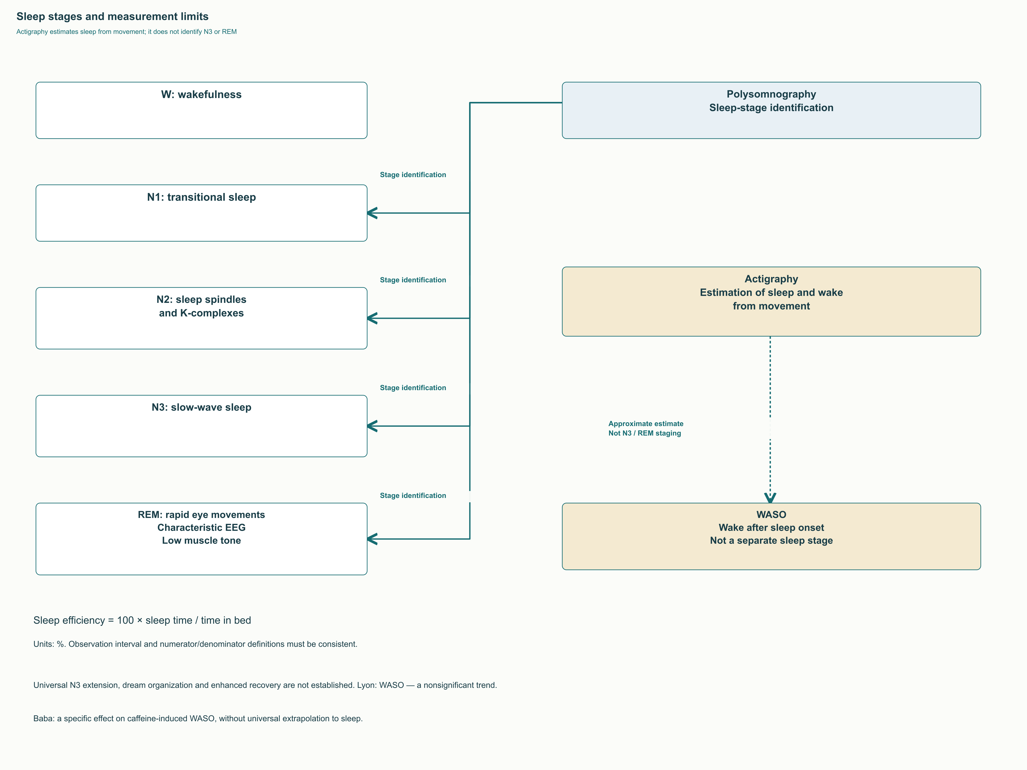
Theanine has no proven mechanism as a classic agonist at the benzodiazepine site of the GABA-A receptor. However, absence of that mechanism does not guarantee an absence of drowsiness, REM effects, or morning sluggishness in every person and combination. Available small studies suggest possible benefits for selected subjective or actigraphic measures, not an established restructuring of sleep with longer N3 after 200–400 mg. If the main problem is chronic insomnia, its causes and interventions with demonstrated clinical effects need assessment.
There is also a separate sleep study involving caffeine: Baba’s 2023 crossover trial in healthy young women compared 50 mg of theanine, 30 mg of caffeine, their combination, and placebo. Sleep stages were determined using EEG; caffeine increased WASO, whereas the combination did not differ from placebo on that measure. Sleep onset latency and the number of awakenings did not change significantly. This is a specific finding with a small caffeine dose, not proof of general protection against evening coffee or guaranteed N3 extension.
Sleep is particularly difficult to interpret when theanine, magnesium, and apigenin are added while caffeine intake is changed. Such a combination may feel helpful but is not an established “deep sleep protocol” based on studies of isolated theanine. Late caffeine can also remain active longer than appreciable plasma theanine concentrations. Absence of unpleasant evening arousal does not guarantee preservation of normal sleep.
Dosing, combined use, and drug interactions
How to interpret studied regimens
Published doses should be related to the task, formulation, and population rather than combined into a universal therapeutic table. An acute experiment using 200 mg before arithmetic, four weeks of 200 mg in the evening, and six weeks of 400 mg daily in children answer different questions. For an adult discussing a trial with a professional, defining the expected outcome and reassessment date matters more than endlessly increasing the dose. Lack of noticeable benefit does not prove that a different “stack” or more elaborate regimen is needed.
| Aim | Example of a studied intervention | Practical boundary |
|---|---|---|
| Brief stress challenge | A single 200 mg dose in small laboratory experiments | Not a confirmed treatment course for an anxiety disorder |
| Selected attention tests | Theanine with caffeine in several dose combinations | No established optimal 2:1 ratio |
| Subjective symptoms and adult sleep | 200 mg/day for four weeks in the Hidese trial | Between-condition results depend on the selected measure |
| Sleep in boys with ADHD | 400 mg/day for six weeks in the Lyon trial | Not a universal pediatric recommendation or proof of ADHD treatment |
| Generalized anxiety during treatment | 450–900 mg/day for eight weeks in the Sarris trial | No advantage on the primary anxiety scale |
The amount in a capsule, stereochemical form, and presence of additional ingredients must be known. When combining it with beverages, total daily caffeine matters rather than just the amount in one tablet. A branded ingredient name identifies product origin but does not automatically make every study independent or every dose equally effective. Some studies were funded by manufacturers or food-industry organizations, which should be considered alongside design and completeness of reporting.
Is an empty stomach necessary?
Amino acid competition for transport systems is a genuine physiological possibility. However, there is no reliable human basis for claiming that protein-containing food reduces brain theanine concentration by 40–60% or delays clinical effects by 2–3 hours. Plasma peak timing cannot be substituted for entry into human brain tissue, which was not directly measured in the trials discussed. Strict intervals such as “15–30 minutes before food or two hours afterward” should therefore not be presented as an established clinical standard.
If tolerability depends on food, that is a separate practical question. When discomfort occurs, taking it fasting for theoretical competition reasons may be less convenient than taking it with food. Keeping conditions consistent when personally observing effects helps avoid mixing the influences of sleep, diet, and caffeine. Such an observation nevertheless does not measure LAT1 or allow individual bioavailability to be calculated.
What is known about combining it with medicines
Antihypertensives, sedatives, stimulants, and NMDA modulators provide different theoretical reasons to discuss combined use, but clinically established interaction regimens are much fewer. A possible blood-pressure effect does not mean proven additivity with every ACE inhibitor, angiotensin receptor blocker, or calcium channel antagonist. Nor is there a confirmed threshold of “monitor blood pressure only above 400 mg”: risk depends on baseline state and all interventions. Symptoms of hypotension require assessment of the whole regimen rather than automatic changes to a prescribed medicine.
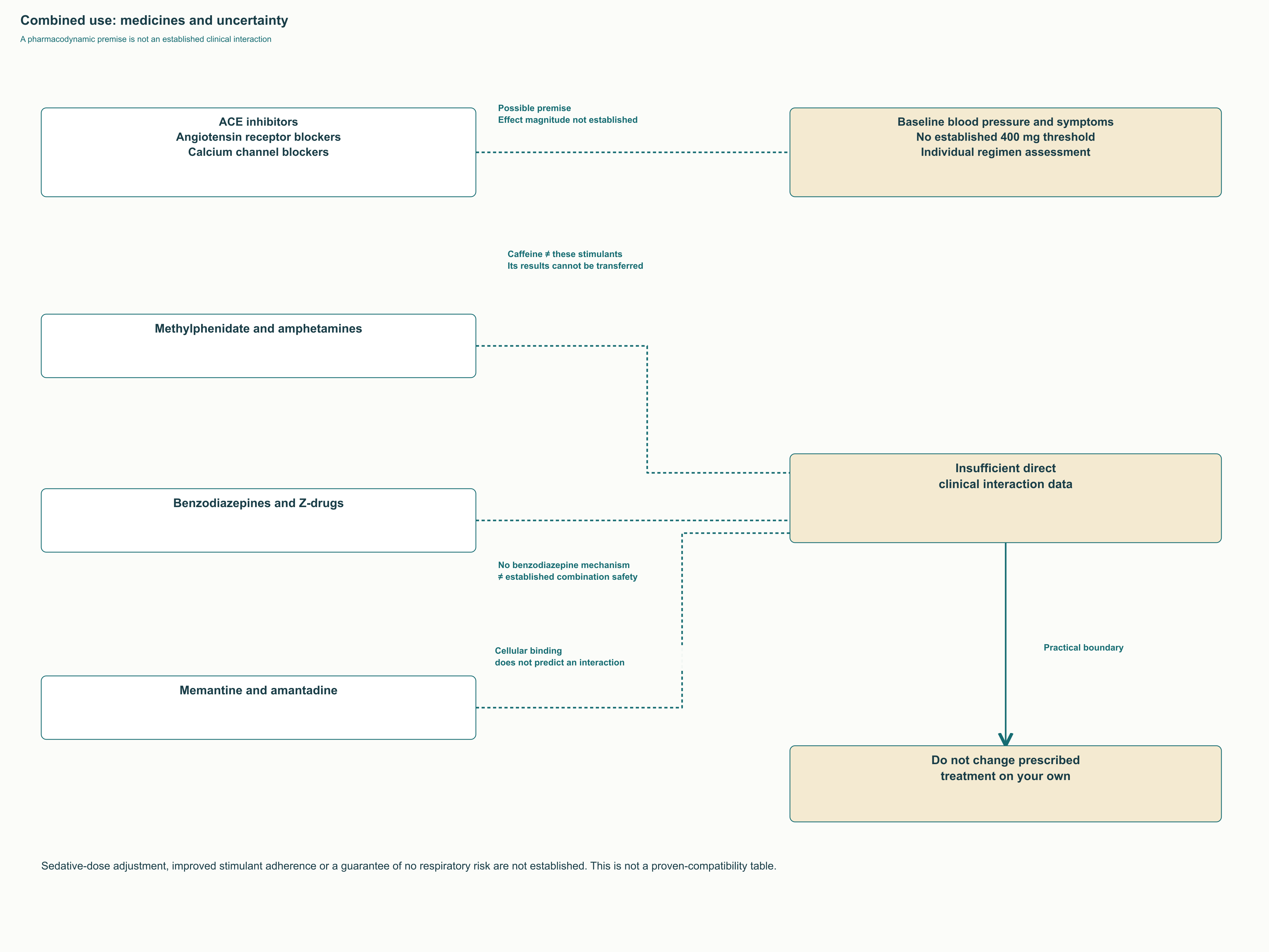
Caffeine findings cannot be transferred to amphetamines and methylphenidate, which act differently and are used in another clinical context. Claims that theanine removes their adverse effects or improves treatment adherence are not supported by the corresponding outcomes. Similarly, absence of a benzodiazepine mechanism cannot guarantee safety with sedatives or an absence of respiratory depression under every circumstance. Prescribed doses should not be independently reduced because theanine has been added.
Memantine and amantadine likewise cannot be connected with theanine through a single arrow labelled “competition for GluN2.” Their pharmacology differs from radioligand binding and partial co-agonism in cell culture. Direct clinical evidence is insufficient to predict the magnitude of such an interaction. For a professional, the correct working note is an unknown interaction with a specific theoretical basis rather than invented established compatibility or incompatibility.
Safety and its place in professional practice
What toxicology and food-ingredient status mean
A 13-week rat study tested doses up to 4,000 mg/kg daily; this highest dose was designated the NOAEL under the study conditions. NOAEL denotes a level at which specified adverse effects were not detected by that design, not a recommended human amount. It does not establish pregnancy safety, the absence of rare reactions, or tolerability in serious coexisting disease. Converting it by simply multiplying by human body weight is incorrect.
The FDA considered GRN 209, a notification concerning food uses of theanine up to 250 mg per serving in specified product categories, and had no questions about the notifier’s submitted conclusion. This is a regulatory context for ingredient use, not approval of a medicine for anxiety or insomnia. It also does not establish a universal safe daily limit for every person and combination. Food status, safety within a particular study, and evidence of therapeutic effectiveness remain separate questions.
Human trials generally describe acceptable short-term tolerability, but small samples detect rare adverse events poorly. Evidence is insufficient for a confident general recommendation in pregnancy, breastfeeding, substantial renal or hepatic impairment, or young children. Failure to find harm is not proof of safety in those groups. The actual formulation also matters when choosing a supplement, because an adverse reaction can arise from other ingredients or product quality.
Tolerance, dependence, and the criterion for benefit
Theanine has no established clinical dependence profile comparable with some psychoactive medicines. However, dedicated long-term studies of tolerance and withdrawal are limited, so claiming these never occur after any duration is too strong. Referring to the absence of a “massive dopamine release” does not replace observation of behavior and symptoms after discontinuation. In a caffeine combination, caffeine withdrawal symptoms can persist regardless of theanine’s presence.
In professional practice, theanine is reasonably considered a substance whose effects on particular attention, stress-response, and sleep measures are being studied. Its most thoroughly described molecular effects come from models, while human results depend on the task and do not combine into universal “neuroprotection.” Practical value should be judged by a clear change in well-being or function, tolerability, and alternatives rather than the number of signaling cascades in advertising. This approach preserves interest in the molecule while helping avoid promises beyond what studies have shown.
FAQ
Does increased alpha activity prove better concentration?
No. Alpha activity’s meaning depends on region, state, and task; functional benefit needs separate behavioral outcomes.
Must theanine be taken on an empty stomach?
There is no reliable evidence for strict intervals or the claimed 40–60% reduction in human brain concentration. Transporter competition in a model does not establish an everyday dosing schedule.
Does theanine remove caffeine’s adverse effects?
It does not guarantee this. Some trials found changes in blood pressure or particular attention tests, but reduced jitteriness was not universal.
Can theanine be considered treatment for an anxiety disorder?
In a small generalized-anxiety trial it did not outperform placebo on the primary scale. Stress-task findings in healthy participants do not replace treatment trials.
Does it lengthen deep sleep?
The human evidence considered does not establish universal N3 extension. Actigraphy does not determine sleep stages as polysomnography does.
Does GRAS status mean safety in pregnancy?
No. Food regulatory status does not prove supplement safety during pregnancy, breastfeeding, or illness.
Sources
- FDA GSRS: Theanine — идентификация вещества
- Scheid et al. (2012). Kinetics of L-theanine uptake and metabolism in healthy participants are comparable after ingestion of L-theanine via capsules and green tea
- Van der Pijl et al. (2010). Human disposition of L-theanine in tea or aqueous solution
- The involvement of L-type amino acid transporters in theanine transport
- An imaging approach for determining the mechanism of enhancement of intestinal absorption of an L-theanine supplement
- Yokogoshi et al. (1998). Effect of theanine, r-glutamylethylamide, on brain monoamines and striatal dopamine release in conscious rats
- Kakuda et al. (2002). Inhibition by theanine of binding of [3H]AMPA, [3H]kainate, and [3H]MDL 105,519 to glutamate receptors
- Kakuda et al. (2008). Theanine, an ingredient of green tea, inhibits [3H]glutamine transport in neurons and astroglia in rat brain
- Ogura et al. (2012). L-Theanine Promotes Neurogenesis by Facilitating the Expression of Slc38a1 in Undifferentiated P19 Cells
- Yamada et al. (2007). Theanine, r-glutamylethylamide, increases neurotransmission concentrations and neurotrophin mRNA levels in the brain during lactation
- Behavioral and molecular evidence for psychotropic effects in L-theanine (2011)
- Sebih et al. (2017). Characterization of L-Theanine Excitatory Actions on Hippocampal Neurons: Toward the Generation of Novel N-Methyl-D-aspartate Receptor Modulators Based on Its Backbone
- Takeda et al. (2011). Facilitated Neurogenesis in the Developing Hippocampus After Intake of Theanine, an Amino Acid in Tea Leaves, and Object Recognition Memory
- Huang et al. (2025). Исследование L-теанина, ферроптоза и оси Nrf2/Keap1 в модели лишения сна и клетках HT22
- Lorincz et al. (2009). Temporal Framing of Thalamic Relay-Mode Firing by Phasic Inhibition during the Alpha Rhythm
- Nobre, Rao, Owen (2008). L-theanine, a natural constituent in tea, and its effect on mental state
- Gomez-Ramirez et al. (2007). The deployment of intersensory selective attention: a high-density electrical mapping study of the effects of theanine
- Kelly et al. (2008). L-theanine and caffeine in combination affect human cognition as evidenced by oscillatory alpha-band activity and attention task performance
- Kimura et al. (2007). L-Theanine reduces psychological and physiological stress responses
- Billman (2013). The LF/HF ratio does not accurately measure cardiac sympatho-vagal balance
- Yoto et al. (2012). Effects of L-theanine or caffeine intake on changes in blood pressure under physical and psychological stresses
- Siamwala et al. (2013). L-Theanine promotes nitric oxide production in endothelial cells through eNOS phosphorylation
- Owen et al. (2008). The combined effects of L-theanine and caffeine on cognitive performance and mood
- Haskell et al. (2008). The effects of L-theanine, caffeine and their combination on cognition and mood
- Giesbrecht et al. (2010). The combination of L-theanine and caffeine improves cognitive performance and increases subjective alertness
- Dodd et al. (2015). A double-blind, placebo-controlled study evaluating the effects of caffeine and L-theanine both alone and in combination on cerebral blood flow, cognition and mood
- Rogers et al. (2008). Time for tea: mood, blood pressure and cognitive performance effects of caffeine and theanine administered alone and together
- Kakuda et al. (2000). Inhibiting effects of theanine on caffeine stimulation evaluated by EEG in the rat
- White et al. (2016). Anti-Stress, Behavioural and Magnetoencephalography Effects of an L-Theanine-Based Nutrient Drink: A Randomised, Double-Blind, Placebo-Controlled, Crossover Trial
- Unno et al. (2013). Anti-stress effect of theanine on students during pharmacy practice: positive correlation among salivary α-amylase activity, trait anxiety and subjective stress
- Hidese et al. (2019). Effects of L-Theanine Administration on Stress-Related Symptoms and Cognitive Functions in Healthy Adults: A Randomized Controlled Trial
- Sarris et al. (2019). L-theanine in the adjunctive treatment of generalized anxiety disorder: A double-blind, randomised, placebo-controlled trial
- Lyon et al. (2011). The effects of L-theanine (Suntheanine®) on objective sleep quality in boys with attention deficit hyperactivity disorder (ADHD): a randomized, double-blind, placebo-controlled clinical trial
- Baba et al. (2023). Theanine maintains sleep quality in healthy young women by suppressing the increase in caffeine-induced wakefulness after sleep onset
- Borzelleca et al. (2006). A 13-week dietary toxicity and toxicokinetic study with L-theanine in rats
- FDA. GRAS Notice No. 209: L-theanine
- EFSA (2015). Scientific Opinion on the safety of caffeine