The NLRP3 Inflammasome: Assembly, IL-1β, and the Limits of the Inflammaging Model
NLRP3 assembly, priming, K⁺ efflux, IL-1β/IL-18 maturation, and clinical evidence are considered separately. Inflammaging is not one sensor, and IL-1β blockade is not direct NLRP3 inhibition.
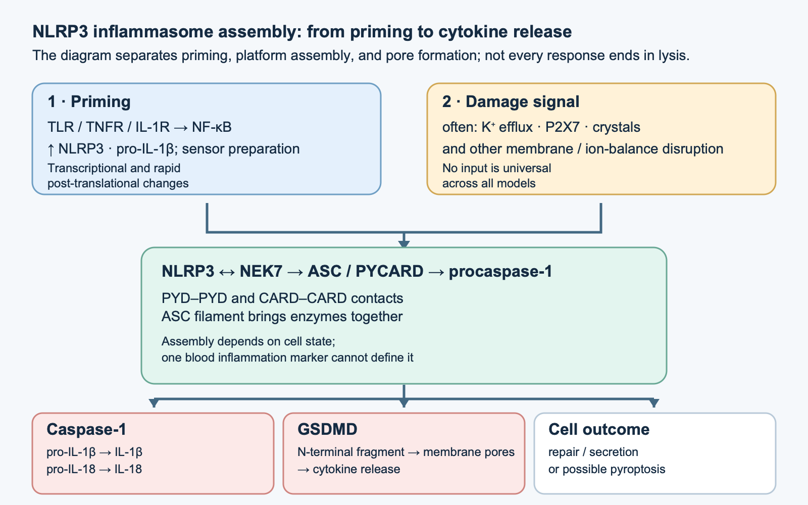
The same macrophage may encounter a urate crystal, extracellular ATP, or a bacterial toxin without needing a separate receptor for the chemical composition of every stimulus. The NLRP3 inflammasome assembles in response to changes in the cellular environment that follow such exposures. Once assembled, the platform activates caspase-1, which matures IL-1β and IL-18; cleavage of gasdermin D can open membrane pores and lead to pyroptosis. This sequence is well described in cells and experimental models. It is much harder to show that this single pathway explains chronic inflammation or human lifespan.
Platform structure and cellular location
NLRP3 is an intracellular NLR-family protein with three principal modules. Its N-terminal PYD mediates homotypic interaction with the PYD of the ASC adaptor. The central NACHT contains a nucleotide-binding domain (NBD) and the neighboring HD1, WHD, and HD2; nucleotide state and the ATP-binding/hydrolysis cycle contribute to conformational change. C-terminal leucine-rich repeats (LRRs) form regulatory contacts, including at the interface with NEK7. The exact location of each domain boundary depends on the structural construct used; a list of amino-acid positions does not replace a map of the actual protein in a living cell.
ASC/PYCARD has its own PYD and CARD. The PYD extends the nucleated NLRP3 network, while the CARD recruits procaspase-1 through CARD–CARD interaction. Many ASC molecules form filaments and a visible “speck”; proximity between procaspases promotes activation and proteolytic processing. Caspase-1 is a cysteine protease with a large p20 and a small p10 subunit. The notation p20₂/p10₂ is useful for the mature enzyme form, but cellular catalytic activity need not wait until fully separated subunits appear. Self-assembly amplifies the signal: one sensor can recruit many adaptors and effectors.
Cryo-EM and biochemistry distinguish different protein states. Work on full-length mouse NLRP3 showed a membrane-associated double-ring “cage” of roughly 12–16 subunits with shielded PYDs. A structure of full-length human NLRP3 with MCC950/CRID3 described a different inactive oligomeric context. These cannot be collapsed into a universal claim that all resting human macrophage NLRP3 freely floats in the cytosol as one decamer. Localization depends on the state of the trans-Golgi network, other membranes, cell type, and preparation. A separate NLRP3–NEK7 structure shows how NEK7 binds LRR and parts of NACHT and supports an assembly-competent configuration; NEK7’s structural role is not the same as simple phosphorylation of NLRP3 by that kinase.
Assembly is constrained by priming, the nucleotide pocket state, ubiquitination, phosphorylation, and availability of membrane sites. Rapid deubiquitination involving BRCC36/BRCC3, roles for IRAK1 and JNK1 in specific models, and PYD phosphorylation have been reported. These observations do not form one obligatory “10–30-minute timer” for every human cell. Nor does a list of specific E3 ligases and amino-acid residues from cell systems let us measure inflammasome activity in a patient without analyzing the relevant proteins.
From priming to the assembly signal
Signal 1: provide substrate and prepare the sensor
In the classical two-step model, bacterial LPS acting through TLR4, other TLR ligands, TNF, or IL-1 activates signaling pathways leading to NF-κB. For TLR4, signaling through MyD88 and/or TRIF depends on compartment and timing; TNFR1 uses its own adaptors and should not be mechanically written as a “MyD88-dependent” pathway. Through IRAK–TRAF6–TAK1–IKK and degradation of IκBα, the NF-κB dimer enters the nucleus. Expression of NLRP3 and especially IL1B, which encodes cytosolic pro-IL-1β, increases. This supplies substrate for caspase-1. Unlike pro-IL-1β, pro-IL-18 is already present in some cells before an inflammatory stimulus, although its expression also depends on tissue and state.
Priming can also be post-translational. Experiments show that, after TLR signaling, removal of some ubiquitin chains from NLRP3 and changes in phosphorylation can increase the response without waiting for complete new protein synthesis. “Signal 1” is therefore a functional category of complex preparation, not only a two-hour rise in mRNA. Transcription and rapid preparation can occur at the same time. A change in IL-1β after LPS in a monocyte does not prove that ionic damage occurred or that the same number of ASC specks assembled as after nigericin.
Signal 2: disruption of ionic and membrane balance
For many classical stimuli, a shared integrator is a fall in intracellular K⁺. At rest, cytosolic K⁺ is high relative to the extracellular environment because of Na⁺/K⁺-ATPase activity. In experiments, high-potassium external buffer suppressed NLRP3 responses to different toxins and particles. This strongly supports a causal role for efflux in canonical models; it does not establish a universal biological threshold of “70–90 mmol/L” for all cells or a rule for every NLRP3 pathway. A change in the gradient, rather than chemical recognition of each molecule, links diverse stimuli.
Extracellular ATP released after tissue damage binds P2X7, an ATP-gated cation channel, and can trigger K⁺ efflux; the concentration required depends on the experimental system, ATP availability, and other purinergic pathways. Nigericin is a laboratory K⁺/H⁺ ionophore that is convenient for reproducibly lowering cellular K⁺. Its effect should not be treated as typical of every human infection. Bacterial pore-forming toxins also damage membranes and alter ionic composition. Pannexin-1 was linked to a P2X7-dependent large-pore response in particular experiments, but should not be depicted as an obligatory sequential intermediate in every ATP-triggered NLRP3 response.
Particles act through another entry point. After phagocytosis, monosodium urate crystals in gout, cholesterol crystals in an atherosclerosis model, silica, and aluminum salts can disrupt the endolysosomal membrane. Release of lysosomal components, including cathepsins, may contribute to the response, but a cathepsin B inhibitor does not prove that B alone is the direct NLRP3 sensor. In these experiments the crystal triggers a chain of damage; it does not demonstrate direct crystal binding to LRR. Fibrillar protein aggregates have been studied in microglia and macrophages; exposure conditions and the stage of neurodegeneration matter.
Mitochondria may add another layer. Some models show changes in membrane potential, mitochondrial reactive oxygen species, oxidized mtDNA appearing in the cytosol, and altered cardiolipin localization. Primary studies support participation of such events; they do not establish that every NLRP3 stimulus must pass through mPTP, VDAC, direct binding of ox-mtDNA to the same NLRP3 site, and cardiolipin. ROS inhibition may affect priming and many other reactions. A persuasive mechanism must distinguish timing, compartment, and specific loss of function.
Ca²⁺ and Cl⁻ ions, cell volume, and disruption of the trans-Golgi network can also change the probability of assembly. However, the available experiments do not imply one “unified obligatory ionic network” in which CLIC channels contribute 100% in every tissue. K⁺ loss is an important and frequent common mechanism, but described exceptions exist; this is especially clear in the alternative response of human monocytes.
Proteolysis: two cytokines and a pore-forming effector
After recruitment to the ASC protein platform, active caspase-1 cleaves pro-IL-1β, about 31 kDa, to form mature IL-1β, about 17 kDa. The principal site in the human precursor is after Asp116. IL-1β lacks a classical signal peptide and does not follow the usual ER–Golgi secretory route; after leaving the cell, it binds IL-1R1 together with the IL-1RAcP co-receptor and activates tissue inflammatory responses, fever, and acute-phase signaling. The presence of pro-IL-1β in a cell does not itself mean that mature cytokine has been secreted. Serum IL-1β, especially at low levels, also depends on the measurement method.
Pro-IL-18 can also be a caspase-1 substrate. Mature IL-18 acts through IL-18Rα/β and can strengthen IFN-γ responses in NK and T cells, especially together with IL-12. It is not merely a “harmful aging cytokine”: this axis has protective functions in infection and tissue immune surveillance. The biological response also depends on IL-18 binding protein (IL-18BP) and tissue context. An antibody that neutralizes IL-1β does not automatically turn off IL-18.
Caspase-1 also cleaves gasdermin D (GSDMD), releasing its N-terminal domain from intramolecular inhibition. N-terminal fragments oligomerize on the inner surface of the plasma membrane and form pores. Small solutes pass through them and IL-1β/IL-18 may exit; osmotic disruption often completes pyroptosis. Cellular mechanical resilience, membrane repair, and pore-forming intensity allow secretion before final lysis in some contexts. Giving one pore diameter and a constant “16-mer” for GSDMD pores would be overprecise: structural parameters depend on gasdermin species, membrane model, and method. The bibliographic GSDMA3 structure is not a direct measurement of a GSDMD pore in a human macrophage.
The pathway can be summarized without falsely assuming obligatory lysis:
\mathrm{pro\!\!\!{-}IL\!\!\!{-}18}\xrightarrow{\mathrm{caspase\!\!\!{-}1}}\mathrm{IL\!\!\!{-}18},$$
$$\mathrm{GSDMD}_{\rm full}\xrightarrow{\mathrm{caspase\!\!\!{-}1}}\mathrm{GSDMD}_{\rm N}+\mathrm{GSDMD}_{\rm C};\quad
\mathrm{GSDMD}_{\rm N}\rightarrow\text{membrane pores}\rightarrow\text{secretion / possible pyroptosis}.$$
These reactions show molecular changes, not the stoichiometry of a tissue-level outcome. ASC specks and inflammasome particles may remain extracellular after cell death and sustain inflammation in experiments. Observing this effect does not prove a self-propagating “prion disease” in humans.
### Canonical, noncanonical, and alternative pathways must not be conflated
| Pathway and model | Proximal signal | What directly cleaves GSDMD | How IL-1β/IL-18 mature | Important boundary |
|---|---|---|---|---|
| Canonical NLRP3 in a primed macrophage | Membrane/ion-gradient damage, crystals, and other stimuli → NLRP3–NEK7–ASC | Caspase-1 | Caspase-1 on the inflammasome platform | K⁺ efflux is required for many stimuli, but not every described context |
| Noncanonical response to cytosolic LPS | Human caspase-4/5; mouse caspase-11 recognizes LPS | Caspase-4/5/11 | Secondary NLRP3–ASC–caspase-1 activation after GSDMD pores and K⁺ efflux | Do not confuse cytosolic LPS recognition with surface TLR4 priming |
| Alternative NLRP3 in primary human monocytes | LPS–TLR4–TRIF–RIPK1–FADD–caspase-8 | Pyroptotic rupture is not an obligatory feature | NLRP3–ASC–caspase-1 can produce mature IL-1β | K⁺ efflux and ASC specks were not required in the original model; do not generalize to all cells |
Noncanonical human caspases 4/5 and mouse caspase-11 can bind cytosolic LPS through their CARD and activate GSDMD. They are not equivalent to caspase-1 as direct processors of pro-IL-1β. GSDMD pores trigger a K⁺-dependent secondary activation of NLRP3 when the cell is appropriately primed. Conversely, work in human monocytes showed an LPS-dependent alternative NLRP3 response without the characteristic ASC specks, K⁺ efflux, or pyroptosis. This explains why two separate sequential injections of LPS and ATP are a useful laboratory protocol, but not a law of all physiology.
## Inflammaging: from models to humans
“Inflammaging” describes an age-related tendency toward a chronic, low-grade inflammatory background. It is a population-level description, not a diagnosis based on one IL-1β, CRP, or “NLRP3 activity” measurement. Causes are multiple: cellular senescence and its secretory phenotype, immune remodeling, chronic infections, changes in barriers and microbiota, tissue injury, adipose tissue, and metabolism. Some of these can engage NLRP3; the converse claim that NLRP3 is the sole or main source of age-related inflammation in every person has not been established.
In mouse adipose tissue with obesity and in cell experiments, saturated fatty acids and related metabolic signals strengthened NLRP3-dependent inflammation; removing selected pathway components altered insulin responses. This supports the hypothesis of metaflammation, but a genetically modified mouse on a defined diet does not reproduce the full long-term history of human diabetes. In people, obesity, insulin resistance, and inflammatory markers correlate through many pathways. The data do not allow an individual “NLRP3 inhibitor” dose to be prescribed from body weight or NLRP3 to be called the established primary cause of every case of type 2 diabetes.
Cholesterol crystals triggered NLRP3 responses in macrophages, and mouse atherogenesis models showed a contribution of this pathway under certain conditions. Human plaques also involve lipoproteins, hemodynamics, a necrotic core, and other immune networks. The CANTOS clinical trial tested blockade of **IL-1β**, not selective shutdown of NLRP3: canakinumab does not establish that NLRP3 itself was necessary for the cardiovascular effect. A fall in CRP confirms a pharmacodynamic response, but cannot be equated with demonstrated slowing of aging.
In mouse models of amyloid deposition, NLRP3 participated in microglial pathology; aggregated α-synuclein triggered an inflammasome response in some experimental systems. These findings connect a cellular response to a neurodegeneration hypothesis, but do not confirm that blocking NLRP3 treats Alzheimer’s or Parkinson’s disease in people. Data for fibrosis of the liver, kidneys, and other organs are more complex: TGF-β, macrophages, metabolic injury, and epithelial responses are intertwined. A reduction in ASC staining or the number of NLRP3 transcripts in a tissue cannot replace evidence on a clinical outcome.
[](https://api.nutrifit.health/api/v2/public/knowledge/files/images/nutrifit-scientific-20261009-nlrp3-inflammasome-il1beta-inflammaging-drivers-nlrp3-evidence-ladder-33968f086afd.png)
### Why “inflammaging” is not the same as a continuously active inflammasome
An age-related rise in average CRP, IL-6, and other markers in some populations does not mean that NLRP3 is continuously assembling in every older person. The term describes a tendency and variation between people; markers are affected by hidden or recent infections, smoking, obesity, chronic diseases, medications, kidney and liver function, physical activity, and cohort composition. Immune aging changes innate and adaptive cells at the same time: older monocytes may prime differently, lymphocytes may regulate responses differently, and damaged tissues may provide more DAMP signals. Measuring circulating IL-1β does not reveal which cell type produced it, which processor generated the mature form, or whether NLRP3 was the cause.
Senescent cells, mitochondrial stress, barrier disruption, crystals, altered microbiota, and metabolic overload are different potential inputs to inflammatory networks. Their relative contribution depends on tissue and disease. In one model, signaling may begin with TLR/NF-κB; in another, with cytosolic LPS, crystal injury, or another inflammasome. Finding an NLRP3-dependent component in an experiment does not remove the contribution of AIM2, NLRC4, caspases-4/5, cGAS–STING, adaptive immune responses, or non-inflammasome cytokines.
Thus, “age → DAMPs → NLRP3 → inflammation → aging” is useful as a testable scheme, but not as an established linear causal chain in humans. A claim of aging prevention would require more than target engagement: it would require a clinically meaningful change in a prespecified outcome in an adequate long-term trial that controls for infection and other risks. Neither a mouse model nor a brief drug-induced fall in IL-1β substitutes for that experiment.
### Disease, model, and strength of inference
| Context | What was studied | What the result supports | What it does not establish |
|---|---|---|---|
| Gout inflammation | Monosodium urate crystals and NLRP3 in cell and animal models | Crystals can trigger an inflammatory response through phagolysosomal damage and subsequent inflammasome activation | That every human flare is caused only by NLRP3 or that a mechanistic scheme determines individual treatment |
| Atherosclerosis | Cholesterol crystals and NLRP3 in plaque models | Innate inflammation contributes to atherogenesis in some models | That CANTOS directly inhibited NLRP3 or that NLRP3 was the sole mediator of clinical benefit |
| Obesity and insulin resistance | Cell and genetic models under high-fat load | NLRP3 can link some metabolic signals to inflammation in a defined model | That eliminating NLRP3 prevents diabetes in people or explains every form of insulin resistance |
| Neurodegenerative diseases | Microglia and amyloid or α-synuclein aggregates, mostly in experimental models | Inflammasomes can respond to aggregates and change local inflammatory processes | That an NLRP3 inhibitor treats Alzheimer’s or Parkinson’s disease or preserves cognition in people |
| Aging | Age-related markers, tissues, and experimental models | Inflammatory pathways may be one part of complex age-related remodeling | That one biomarker measures aging rate or that blocking one pathway extends human life |
In atherosclerosis models, cholesterol crystals activated inflammatory responses, while in human coronary disease a different intervention point—IL-1β—was tested clinically. These evidence levels cannot be fused into one proof. Experimental microglia and aggregate studies also make neuroinflammation biologically plausible, but do not establish clinical efficacy. For a separate look at this literature, see the articles on [neuroinflammation and microglia](/knowledge/neuroinflammation-microglia-kynurenine-pathway) and [baicalin and neuroinflammation](/knowledge/skullcap-baicalin-neuroinflammation-gaba); their existence does not mean botanical products block the inflammasome in humans.
## What clinical interventions show
### IL-1β blockade in CAPS: treatment of a rare monogenic syndrome
Cryopyrin-associated periodic syndromes (CAPS) are linked to pathogenic NLRP3 variants that can lead to excessive IL-1β production. This is an example of a rare disease with a genetically specified connection between a sensor and a cytokine. In a randomized canakinumab study, 35 patients with CAPS initially received the drug subcutaneously; 34 achieved complete response in the initial phase. Among the 31 participants who entered the randomized withdrawal phase, all 15 who continued canakinumab remained in remission, whereas 13 of the 16 receiving placebo had a flare. Infections were reported; serious adverse events included urosepsis and vertigo.
This result supports a clinical role for IL-1β in specific rare syndromes and efficacy of cytokine blockade in this population. It does not prove that the same drug is appropriate for nonspecific age-related symptoms, ordinary low-grade inflammation, or neurodegeneration. Moreover, canakinumab binds IL-1β downstream of the inflammasome; this is not a direct experiment selectively switching off NLRP3 in every tissue.
### CANTOS: testing an inflammatory hypothesis, not an NLRP3 drug
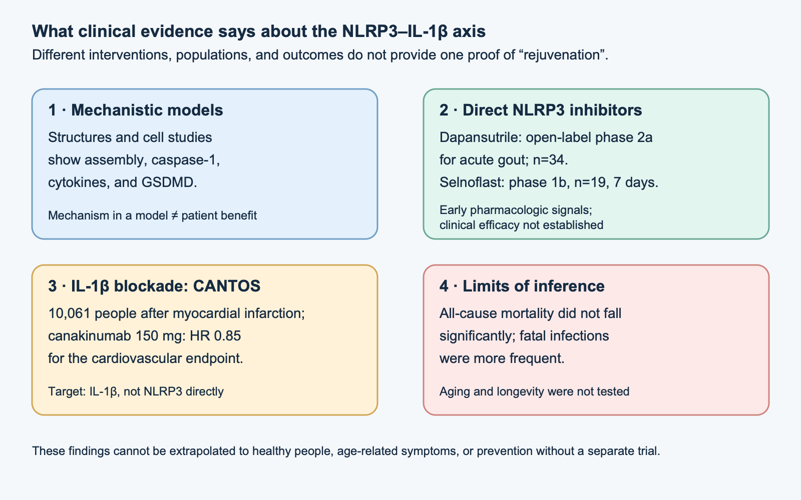
CANTOS enrolled 10,061 people with stable disease after a prior myocardial infarction and hsCRP of at least 2 mg/L. Participants were randomized to subcutaneous placebo or canakinumab at 50, 150, or 300 mg once every three months; median follow-up was 3.7 years. The primary composite endpoint included nonfatal myocardial infarction, nonfatal stroke, or cardiovascular death. Event rates were 4.50 per 100 person-years in the placebo group and 3.86 in the 150-mg group; for this dose the hazard ratio was 0.85 (95% CI 0.74–0.98; p=0.021). Only the 150-mg dose crossed the prespecified adjusted significance threshold for the primary endpoint. The differences were not accompanied by lower lipid levels; all-cause mortality did not change significantly. Fatal infections were more frequent in the pooled canakinumab groups.
CANTOS is an important trial of an innate immune pathway intervention and recurrent cardiovascular events in a specific high-risk group. The drug targeted mature IL-1β, not NLRP3, ASC, caspase-1, or GSDMD. Because IL-1β may arise from different cellular sources and processors, the antibody’s effect alone does not identify the inflammasome as the necessary mechanism. The trial also did not measure biological age and was not a lifespan-extension study. Saying “CANTOS proved rejuvenation” goes far beyond its design and outcomes.
### Direct NLRP3 inhibitors: early clinical signals
Dapansutrile (OLT1177) is an oral selective NLRP3 inhibitor. In an open-label adaptive phase 2a study during a gout flare, 34 adults received 100, 300, 1,000, or 2,000 mg per day for eight days; 29 were included in the per-protocol analysis. Mean self-reported pain fell from baseline, but there was no placebo comparison, groups were small, doses were assigned sequentially, and gout flares may improve over time and with concomitant care. The observed improvement therefore does not define a causal effect size. Adverse events were reported in 25 of 34 participants; 45 events and two serious events considered unrelated by investigators were reported. These data cannot be summarized as “there were no side effects” or as confirmation of long-term gout prevention.
In a randomized phase 1b study, selnoflast was studied in 19 adults with moderately to severely active ulcerative colitis. Participants were assigned 2:1 to selnoflast or placebo for seven days; the selnoflast dose was 450 mg once daily. Plasma and sigmoid-biopsy concentrations reached a calculated range associated with IL-1β inhibition in experimental measurements. After ex vivo LPS stimulation, IL-1β production in whole blood decreased in the drug group, but plasma IL-18 did not change, and there were no convincing between-group differences in the intestinal-tissue IL-1 signature or fecal markers. The study was short and small, and tissue target measurement was indirect; it examined safety, pharmacokinetics, and pharmacodynamics, not clinical remission or prevention of complications.
Neither of these early signals justifies using a drug in healthy people to “reduce age.” A direct NLRP3 inhibitor and an antibody to IL-1β are different interventions: the former limits sensor assembly/activity within the molecule’s pharmacological profile; the latter neutralizes one cytokine after it is produced. Their effects, tissue distribution, reversibility, and risks cannot be treated as interchangeable.
## Practical limits of biomarkers and interventions
Elevated CRP, IL-6, or a single IL-1β measurement is not a specific test of NLRP3 activity. In clinical practice a marker may help assess inflammation or follow an established disease, but a direct conclusion about the inflammasome would require evaluation of the relevant cell, mature cytokines, caspase processing, or other pathway components. Even an ASC speck assay or stimulated-cell analysis does not become a validated aging test merely because it measures a mechanism.
When an autoinflammatory disease is suspected, the pattern of recurrent episodes, age at onset, fever, skin and joint manifestations, family history, acute-phase tests, and consultation with a clinical immunologist or rheumatologist matter. A genetic variant must be interpreted alongside the clinical phenotype: a rare NLRP3 substitution is not automatically a pathogenic mutation, and a negative panel does not rule out every inflammatory cause. Independently interpreting a genotype as a prescription for an inhibitor is unsafe.
For chronic inflammatory disease, treatment is selected by diagnosis and trials in that population, not by one presumed cell-biology pathway. Neutralizing IL-1, suppressing NLRP3, inhibiting caspase-1, and changing GSDMD act at different nodes. Broadly weakening innate defense may alter responses to infection; fatal infections were a real safety signal in CANTOS. At the same time, not all inflammation is harmful: IL-1β and IL-18 contribute to infection control and tissue immunity, and a transient injury response differs from a chronic process.
The source material listed high doses of resveratrol, melatonin, magnesium, omega-3, acetyl-L-carnitine, and alpha-lipoic acid as though they formed an NLRP3-suppression regimen. A cellular mechanism or preclinical publication about a substance does not establish a human dose, clinical effect, or safety for that purpose. This article does not propose a supplement protocol or replace treatment for gout, CAPS, cardiovascular disease, or inflammatory bowel disease. Nutrition, sleep, movement, and smoking cessation can be discussed on their own medical grounds; they should not be called proven pharmacological blockade of the inflammasome.
## How to read the evidence
First check what the intervention engaged: NLRP3, IL-1β, IL-1R, caspase-1, or another component. Then separate a cell or animal model from a human trial, short-term target engagement from a change in symptoms, and a symptom outcome from complications, mortality, and longevity. A clinical report should be assessed for population, control, duration, dose, prespecified endpoint, missing participants, adverse events, and co-funding. In a small study, absence of a signal does not prove absence of an effect, but a biologically plausible mechanism does not become a positive clinical result.
| Observation | Appropriate level of inference |
|---|---|
| NLRP3 assembly, cytokine cleavage, or GSDMD pores in a cell | Supports the mechanism in that model |
| Genetic knockout or inhibitor in an animal disease model | Supports a causal contribution of the target under those model conditions |
| Lower IL-1β during brief pharmacological exposure | Pharmacodynamic signal; not necessarily clinical benefit |
| CANTOS with canakinumab after prior myocardial infarction | Effect of IL-1β blockade on some cardiovascular outcomes in the selected population |
| Any of the above on its own | Not proof of rejuvenation, longer life, or universal prevention of age-related disease |
## Method and limits of this review
This is a narrative review of structural and functional studies, disease models, and clinical trials, not a systematic search. On 9 October 2026, targeted checks were made of primary publications on NLRP3 structures, alternative activation in monocytes, the CANTOS and dapansutrile clinical trials, and selnoflast pharmacokinetics and pharmacodynamics. No systematic protocol, formal risk-of-bias assessment, GRADE, or external peer review was conducted. The main limitations are the predominance of cell and animal evidence linking NLRP3 to inflammaging and the absence of a direct NLRP3-inhibitor trial with human aging or longevity outcomes. Mechanistic and clinical conclusions above are separated by evidence type and intervention target.
FAQ
Does CANTOS prove that NLRP3 causes inflammaging?
No. CANTOS tested IL-1β neutralization in high-risk people after myocardial infarction. It was not a direct study of an NLRP3 inhibitor and did not measure aging or longevity.
Does a rise in CRP mean NLRP3 is active?
No. CRP reflects systemic inflammation and is not a specific test of NLRP3 assembly. Clinical context and assessment of the particular disease are needed.
Is dapansutrile established for self-treatment of gout?
No. The early study was open-label and had no placebo control. It does not establish standard treatment or flare prevention.
Did selnoflast show remission of ulcerative colitis?
No. The short phase 1b assessed safety, pharmacokinetics, and pharmacodynamics in a small group, not clinical remission.
Does NLRP3 always lead to pyroptosis?
No. GSDMD pore formation may accompany cytokine release, but cell fate depends on the extent of damage and membrane repair.
Are there supplements with a proven dose to suppress NLRP3 and rejuvenate?
No such protocol is established in the clinical evidence reviewed. Cellular mechanisms and preclinical results do not define a safe and effective dose for that goal.