Telomeres, p53–PGC and mitochondrial energy — NutriFit
Telomeres, p53 and mitochondria: linking genome protection to cellular energy
Telomere protection, DDR and p53–PGC connect genome status with mitochondrial energy. Shelterin, BER, mouse experiments, telomere disorders and the limits of intervention claims.
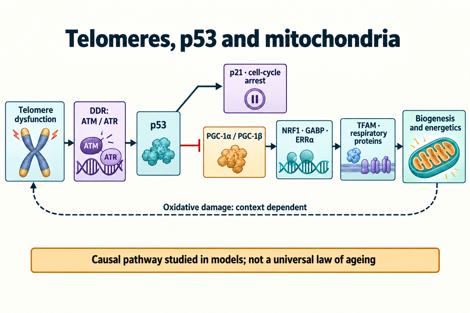
A chromosome end is more than a stretch of DNA whose length can be compared with age. Telomeric repeats, end architecture and shelterin proteins together determine whether a cell treats that end as a normal structure or as damage. Loss of protection activates the DNA damage response, which can restrict division, induce senescence or apoptosis and remodel metabolism. In certain experimental settings, p53 connects this signal to repression of PGC-1α and PGC-1β, coactivators involved in maintaining mitochondria.
Sahin and colleagues and Jaskelioff and colleagues established causal relationships in particular mouse models of telomerase deficiency. Their findings illuminate telomere dysfunction; they do not establish a single explanation for all human ageing or make telomere extension an established treatment. Telomere length, end protection, telomerase activity, metabolic function and a clinical outcome are connected measurements, but they cannot substitute for one another.
1. Why copying a genome has limits
From Carrel's cultures to replicative senescence
Early ideas about immortal cell cultures drew partly on Alexis Carrel's experiments. Leonard Hayflick and Paul Moorhead's 1961 study demonstrated a limited replicative capacity of normal human diploid cells in culture, leading to the concept of the Hayflick limit. The number of population doublings depends on starting material, culture conditions and the definition of growth arrest. Several dozen doublings describe particular systems, not a universal constant for every cell. WI-38 and MRC-5 also belong to different stages of this history and should not both be assigned to the same 1961 experiment.
A senescent cell may remain alive and metabolically active despite a sustained restriction of proliferation. SA-β-gal measured at pH 6, chromatin changes including SAHF in some models, and the senescence-associated secretory phenotype, SASP, help characterize this state. None alone diagnoses senescence universally. Replicative senescence, stress-induced premature senescence and responses to oncogenic signals such as HRASV12 can converge on DDR and p53/p21, while differing in the initiating lesion and cellular setting.
End replication and processing newly formed ends
DNA polymerase extends an existing 3′-OH and synthesizes DNA in the 5′→3′ direction. On the lagging strand, polymerase α–primase generates primers for Okazaki fragments. Removing the final RNA primer can leave no suitable neighbouring 3′ end from which to replace the terminal region. This underlies the end-replication problem discussed by Watson and Olovnikov in the early 1970s.
The modern mechanism includes more than loss of the last primer. RNase H and FEN1 participate in primer processing, while formation of the postreplicative 3′ overhang requires regulated end processing involving factors such as Apollo/DCLRE1B and EXO1. Telomerase adds repeats to the G-rich strand, and CST coordinates complementary C-strand fill-in. Losses of tens or hundreds of base pairs per division in selected cultures therefore cannot be assigned to every division in an organism. Sufficient telomerase activity can substantially alter the balance between extension and processing.
TTAGGG repeats, the overhang and G-quadruplexes
Human telomeres contain arrays of repeats, schematically:
5′3′−(TTAGGG)n−3′−(AATCCC)n−5′
The G-rich strand forms a single-stranded 3′ overhang. Its length, like the length of the entire telomeric tract, varies with cell type and cell-cycle stage. Tracts of several to more than ten kilobases and overhangs of tens to hundreds of nucleotides are ranges from particular measurements, not mandatory dimensions of each end. Mean leukocyte telomere length is also affected by the composition of circulating cell populations and does not directly reveal every chromosome end in the heart or brain.
Sequences containing neighbouring guanines can form G-quadruplexes. Guanine quartets use Hoogsteen bonding and are stabilized by cations, especially K⁺. These structures affect enzyme access, replication and protein binding. The ability of an isolated sequence to form a quadruplex does not mean all telomeres permanently occupy that conformation.
T-loops and D-loops: protecting an accessible end
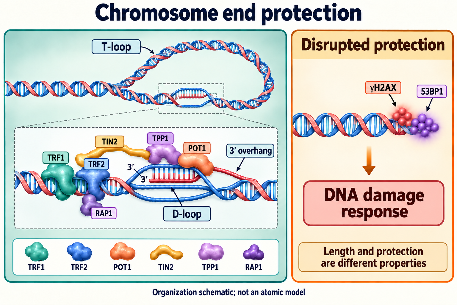
Griffith and colleagues' 1999 electron-microscopy study showed large T-loops in telomeric DNA. A single-stranded 3′ overhang invades a duplex telomeric region and pairs with its complementary strand; local displacement of the other strand forms a D-loop. This arrangement helps distinguish a natural chromosome end from a free double-strand break.
Protection is dynamic. The end must undergo replication, maturation and, where appropriate, telomerase-mediated extension. T-loops and their associated proteins change over time. Here, D-loop describes local strand displacement within a telomere; it should not be confused with the mitochondrial DNA regulatory D-loop region discussed later.
2. Shelterin organizes chromosome-end protection
Six components and what structural records establish
Shelterin comprises TRF1, TRF2, POT1, TIN2, TPP1 and RAP1. TRF1 and TRF2 recognize duplex telomeric repeats through DNA-binding domains and contain TRFH domains involved in dimerization and partner interactions. TRF1 helps replication proceed through a difficult telomeric substrate with helicases and other factors. TRF2 is particularly important for end protection and restricting ATM signalling and classical end joining.
POT1 binds single-stranded telomeric DNA. TPP1 connects it to the wider system and contributes to telomerase recruitment and processivity, including through its TEL patch. TIN2 bridges components, while RAP1 associates with TRF2 and affects selected recombination and protection pathways. These functions overlap and depend on the model; a rigid assignment of one entirely separate task to each protein is inadequate.
Structural records represent particular experimental constructs. PDB 1XJV contains human POT1 bound to single-stranded telomeric DNA, whereas 2I46 describes human TPP1. 3BQO contains a TRF1 TRFH domain with a TIN2 peptide, and 5XYF is a TIN2–TPP1–TRF2 complex. None is an atomic model of an entire T-loop. A binding affinity measured for an isolated domain is not a fixed affinity of the complete in vivo system.
Why a normal end should not activate ATM or ATR
TRF2 loss can activate ATM while a repeat tract remains present. Loss of POT1 protection at single-stranded DNA facilitates RPA binding and an ATR response. These observations separate DNA quantity from protection quality. Concealing the end in a loop matters, but restriction of DDR also depends on protein interactions, chromatin access and end processing.
Shelterin does not eliminate all repair. Replication intermediates must be resolved, damaged bases processed and newly replicated ends matured. The system permits necessary reactions while limiting hazardous outcomes, especially joining different chromosomes.
Classical end joining, alternative pathways and recombination
Classical non-homologous end joining, c-NHEJ, uses Ku70/Ku80, DNA-PKcs and XRCC4–ligase IV. Treating a normal telomere as a break can cause chromosome fusions and breakage–fusion–bridge cycles; shelterin restricts these events. Alternative joining, including POLQ-dependent microhomology-mediated reactions, has different participants. PARP1 and ligase III contributions are context dependent and should not be equated with c-NHEJ.
Homologous recombination is regulated rather than erased from telomere biology. It helps process some intermediates, and alternative lengthening of telomeres, ALT, in a subset of tumours uses recombination-based mechanisms. RAD51, BLM, SLX4 and other factors perform distinct jobs. SLX4 is not simply a switch that blocks RAD51, and a protected telomere does not permanently prohibit every homologous reaction.
3. From a dysfunctional end to p53
The shortest end, uncapping and TIFs
One or a few critically compromised ends can matter more than the mean length of all telomeres. Damage or loss of protection can also cause uncapping without appreciable prior shortening. There is no universal threshold of 1.5 or 3 kb at which every end in every tissue becomes irreparable.
Telomere dysfunction-induced foci, TIFs, are identified by colocalization of telomeric signals and DDR markers such as γH2AX or 53BP1. Telomere FISH can be combined with immunofluorescence, or telomeric protein markers used with their methodological limitations. A TIF indicates a response at a telomeric region; it does not independently measure length, identify the initiating damage or establish an irreversible cell fate. Persistent foci should be distinguished from dynamic signals during replication and end processing.
MRN–ATM and RPA–ATR respond to different substrates
MRN comprises MRE11, RAD50 and NBS1 and participates in recognizing and processing double-strand damage and recruiting ATM. ATM Ser1981 autophosphorylation is commonly measured as an activation marker. Regulation cannot, however, be reduced to obligatory dissociation of a single dimer following one modification; break architecture, MRN, chromatin and additional modifications matter.
ATR responds chiefly to RPA-coated single-stranded DNA and interacts with ATRIP. The RAD9–RAD1–HUS1, or 9-1-1, complex, loading by RAD17 and TOPBP1-mediated activation support signal transmission. Other activation routes include ETAA1. ATM and ATR therefore provide useful pathway outlines rather than two entirely independent linear chains. End resection, fork stalling and cell-cycle stage can alter their relative contributions.
γH2AX, MDC1 and recognition of damaged chromatin
Phosphorylation of histone variant H2AX at Ser139 produces γH2AX. This is a posttranslational chromatin modification, not by itself evidence for inherited epigenetic memory. MDC1 recognizes the relevant phosphorylated region through BRCT domains and supports expansion of the DDR platform. RNF8 and RNF168 organize ubiquitin signals involved in recruiting downstream factors.
53BP1 reads a combination of marks: its Tudor domain recognizes H4K20me2, while its UDR region recognizes ubiquitinated H2A, particularly H2AK15ub. The Tudor domain should not be assigned K63-polyubiquitin as its principal specific ligand. Replicated chromatin, accessibility of marks and opposition by BRCA1-associated processes influence repair choices. The presence of 53BP1 in a focus does not make p53 a direct physical constituent of every such focus.
Chk2, Chk1 and cell-cycle restriction
ATM-dependent Chk2 phosphorylation at Thr68 promotes FHA-mediated interactions, dimerization and activating modifications including Thr383/Thr387. ATR can phosphorylate Chk1 at Ser317 and Ser345, with mediators such as Claspin linking this response to replication stress. These effector kinases act on cell-cycle regulators and help coordinate repair with a pause in division.
They do not dictate one unavoidable fate. Limited transient damage can be followed by resumed proliferation, whereas prolonged or severe signalling can lead to senescence, apoptosis or other outcomes. p53, p21, p16/RB, cell identity, mitochondrial condition and survival factors affect the decision. Identical Chk1 or Chk2 phosphorylation sites do not guarantee identical outcomes in cultured fibroblasts and intact tissues.
p53 and MDM2: stabilization is a regulated process
Under basal conditions, E3 ligase MDM2 binds the p53 transactivation region, restricts its activity and promotes ubiquitination and proteasomal removal. PDB 1YCR contains MDM2 with a p53 fragment; Phe19, Trp23 and Leu26 form important hydrophobic contacts. It does not represent a complete stress-signalling complex. C-terminal p53 lysines, including positions 370, 372, 373, 381, 382 and 386, undergo modifications with functions extending beyond one MDM2 reaction.
During DNA damage, ATM, checkpoint kinases and other regulators modify both p53 and MDM2. p53 Ser15/Ser20 and MDM2 Ser395 phosphorylation are parts of this network. Acetylation and feedback dynamics also change stability and activity. Protein lifetime increases under particular conditions, but a fixed four-to-six-hour lifetime or complete irreversible dissociation is not a universal rule.
p53 functions as a tetramer; PDB 1C26 describes its tetramerization domain. Through p21 it restricts the cycle, while in other settings it regulates apoptotic and metabolic genes. Normal p53 can support aspects of metabolic adaptation and respiration, including through SCO2 and TIGAR. PGC repression in the context below therefore does not make p53 universally harmful to mitochondria or justify switching it off therapeutically.
4. What the telomere–p53–PGC experiments established
Why telomerase-deficient mice develop different phenotypes
Deleting Tert or Terc does not immediately remove every telomere. Many laboratory mice begin with longer telomeres than humans, so severe phenotypes accumulate across successive telomerase-deficient generations. Timing depends on genetic background, starting lengths and breeding design. A rule that the first three generations are always normal and the fourth always collapses is too categorical.
Defects are readily expected in rapidly renewed tissues because regenerative capacity becomes limited. Findings in less proliferative organs raised a further question: how can nuclear stress weaken heart or liver function beyond reducing the number of dividing cells? Investigators addressed transcriptional programmes, mtDNA and functional measurements rather than relying on length alone.
Sahin 2011: causal evidence with a defined scope
Sahin and colleagues linked telomere dysfunction in telomerase-deficient mouse tissues and cells with reduced PGC-1α/β and mitochondrial and metabolic changes. Genetic and functional interventions supported a causal contribution: delivery of Tert or PGC-1α and changes in p53 status improved selected measurements. Recovery varied by endpoint, and the paper also described partial rescue.
Complete recovery of expression in one comparison does not establish complete organismal restoration. The cardiac experiment included doxorubicin-induced cardiomyopathy, so improved contractile function cannot be generalized to elimination of all age-related heart failure. Other consequences of telomere dysfunction retain independent contributions.
PPARGC1A and PPARGC1B promoter repression
PPARGC1A and PPARGC1B encode PGC-1α and PGC-1β. Reporter assays and chromatin immunoprecipitation, ChIP, supported p53-dependent repression and p53 binding to regions of the corresponding mouse promoters. This was not a ChIP-seq demonstration and does not assign a single percentage of repression to an entire human body. Experimental coordinates describe particular promoter fragments relative to the chosen transcriptional start.
A classical p53 response-element consensus can be written:
5′−RRRCWWGYYY−(N)0–13−RRRCWWGYYY−3′
R denotes a purine, Y a pyrimidine and W either A or T. This is a degenerate motif rather than the exact sequence of every functional site. Chromatin accessibility, site quality and number, and p53 partners affect regulation. Sin3/HDAC-containing corepressor interactions exist in p53 biology, but an obligatory mSin3A–HDAC1/2 sequence should not be presented as the single directly demonstrated mechanism of this study.
PGC coactivators do not switch all mitochondria on or off
PGC-1α was described in the late 1990s; PGC-1β was identified later. They interact with transcription factors and activation machinery including p300/CBP and Mediator, rather than recognizing DNA sequences as independent sequence-specific factors do. Partners include NRF1, GABP, ERRα and PPAR proteins, with tissue and cell state determining the programme.
NRF2 requires careful naming. In the classical nuclear respiratory-factor literature, NRF-2 means the GABP complex containing GABPA and GABPB. The antioxidant regulator NRF2 encoded by NFE2L2 is a different protein. Conflating them merges two connected but distinct programmes. Lower PGC reduces coactivation of selected targets; it does not necessarily eliminate all NRF proteins, ERRα or respiratory components.
TFAM, mtDNA packaging and its regulatory D-loop
TFAM contains HMG domains, binds and bends mtDNA and participates in packaging and transcription. Its ratio to mtDNA matters: deficient maintenance and excessive packaging can have different effects. Transcription also requires POLRMT and TFB2M, with TEFM supporting elongation. Replication uses POLG, TWINKLE and other factors, while RNA primers and the regulatory region connect transcription with replication.
Reduced TFAM can accompany changes in mtDNA copy number and gene expression, but “D-loop replication is completely blocked” is an inadequate description. D-loop denotes a regulatory region and mitochondrial-genome structure, not an independent enzymatic step. Copy number, nucleoid quality, transcription rate and respiratory capacity require separate measurements.
5. Respiratory function and energy balance
Oxidative phosphorylation uses two genomes
Human mtDNA encodes 13 oxidative-phosphorylation proteins: seven complex I components, complex III cytochrome b, three complex IV subunits and two ATP-synthase components. Most respiratory proteins are nuclear encoded, and complex II is entirely nuclear encoded. Coordination of both programmes supports assembly and renewal, so mtDNA depletion and changes in nuclear transcription can act together.
Within complex I, membrane ND subunits including ND1, ND4 and ND6 cooperate with nuclear components. NDUFS3 belongs to the core hydrophilic region, whereas NDUFB8 belongs to the membrane arm; describing both as equivalent peripheral subunits is incorrect. Complex III includes UQCRC2, Rieske iron–sulfur protein UQCRFS1 and mtDNA-encoded cytochrome b. Complex IV combines MT-CO1 and MT-CO2 with nuclear subunits such as COX4; CuA and haem a3–CuB centres have distinct functions.
ATP synthase's catalytic sites are chiefly associated with F1 β subunits, while α subunits have another role. MT-ATP6 encodes membrane subunit a, involved in the proton pathway; c subunits are encoded by nuclear ATP5MC genes. MT-ATP8 is also mitochondrial encoded. These distinctions matter: loss of one programme does not directly remove every subunit or establish the same defect in all five complexes.
Supercomplexes, potential and respiratory control
Complexes I, III and IV can assemble into supercomplexes, including I–III₂–IVₙ organizations. Their arrangement can influence stability and interactions, but individual complexes also function. The extent of substrate channeling and the role of supercomplexes in limiting ROS remain research questions. Every electron need not move through one closed channel, and disassembly need not produce an identical explosive leak.
For a proton moving from the intermembrane space to the matrix, define Δψ as matrix potential relative to the outer side of the inner membrane and ΔpH as matrix pH minus outside pH:
ΔGH+,in=FΔψ−2.303RTΔpH
A negative electrical term and a more alkaline matrix generally favour proton entry. Loss of potential can reflect lower respiratory capacity, uncoupling, increased ion conductance or combined causes; potential alone cannot distinguish them. In a classical isolated-mitochondria experiment, respiratory control ratio is:
RCR=JO2,state4JO2,state3
State 3 denotes ADP-stimulated respiration, while state 4 needs a specified protocol, for example after ADP consumption. Substrates, isolation quality and how resting respiration is established influence the ratio. Lower RCR identifies neither one unique cause nor a universal clinical threshold.
ATP, adenylate kinase and energy charge
Impaired oxidative phosphorylation can reduce ATP availability, but cells may change consumption, increase glycolysis and redistribute substrates. Compensation differs between working muscle, resting fibroblasts and liver cells. Total ATP measurement also differs from measuring synthesis flux and local energy availability.
Adenylate kinase links adenine nucleotide pools:
2ADP⇌ATP+AMP
Adenylate energy charge, AEC, normalizes their composition:
AEC=[ATP]+[ADP]+[AMP][ATP]+0.5[ADP]
With nonnegative concentrations, AEC lies between 0 and 1. Rising AMP and ADP can signal energetic stress, including through AMPK, but this ratio does not show the size of the entire pool. Lactate may increase with glycolysis or altered redox balance; it is not specific to telomere dysfunction. Severe ATP defects in a model do not imply mandatory systemic lactic acidosis in every person with short telomeres.
Antioxidant enzymes form a distributed network
PGC-related programmes can support antioxidant expression; St-Pierre and colleagues' 2006 study demonstrated contributions to oxidative-stress responses in experimental systems. The network includes SOD2, glutathione peroxidases, PRDX3/PRDX5 and other enzymes also regulated by FOXO, NFE2L2 and tissue-specific factors. Catalase is predominantly peroxisomal and should not be depicted as the main matrix enzyme in every cell.
Protection also depends on regeneration of glutathione and thioredoxin and reducing equivalents including NADPH. Raising one enzyme does not guarantee improvement of the whole network; peroxide production, removal and compartment all matter. Reduced PGC likewise does not imply disappearance of all antioxidant activity.
ROS sites depend on the respiratory state
Sites in complexes I and III can transfer electrons to oxygen. Their contributions depend on reduction state, substrates, potential, flow direction and inhibitors. High-potential reverse electron transport and defective forward transport provide different settings. A damaged mitochondrion does not necessarily produce more ROS in every condition; severely reduced flux can diminish some sources.
For selected semiquinone states, balanced electron-transfer schemes are:
FMNH∙+O2Q∙−+O2⟶FMN+H++O2∙−⟶Q+O2∙−
These are simplified chemical expressions; enzyme microstates need not share identical protonation. Where iron is available, hydrogen peroxide can participate in the Fenton reaction:
Fe2++H2O2⟶Fe3++HO∙+OH−
Superoxide also reacts with NO to form peroxynitrite. Oxidation, nitration and damage to proteins, lipids or DNA depend on local chemistry and defence. The hydroxyl radical reacts near its formation site; depicting it travelling from the matrix across the cell to a telomere is inappropriate.
6. Telomere oxidation and feedback
Guanine vulnerability is not a genomic lightning rod
Guanine is more readily oxidized than other canonical bases under comparable conditions. Adjacent guanines can affect lesion distribution and charge transfer within DNA. Oxidation potentials nevertheless depend on environment, measurement method and molecular context. They should not be relabelled as gas-phase ionization potentials and used to imply that every oxidative equivalent must migrate to telomeres.
TTAGGG repeats make telomeric DNA an important oxidation substrate. Replication, DNA conformation and protein contacts also affect vulnerability. Hydrogen peroxide can transmit signals and move between compartments, but removal and local reactions constrain its path. Nuclear damage depends on these processes rather than solely on how much mitochondrial ROS is formed.
8-oxoG and TRF binding: damage can precede shortening
8-Oxoguanine, 8-oxoG, is a damaged base; 8-oxo-2′-deoxyguanosine, 8-oxodG, is its corresponding nucleoside. Their names are related but chemically distinct. Conformation and pairing of the lesion influence copying accuracy, including the risk of adenine misincorporation. Oxidation does not inevitably force all DNA into one syn conformation.
Opresko and colleagues in 2005 examined how oxidative damage of telomeric substrates affects TRF1 and TRF2 binding in vitro. The response depends on lesion position, density and substrate construction. It does not establish immediate eviction of 90% of all shelterin proteins in a living cell or inevitable unfolding of every T-loop. Oxidative damage can impede replication and trigger DDR before appreciable length loss, reinforcing the need to distinguish damage from shortening.
OGG1 and BER operate at telomeres
OGG1 recognizes and removes 8-oxoG through base excision repair, BER. Subsequent steps process the abasic site, fill the gap and ligate DNA. Short-patch repair involves APE1, polymerase β, XRCC1 and a ligase, while alternative routes have different participants. Telomeric context affects access and completion, but OGG1 is not intrinsically unable to operate there.
G-quadruplexes, protein binding, chromatin and replication can change the efficiency of individual steps. Abasic sites and single-strand breaks produced during BER can themselves promote replication stress when processing is poorly coordinated. Persistent telomeric DDR therefore does not prove that base repair is absent. A repair slowdown or persistence time must be assigned to a specific experiment rather than an obligatory weeks-long lesion in every telomere.
A possible amplification loop, not one law of ageing
Under some conditions, weakening selected mitochondrial and defence functions increases oxidative injury that can affect telomeres again. PGC's arrow indicates its normal support of the programme; the inhibitory sign indicates p53-dependent repression. Direction and strength of feedback depend on tissue, substrates, repair and compensation.
The circuit is useful for experimental design: intervene at different points and measure whether predicted outcomes change. It does not establish that every ageing cell follows the same loop or that this circuit explains exponential increases in all age-related risks. Mitochondrial injury can begin independently, and DDR can affect tissues through inflammation, cell loss and impaired regeneration.
7. Tissue-specific consequences
Heart: reserve capacity and calcium cycling
Cardiomyocytes continually require ATP for contraction, ion pumps and restoration of calcium cycling. Fatty-acid, carbohydrate and other substrate use changes with workload and systemic state. Impaired PGC-related programmes can restrict metabolic flexibility and respiratory reserve. Limited energy availability can weaken SERCA2a/ATP2A2 and other ATP-dependent functions, but cardiac dysfunction is not reducible to one pump.
Calcium overload, oxidation and altered mitochondrial permeability can amplify injury. Cyclophilin D affects permeability-transition sensitivity, whereas BAX/BAK-associated outer-membrane changes participate in other death pathways; these are not one obligatory sequence for all apoptosis. A telomere-dysfunction model identifies a possible susceptibility mechanism, not a fixed ejection fraction or inevitable clinical cardiomyopathy for every patient.
Muscle: fibre identity, proteostasis and satellite cells
PGC-1α contributes to oxidative programmes and adaptation of muscle fibres to exercise. Creatine kinase, including CKM, and phosphocreatine buffer energy demand. Energetic impairment can change protein quality and the balance of synthesis and breakdown. FOXO-linked pathways, MuRF1/TRIM63 and atrogin-1/FBXO32 participate in selected atrophy models, but are not exclusively downstream of short telomeres.
Human age-related sarcopenia is not universally selective loss of type I fibres. Muscle, innervation, activity and disease influence fibre patterns, and vulnerability of type II fibres is often important. Satellite cells, commonly characterized with PAX7, contribute to repair; their regenerative defects can follow a different course from those of mature fibres. Biogenesis must accompany removal of damaged mitochondria. PINK1/Parkin-dependent mitophagy is one such quality-control route rather than the only renewal system.
Liver: lipid oxidation and glucose production
In liver, PGC-related programmes support both mitochondrial function and regulation of glucose production. PPARα, CPT1A and β-oxidation enzymes affect fatty-acid use, while HNF4A and FOXO1 interact with coactivators in regulating PCK1, G6PC and other gluconeogenic processes. Sahin's experiments connected changes in this programme with fasting responses and glucose production.
Lipid accumulation, diacylglycerol and ceramide signalling, insulin resistance and inflammation can intersect with mitochondrial impairment. This does not make “short telomere–steatohepatitis–hypoglycaemic coma” an obligatory sequence. Steatosis, fibrosis and metabolic disease have multiple causes; glucose production can instead be excessive in some diabetes settings. Tissue identity therefore also changes the meaning of manipulating PGC.
Haematopoiesis and neural progenitors
Haematopoietic stem cells must combine quiescence, genome preservation and the ability to regenerate blood cells. Persistent telomeric DDR can restrict proliferation, induce death or change function, particularly in telomere disorders. Metabolic and niche signals add complexity: cell count, repopulating activity and transcriptional profile require separate assessments.
Mouse neural progenitors are studied in the subventricular zone, SVZ, and subgranular zone, SGZ, using markers such as SOX2, Nestin and DCX. These mark different states and are not interchangeable. Recovery of selected neural and glial features in a mouse model does not promise restoration of human neurogenesis, memory or every brain connection. Species differences, regional organization and age are especially relevant when transferring findings about the SVZ and olfactory pathways.
8. Telomerase reactivation: the reversible defect
Jaskelioff's inducible TertER/ER system
Mariela Jaskelioff and colleagues used a genetic system in which TERT is fused to a modified receptor domain and regulated by 4-hydroxytamoxifen. TertER/ER allowed telomerase function to be restored after prolonged limitation. Online publication in 2010 and the 2011 Nature issue refer to the same study, not two independent experiments.
Later generations had marked telomere dysfunction because of this particular genetic design. They were not simply normally aged mice given an over-the-counter product. Restoring an experimentally deficient function tests consequences and reversibility of that defect. It does not establish that ordinary ageing is entirely a telomerase deficiency, and it provides no human 4-hydroxytamoxifen protocol.
Damage reduction and functional recovery are distinct experiments
Reactivation reduced signs of telomeric damage and improved selected cellular and tissue functions in this model. Less DDR can reduce p53 stabilization and relieve some restrictions, but a complete sequence in which all TIFs vanish, all p53 is removed and all PGC/TFAM expression returns cannot be assumed to have been measured in every organ.
Jaskelioff's study concerns inducible reactivation and tissue recovery; Sahin's separately examines p53–PGC and mitochondrial effects. They support compatible biology, but assembling them into one fully demonstrated pathway requires additional evidence. Obligatory HDAC-dependent reactivation of every PGC promoter does not follow directly from improved regeneration.
Partial reversibility and the tumour barrier
Reported improvements involved intestine, spleen, testes and nervous-system features, including selected progenitor, myelination and olfactory outcomes. A period of several weeks showed that some consequences of a chronic telomeric defect remained reversible. This is a substantial mechanistic result, not elimination of every fibrosis, mutation, tumour clone or age-related change.
Calling the connection a rheostat can express regulated function, but does not establish a single control that turns organismal age backwards. Telomerase supports division of normal progenitors and many tumour cells. p53 both constrains damaged cells and protects against cancer. Genetic p53 removal to test a mechanism cannot be presented as a safe rejuvenation strategy.
9. Humans: telomere disorders and biomarker limits
Inherited disturbances of telomere biology
Dyskeratosis congenita and the wider telomere-disorder spectrum involve variants in DKC1, TERC, TERT, TINF2 and other genes. DKC1 contributes to H/ACA ribonucleoproteins and telomerase RNA stability, as well as other RNA processes. NOP10, NHP2 and WRAP53/TCAB1 affect complex assembly or localization; RTEL1 and PARN introduce further mechanisms. Variants in all these genes should not be treated as the same defect in one enzymatic step.
The classic triad of nail dystrophy, abnormal skin pigmentation and mucosal leukoplakia is neither present in every patient nor the whole clinical spectrum. Hoyeraal–Hreidarsson syndrome may involve developmental abnormalities, cerebellar hypoplasia, immune and haematopoietic problems. Revesz syndrome includes characteristic retinal disease. Coats plus involves CST components including CTC1 and complex replication and telomere-maintenance functions. These syndromes require differentiation rather than one nutrigenetic explanation.
Pathogenic telomere-gene variants also occur in familial pulmonary fibrosis and bone-marrow or liver disease. Frequencies depend on family selection, phenotype and the genes tested. A percentage from a pulmonary-fibrosis series cannot be assigned to all cirrhosis or every age-associated organ impairment.
A mitochondrial endophenotype is not a diagnosis
Cellular telomere-defect models can be assessed for respiration, mtDNA copies, membrane potential, metabolites and oxidative lesions. Combinations illuminate mechanisms but need not appear identically in every patient. Lactate, 8-oxodG or reduced fibroblast respiration is not a specific telomere-disorder test.
Methods for length include qPCR, terminal restriction-fragment analysis, FISH approaches and assays of individual short telomeres. Clinical Flow-FISH uses age-related distributions and specific leukocyte populations in specialized laboratories. Thresholds from one assay should not be mechanically transferred to another. Very short telomeres may support a diagnosis in the appropriate phenotypic setting, but “below 3 kb means one diagnosis and one correction regimen” is not a universal rule.
SNPs do not prescribe a nutritional intervention
TERT rs2853669 is studied in contexts including ETS binding and tumour promoter alterations. Effects depend on genetic and tissue background; findings do not establish one fixed reduction of telomerase activity for every carrier or validate a supplement programme. A common polymorphism also differs from a pathogenic variant causing a defined inherited disorder.
PPARGC1A rs8192678 is usually termed Gly482Ser, not Gly480Ser. Ling and colleagues' human-islet study examined genetic and epigenetic contributions to PPARGC1A expression and insulin secretion. The result does not assign the same functional effect to every tissue or population. An SNP association cannot determine an individual's NMN, NR or antioxidant dose.
Comorbidities, inflammation and different clocks
Observational studies associate shorter leukocyte telomeres with some cardiovascular and metabolic conditions. Smoking, inflammation, blood-cell composition, age, social factors and metabolic state affect interpretation. Genetic evidence and rare telomere syndromes help investigate causality, but different diseases and tumour risks can show different directions of association. Longer is not invariably better.
PGC's role differs among muscle, liver and pancreatic β cells. Reduced oxidative programming in one tissue does not justify unconditional stimulation of glucose production in another. In chronic obstructive pulmonary disease, basal and type II alveolar-cell injury intersects with smoking, inflammation and epithelial renewal. Microglial and SASP-associated signals, including IL-6, TNF and IL-1, interact with other neurological processes; they do not establish a single cause of Alzheimer's or Parkinson's disease.
Horvath, Hannum, PhenoAge and GrimAge models use DNA methylation and were trained for different tasks. They do not directly measure telomere length. Metabolic links to SAM and energy can connect with epigenetic regulation but do not establish one linear ATP-to-clock pathway. Differences among a telomere assay, epigenetic age and function are expected and require interpretation.
10. Experimental interventions: target, measurement and outcome
TA-65 and cycloastragenol: biomarkers are not rejuvenation
Cycloastragenol and TA-65 preparations are discussed as potential telomerase modulators. MAPK/ERK, Sp1 and c-Myc can participate in experimental responses; formulation, exposure and cell identity influence results. Animal pharmacokinetic measurements should not be assigned as universal human bioavailability.
Harley and colleagues' 2011 report examined a multicomponent programme observationally. It differs from Salvador and colleagues' randomized 2016 study, which enrolled 117 CMV-positive adults aged 53–87, with 97 completing one year. PBMC telomere measures were assessed using FISH; low-dose signals differed from inconsistent high-dose results. The study did not demonstrate restoration of p53–PGC in all tissues, longer lifespan or reduced clinical disease incidence.
No clinically significant change in selected laboratory safety measures over a year is not proof of long-term oncological safety in every population. Many tumours maintain telomeres through telomerase and some use ALT. This warrants contextual investigation, but also does not prove every telomerase activator causes cancer. Arbitrary eight-to-twelve-week cycles and nonspecific tumour-marker monitoring do not create a validated preventive protocol.
NAD⁺, NMN, NR and sirtuins: activity versus abundance
NAD⁺ participates in redox reactions and is a substrate for sirtuins and other enzymes. NMN and NR enter NAD-replenishment pathways; uptake, extracellular metabolism and interconversion depend on tissue and route. SLC12A8 has been proposed as an NMN transporter in certain models, but is not the single established explanation for oral NMN uptake in humans. NR transport and phosphorylation by NR kinases also matter, and available precursor forms change during metabolism.
SIRT1 modifies acetylation of PGC-1α and other proteins, while SIRT3 regulates selected mitochondrial targets including SOD2 and metabolic components. Deacetylating an existing protein does not equal restoring DDR-repressed PPARGC1A transcription. Increasing NAD⁺ therefore guarantees neither bypass of p53 restriction, removal of every TIF nor telomere extension. Cellular and mouse NAD–mitochondrial findings, including Gomes and colleagues' 2013 work, do not establish a universal human anti-ageing dose.
AMPK, metformin and AICAR
AMPK integrates adenine nucleotides, activation-loop phosphorylation and upstream regulators such as LKB1. It can affect PGC-1α, substrate use and energetic balance. Direct PGC modifications, SIRT1 interactions and HDAC4/5–MEF2 regulation contribute in some systems; these are not the sole obligatory route in every tissue.
Metformin can alter energetic and redox processes. Complex I inhibition matters in some experimental conditions but depends on concentration, accumulation and model; lysosomal and other signalling routes are also discussed. AICAR usually refers to AICA riboside, converted to the AMP analogue ZMP, rather than a universal direct ageing switch. ACC1/ACC2, malonyl-CoA and CPT1 connect AMPK to fatty-acid use, but systemic consequences depend on tissue. This mechanism alone does not justify medication based on telomere length.
MitoQ, astaxanthin and R-ALA
MitoQ combines a lipophilic triphenylphosphonium cation, TPP⁺, with a quinone moiety. A negatively charged matrix can favour cation accumulation. For an idealized equilibrium of a freely distributed monovalent cation:
Δψ=−FRTln([C+]outside[C+]matrix)
At 37 °C a tenfold concentration change corresponds to approximately 61.5 mV. At about −180 mV the ideal ratio is of order 10³, but does not guarantee a tissue drug concentration: lipid partition, binding, delivery and nonequilibrium behaviour change the interpretation. Depolarization reduces the driving force. Reduced mitoquinol influences selected oxidative reactions; it is neither a universal SOD2 substitute nor a demonstrated restorer of all telomeric processes.
Astaxanthin interacts with lipid environments, with orientation and effects depending on membrane and exposure. That does not prove direct protection of every telomere after oral intake. R-α-lipoic acid connects lipoyl reactions with redox biology, but a covalently bound enzyme cofactor and a free oral supplement are distinct states. Possible NFE2L2/NRF2 effects must be distinguished from GABP/NRF-2 in the biogenesis programme.
Comparing interventions and choosing measurements
Combining plausible targets does not establish synergy. Components and the combination must be compared within an appropriate design, exposure measured, and toxicity or altered cell composition excluded. A morning NAD precursor and evening antioxidant schedule, with doses derived from a biochemical diagram, is not validated correction of this circuit. The comparison preserves the distinction between mechanism and established outcome.
Intervention
Proposed target
What can be measured
Main inference limit
TA-65 / cycloastragenol
Telomerase activity and cellular signals
Enzyme activity, length distributions and cell properties
A telomere biomarker does not establish longer life or long-term safety
NMN / NR
NAD⁺ replenishment and dependent enzymes
NAD pools, metabolism, target acetylation and tissue function
Not restoration of every p53-repressed gene
Metformin / AICAR
Energy signals and AMPK-related processes
Energy flux, AMPK, substrate use and a defined functional endpoint
Exposure and clinical use cannot be derived from telomere length
MitoQ
Potential-dependent quinone delivery
Accumulation, redox reactions, respiration and oxidative damage
Delivery depends on potential; telomere-specific clinical benefit is separately unestablished
Astaxanthin
Lipid environments and selected oxidative processes
Exposure, membrane changes and defined endpoints
A membrane mechanism does not establish targeted telomere protection
R-ALA
Lipoyl and redox biology
Metabolic and redox measures with the chemical form specified
Free supplementation is not equivalent to an enzyme-bound cofactor
Length and telomere damage should be distinguished from mtDNA copy number, respiratory capacity and total ATP. Urinary or circulating 8-oxodG reflects heterogeneous sources and processing rather than a specific burden at every telomere. Lactate/pyruvate requires controlled collection and interpretation; inflammatory cytokines and SASP measures are also nonspecific. Clinical benefit requires functional and clinical endpoints, not only a changing surrogate.
11. Reading causality in this model
A telomeric end can connect genome status with metabolism through DDR and p53, with PGC coactivators providing one mechanism. Shelterin protection, replication, BER, mitochondrial redox balance and tissue renewal interact, but no one component describes all ageing. Ask what the initiating defect was, which tissue was studied, what intervention changed the outcome and what was actually measured.
Telomere extension, lower DDR, PGC recovery and improved tissue function are separate outcomes that can align in one experiment and diverge in another. Genetic mouse models provide strong mechanistic tests; human observations require attention to confounding and population differences. This article explains mechanisms and experimental limits rather than prescribing a universal supplement or drug course.
Method note. The supplied scientific material was rewritten while retaining its thematic coverage, chemical and energetic models and comparison table. Specific checks addressed Sahin's experiments, Jaskelioff's model, PDB records, Gly482Ser and the TA-65 design. GABP/NFE2L2 confusion, ATP-synthase subunit assignments and categorical BER claims were corrected. This is not a systematic review or a complete repeat audit of every bibliography entry. Some primary pages were unavailable for full reading; those gaps were not filled with assumed numbers, efficacy or safety. Illustrations are conceptual schematics rather than atomic structures or a universal sequence of ageing.
FAQ
Are telomere length and protection the same?
No. Shelterin loss or damage can cause DDR without appreciable shortening. Mean length also does not identify the condition of every individual end.
Can p53 be switched off to restore mitochondria?
Genetic deletion tests mechanism, but p53 protects against tumours and participates in normal adaptation. These experiments do not justify therapeutic inactivation.
Does telomerase reactivation demonstrate human rejuvenation?
No. Jaskelioff studied a particular telomerase-deficient mouse genotype. Reversing part of that phenotype does not establish a universal human mechanism or clinical benefit.
Can a telomere test choose TA-65, NMN or MitoQ?
The cited studies do not validate that regimen. Exposure, biomarkers, tissue function and clinical outcomes must be distinguished; length alone is insufficient.