R-ALA, lipoamide, and pyruvate dehydrogenase: what is established — NutriFit
R-alpha-lipoic acid: PDC lipoamide, redox chemistry, and the limits of clinical evidence
Mitochondrial lipoylation and the pyruvate dehydrogenase catalytic cycle, distinguished from the pharmacology of free thioctic acid; clinical neuropathy trials, exposure, and safety.
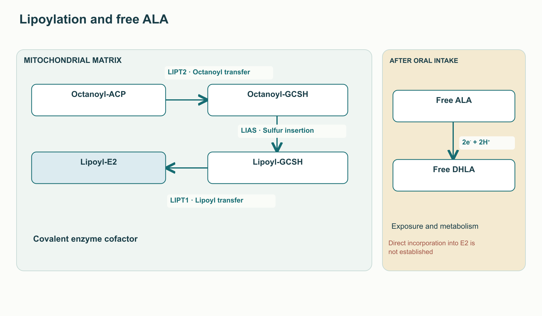
Lipoic acid occurs in human biochemistry in two fundamentally different states. Inside mitochondria, the R-lipoyl group is covalently attached to lysine residues on multienzyme proteins and carries intermediates between their active sites. Oral alpha-lipoic acid—either the free R-enantiomer or the R/S racemate—is introduced from outside the body, undergoes absorption and metabolism, and has been studied primarily as a pharmacological intervention. Conflating these states is the central error in the source material: the fact that lipoamide is essential to the pyruvate dehydrogenase complex (PDH/PDC) does not mean that a supplement directly rebuilds its E2 subunit, corrects a genetic lipoylation defect, or relieves every restriction on pyruvate oxidation.
From cofactor discovery to the question of chirality
Lipoate was identified in bacterial-growth studies as a factor required for certain metabolic reactions; its isolation and structural characterization in the mid-twentieth century opened the way to studying its role in dehydrogenase complexes. The historical names “acetate factor” and “protogen” described observations in bacterial cultures, not recognition of alpha-lipoic acid as a human vitamin. In tissues, the main functional pool is protein-bound lipoamide, so a small amount of free acid does not indicate that the cofactor has a minor role. Terms such as “universal antioxidant” and “antioxidant of antioxidants” refer to historical hypotheses about free lipoic acid and dihydrolipoic acid; they should be evaluated against achievable concentrations and measured endpoints, rather than applied to enzyme-bound lipoamide.
The formula of free alpha-lipoic acid is C8H14O2S2, with a molar mass of about 206.33g/mol. Its five-membered 1,2-dithiolane ring contains a disulfide bond, and its aliphatic chain terminates in a carboxyl group. The asymmetric C3 carbon defines the R and S enantiomers. Mitochondrial biosynthesis produces the natural R configuration; many industrially produced thioctic acid preparations are racemic. The sign of optical rotation depends on solvent, temperature, and wavelength, whereas R/S configuration is assigned by substituent-priority rules: the two designations cannot be equated without specifying the experimental conditions.
Reduction of the disulfide ring yields the two thiol groups of dihydrolipoic acid (DHLA):
R−LA+2e−+2H+⇌R−DHLA.
Stereoselectivity at active sites matters to enzymology, but it does not mean that the S form is always antagonistic, blocks human LIPT1, or worsens the clinical outcome of a racemate. Claims in the source about a three- or fivefold difference in Km, competitive saturation of SMVT, or a fixed-percentage reduction in glucose uptake cannot be turned into general pharmacokinetic rules without specifying the enzyme, substrate, medium, and concentrations. Studies of the pharmacological effects of pure R-enantiomer and racemate should be compared as different interventions, not used to label every gram of the S-enantiomer “toxic ballast.”
The five-membered disulfide ring can undergo ring opening and polymerization, particularly in pure substances during manufacture, heating, and storage. Salt forms, including sodium R-lipoate, differ from the free acid in solubility and physical stability, and dissolution in the gastrointestinal environment changes the chemical state. A technological advantage for a particular form requires specification of temperature, humidity, excipient composition, storage time, and analytical method. The source matrix’s claims of “absolute” solubility, inevitable polymerization at room temperature, fixed Tmax, and a multiple-fold advantage in Cmax conflate experiments on a substance in a vial with clinical pharmacokinetics across different formulations; without a comparative study, these figures are not universal properties of R-lipoate.
How cells assemble lipoamide inside mitochondria
The mitochondrial type II fatty acid synthesis pathway supplies an octanoyl group attached to acyl carrier protein (octanoyl-ACP). LIPT2 transfers it to a specific lysine on the H-protein of the glycine-cleavage system, producing octanoyl-H-protein. LIAS, a radical-SAM enzyme containing iron–sulfur clusters, then forms the disulfide dithiolane scaffold by inserting sulfur atoms into the octanoyl chain. LIPT1 transfers the completed lipoyl group from H-protein to the E2 subunits of dehydrogenase complexes, including DLAT in PDC, DLST in the alpha-ketoglutarate dehydrogenase complex, and DBT in branched-chain alpha-ketoacid dehydrogenase. This pathway is mitochondrial and depends both on Fe–S-cluster assembly and on the availability of protein acceptors.
In the LIAS reaction, a radical generated by cleavage of S-adenosylmethionine activates C–H bonds in the octanoyl residue; an auxiliary cluster supplies sulfur. Because this cluster is destroyed during catalysis, repeated turnover depends on cellular repair and Fe–S-system biogenesis, not merely on the presence of free acid. Disruption of LIAS, LIPT1, LIPT2, or cluster-assembly proteins can cause severe congenital metabolic syndromes with inadequate lipoylation of several enzymes. These disorders demonstrate the vital importance of the endogenous pathway; they do not show that oral R-ALA bypasses a genetic block. In mammalian cells, direct incorporation of free lipoate into target proteins depends on an appropriate salvage mechanism and should not be assumed by analogy with bacteria.
Chemical form
Location
Function that is established
A separate open question
Octanoyl-ACP and octanoyl-GCSH
Mitochondrial biosynthesis
Precursors of the lipoyl group.
Fe–S-cluster availability and LIAS activity in a particular tissue.
E2 or H-protein lipoyl-lysine
Protein complex in the matrix
Covalent transfer of an intermediate between active sites.
Degree of lipoylation in a particular disease.
Free R-ALA, S-ALA, and DHLA
Plasma and accessible cellular compartments after administration
Pharmacological exposure, dependent on exposure.
Protein incorporation, the required tissue concentration, and clinical effect.
The lipoyl arm in the catalytic cycle of pyruvate dehydrogenase
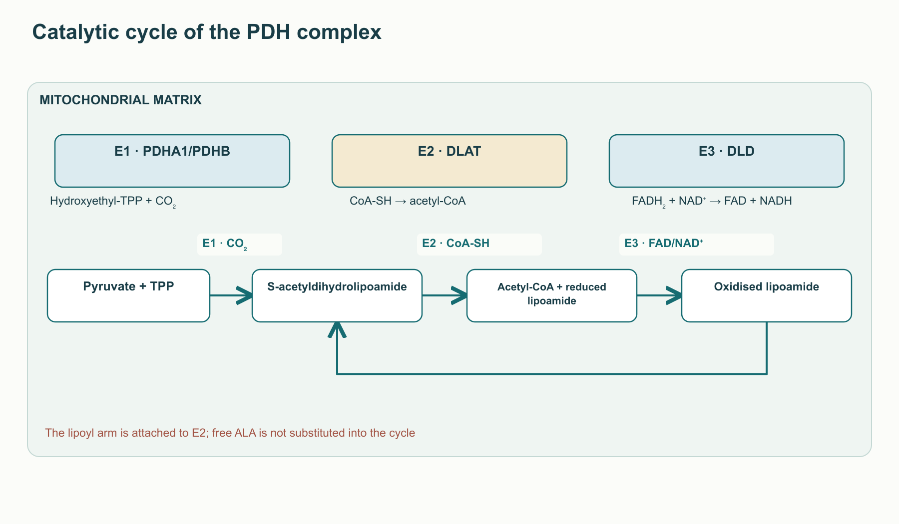
The pyruvate dehydrogenase complex connects cytosolic glycolysis to the mitochondrial tricarboxylic acid cycle: pyruvate enters the matrix and is converted to acetyl-CoA. The complex’s organization limits the loss of reactive intermediates into the aqueous phase. E1 (pyruvate dehydrogenase; the human subunits are PDHA1 and PDHB) binds thiamine pyrophosphate (TPP) with Mg²⁺; E2 (DLAT) carries lipoyl domains and catalyzes acetyl transfer to CoA; E3 (DLD) uses bound FAD to reduce NAD⁺. E3BP/PDHX helps attach E3 to the multimeric E2 core. Complex architecture and subunit copy number in a preparation depend on species, assembly state, and structural method; a fixed model of “exactly 60 E2 and 30 E1” is not needed to understand the chemistry.
The lipoyl group is attached by an amide bond to the ε-amino group of a specific lysine in a protein domain. Flexible linkers allow that domain to reach the separated E1, E2, and E3 active sites; “swinging arm” describes this geometry. It does not mean that a free R-ALA molecule independently shuttles between enzymes or that simply raising its concentration accelerates a properly lipoylated complex that is already saturated. Catalytic turnover requires pyruvate, CoA, NAD⁺, TPP, FAD, a functioning protein scaffold, and an accessible lipoyl domain at the same time.
At E1, the TPP carbanion/ylide attacks the carbonyl carbon of pyruvate; decarboxylation produces hydroxyethyl-TPP and releases CO₂. The oxidized disulfide ring of protein-bound lipoamide accepts reducing equivalents from this intermediate as the hydroxyethyl group is oxidized to an acetyl group. This yields an S-acetyldihydrolipoyl domain, which carries the acetyl group to the E2 catalytic site. CoA-SH attacks the thioester to form acetyl-CoA, leaving reduced dihydrolipoamide on E2. In the third stage, E3 oxidizes its two SH groups back to a disulfide, passing reducing equivalents through a protein disulfide pair and FAD to NAD⁺. The reduced lipoyl domain is not the final product: it is regenerated and ready for the next transfer.
The net reaction is written as pyruvate+CoA−SH+NAD+→acetyl−CoA+CO2+NADH+H+. This equation alone does not report tissue flux, which also depends on substrates, acetyl-CoA product, the NADH/NAD⁺ ratio, E1 phosphorylation, oxygen availability for NADH reoxidation, and downstream cycle activity. The source presents an exact standard ΔG∘′ and “arm” length as universal mitochondrial values; in living cells, chemical potential and complex conformation depend on the local environment, so these figures cannot be translated into a supplement dose.
PDC segment
Protein and required cofactors
Role of the lipoyl group
Possible cause of reduced flux not automatically corrected by free ALA
Transfers acetyl to CoA and changes disulfide to dithiol.
DLAT or LIPT1/LIAS disruption, impaired core assembly, or limited CoA.
E3
DLD, FAD, NAD⁺; E3BP/PDHX for organization
Reoxidizes the dihydrolipoyl arm.
DLD defect, high NADH load, or impaired E3 binding.
Similar chemistry is used by the α-ketoglutarate dehydrogenase complex (E2: DLST, formation of succinyl-CoA), branched-chain α-ketoacid dehydrogenase (E2: DBT, catabolism of leucine, isoleucine, and valine), and 2-oxoadipate dehydrogenase. In the glycine-cleavage system, lipoamide is attached not to E2 but to H-protein GCSH, which transfers an aminomethyl group among the P, T, and L components; with tetrahydrofolate, 5,10-methylenetetrahydrofolate is formed. These distinctions matter clinically: reduced lipoylation can affect several complexes and produce lactate, amino-acid, and one-carbon metabolic features together. But each inherited disorder depends on its own affected gene, not on a generic label of “lipoic acid deficiency.”
Regulation of pyruvate entry: PDK, PDP, and redox state
Pyruvate oxidation is regulated by phosphorylation of E1α. Pyruvate dehydrogenase kinase isoforms PDK1–PDK4 reduce complex activity by phosphorylating specific serine sites on PDHA1; pyruvate dehydrogenase phosphatases PDP1/PDP2 remove these phosphates. In working muscle, calcium signalling can activate PDP, whereas excess acetyl-CoA and NADH alter energy state and help limit entry of additional carbon into the cycle. The contribution of each isoform and site depends on tissue, nutrition, and workload, so the source’s single rule that “Ser293 fully switches off PDC” is too categorical.
When fatty-acid availability is high, the Randle cycle describes competition for oxidative resources and a shift in substrate selection. Cytosolic lactate dehydrogenase reduces pyruvate to lactate while regenerating NAD⁺ for glycolysis: pyruvate+NADH+H+⇌lactate+NAD+. Lactate may rise for many reasons, including hypoxia, high glycolytic flux, impaired pyruvate utilization, medications, or liver disease; it is not a specific test for inadequate free ALA. The source’s claim that R-ALA directly inhibits all PDKs, activates PDP1, and “eliminates” hyperlactatemia is not supported by a mechanistic link to E2 lipoylation and cannot serve as a clinical protocol. Establishing an effect on PDC would require measurements of PDHA1 phosphorylation and isotope flux from pyruvate to acetyl-CoA, while accounting for other causes of lactate change.
Acetyl-group transfer in mitochondria and the role of acetylcarnitine are discussed separately in the article on acetyl-L-carnitine. This is a related topic in acetyl metabolism, but an acetylcarnitine buffer does not replace the PDH reaction or its covalent lipoamide.
Free ALA as a redox reagent: possibilities and limits
Oxidized cyclic ALA and reduced DHLA form a redox pair that accepts and donates two electrons and two protons. Its standard biochemical potential is reported under reference conditions, but the potential in tissue depends on the ratio of reduced to oxidized forms, pH, protein binding, and compartment. The Nernst equation predicts direction at given concentrations; a list of standard potentials does not show that administered free ALA sequentially reduces every antioxidant in the body. Electron transfer also depends on substrate availability, reaction rate, and where the molecules are located.
Reduction of GSSG by dihydrolipoic acid is chemically possible: DHLA+GSSG→ALA+2GSH. In cells, glutathione reductase using NADPH is the main sustained route for GSH regeneration; de novo GSH synthesis depends on glutamate-cysteine ligase and cysteine supply. Ascorbate can reduce a tocopheroxyl radical at a membrane interface, and GSH helps maintain the ascorbate pool. However, the source’s “closed, NADPH-independent chain” is not an established autonomous circuit: DHLA itself must be reduced using reducing equivalents, while free-acid concentrations after supplementation are limited by metabolism and excretion. References to ferroptosis should be read as a mechanistic hypothesis involving lipid peroxidation and GSH-dependent systems, not as evidence that this form of cell death is prevented in humans.
The KEAP1–NRF2–ARE pathway shows how electrophilic and oxidative signals can alter protective-gene transcription. Under basal conditions, KEAP1 with CUL3/RBX1 promotes NRF2 ubiquitination; when sensor-cysteine function changes, available NRF2 forms a complex with small Maf proteins and binds ARE sequences. Potential responses include changes in GCLC/GCLM, NQO1, HMOX1, and SLC7A11 expression, but the direction and magnitude depend on the model. The source’s claim that free R-ALA itself uniformly modifies KEAP1 Cys151, Cys273, and Cys288, separates KEAP1 from CUL3, and produces a sustained several-fold increase in GSH would require direct adduct and transcription experiments in a defined tissue. NRF2 activation after cell treatment cannot automatically be attributed to direct R-ALA binding to those residues; altered redox conditions and intermediate signals are also possible.
Iron and copper ions can catalyze the formation of reactive species. The classic Fenton reaction is written as Fe2++H2O2→Fe3++OH−+∙OH; superoxide and peroxide transformations involving iron form a connected cycle. The two thiol groups of DHLA can form metal complexes in vitro, but complex formation is not equivalent to safely “switching off” the Fenton reaction. Depending on ligand, pH, and reductant availability, chelation may alter iron redox activity, while free DHLA concentrations in a given compartment remain an open question. The source’s conclusion that mercury, cadmium, and arsenic are detoxified and guaranteed to be excreted in bile or urine does not follow from demonstrating chemical binding. Suspected poisoning requires clinical toxicology, not extrapolation from this reaction to a supplement.
Chemical and cellular systems are used to study ALA/DHLA interactions with peroxynitrite, hypochlorite, singlet oxygen, and radicals. Tyrosine nitration, lipoprotein oxidation, and membrane peroxidation are measurable outcomes in such experiments. However, these agents differ in reactivity, and the hydroxyl radical reacts close to where it is formed: a high rate constant with this radical does not guarantee biological protection at an achievable concentration. The source applies a single rate of 1.4×10⁶M−1s−1 and the phrase “lightning-fast neutralization” to different reagents without measurement conditions; these are not treated here as therapeutic kinetic constants.
Insulin signalling and glucose transport
In lipotoxicity and inflammation, lipid metabolites, mitochondrial reactive oxygen species (ROS), and stress kinases can alter insulin-receptor and substrate function. Activation of some PKC isoforms, JNK, and IKK is associated with weakened insulin responses in experimental models, but serine phosphorylation of IRS1 involves many sites, time courses, and physiological roles. Thus, the source’s Ser307, Ser616, and Ser1101 are not universal “pathological switches,” and the figure for the share of all postprandial glucose taken up by muscle depends on method, population, and workload. Even a reduction in phosphorylation at a selected site in a cell line does not demonstrate reversal of systemic insulin resistance in humans.
After insulin binds its receptor, receptor tyrosine-kinase activity increases tyrosine phosphorylation of IRS proteins; the regulatory p85 subunit of class IA PI3K recognizes suitable phosphotyrosine motifs, while catalytic p110 produces PIP3 from PIP2. PIP3 recruits proteins with PH domains, including PDK1 and AKT; phosphorylation of AKT in its activation loop and hydrophobic motif, with mTORC2 involvement, supports downstream signalling. This is an established pathway map, but the map alone does not show where exogenous ALA acts. The source’s claim that R-ALA directly induces ligand-independent insulin-receptor autophosphorylation at specific required IRS1 residues requires direct binding, dose, and insulin-independence controls; such an effect in humans does not follow from general redox chemistry.
PTP1B, located on the cytosolic side of the endoplasmic reticulum, dephosphorylates components of insulin signalling. Its catalytic Cys215 is sensitive to reversible oxidation, making the enzyme a potential target of local redox signals. But the sensitivity of Cys215 to oxidants does not show that oral R-ALA selectively and fully inhibits muscle PTP1B. That claim would require enzyme and cell measurements at achievable exposure and separation of direct enzyme modification from secondary changes in peroxide concentration. The source’s pKa values and list of specific serine/tyrosine sites are not used here as universal clinical mechanisms.
Phosphorylated AKT regulates TBC1D4/AS160, a GAP for Rab proteins: after its activity changes, GLUT4 storage vesicles can move and fuse with the membrane of a myocyte or adipocyte. Rab proteins, the cytoskeleton, and SNARE proteins including syntaxin-4 participate in this machinery; a parallel pathway during muscle contraction involves AMPK and TBC1D1. Cell and animal models have reported effects of lipoic acid on glucose uptake and GLUT4; the relative contributions of AKT, AMPK, p38 MAPK, and changes in energy state depend on dose and system. The source leaps from this signalling pathway to a claim of massive insulin-independent glucose uptake even when insulin is nearly absent or the receptor is defective. ALA has not been clinically shown to replace insulin or repair a receptor defect.
Clinical assessment: symptoms, nerve function, and metabolic outcomes
Distal symmetric diabetic polyneuropathy combines pain and paraesthesia with objectively measurable sensory, motor, and nerve-conduction abnormalities. Its mechanisms include chronic hyperglycemia, nerve-fibre injury, and vascular changes; a single redox mechanism cannot describe the entire course of disease. When evaluating thioctic acid, it is therefore important to distinguish a short-term symptom effect on the TSS, a change on neurological examination, nerve conduction, and prevention of disabling complications. Trials used different formulations and routes, predominantly racemic ALA; their results do not directly assess sodium R-lipoate.
ALADIN I studied a short course of intravenous treatment at several doses in symptomatic neuropathy. A symptom-improvement signal in a dose group does not mean remyelination or a change in long-term disease course. ALADIN II assessed electrophysiological parameters during prolonged oral exposure in a small sample, but participant numbers and heterogeneous measurements limit generalization. ALADIN III used an intravenous phase followed by oral treatment; its design cannot be reduced to the effect of tablets alone. These differences explain why the trial programme cannot be represented as a single ladder of “increasingly proven efficacy.”
The primary SYDNEY study reported improved TSS after 14 intravenous infusions of 600 mg racemic ALA compared with placebo in 120 patients; mean change from baseline was 5.7 versus 1.8 points. In SYDNEY 2, 181 patients received oral ALA at 600, 1200, or 1800 mg, or placebo, for five weeks. Mean TSS reductions were 4.9, 4.5, and 4.7 points, respectively, versus 2.9 points with placebo; the primary publication also described dose-related increases in nausea, vomiting, and dizziness. The authors considered 600 mg the most reasonable balance of benefit and tolerability in that study. This is a conclusion about short-term symptoms in the studied population, not a universally equivalent dose for all chemical forms. The source gives an incorrect PMID, 12610024, for SYDNEY I; the publication involving 120 participants is PMID 12610036.
The four-year NATHAN 1 trial matters particularly for its negative result on the prespecified primary composite endpoint, NIS-LL+7: the difference between oral ALA 600 mg/day and placebo did not reach statistical significance (P=0.105). Some secondary neurological examination measures and responder analyses were more favourable in the ALA group, but nerve-conduction parameters and TSS did not differ significantly. Objective nerve conduction changed little in the placebo group, making it harder to detect prevention of progression. The source incorrectly identifies NIS-LL as the primary endpoint and interprets its secondary result as proof that disease progression was slowed. The warranted conclusion is narrower: some signs may have improved, while the primary composite outcome was not confirmed; the trial does not prove remyelination, prevention of ulcers, or “absolute safety.”
Trial programme
Route and duration
What was measured
What the results support
ALADIN I and SYDNEY
Intravenous ALA, weeks
TSS symptoms and additional signs
A short-term symptom signal; not proof of long-term neuroprotection.
SYDNEY 2
Oral ALA, five weeks
Primary TSS endpoint and tolerability
Symptom improvement versus placebo; higher doses showed no clear advantage and were less well tolerated.
NATHAN 1
Oral ALA, four years
Primary composite NIS-LL+7; secondary measures
Primary outcome was negative; some secondary neurological scores were favourable.
Clamp studies in type 2 diabetes assessed short-term changes in glucose disposal after intravenous ALA, while small oral-treatment courses assessed insulin sensitivity and selected metabolic markers. A clamp measures a physiological response under controlled insulin and glucose infusion; it does not demonstrate fewer cardiovascular events, diabetes remission, or efficacy of a particular salt. Koh and colleagues studied body weight in people with obesity, but the source incorrectly attributes their article to the American Journal of Clinical Nutrition: the cited work appeared in the American Journal of Medicine. Changes in weight and biomarkers in such a study do not justify treating ALA as obesity therapy. For MASLD, cellular mechanisms involving AMPK–ACC, β-oxidation, and SREBP-1c are of interest, but lower ALT/AST or a change in one marker is not equivalent to histological resolution of steatohepatitis or lower fibrosis risk.
The ISLAND trial compared irbesartan, ALA, their combination, and placebo for four weeks in people with metabolic syndrome, assessing flow-mediated dilation of the brachial artery and inflammatory markers. FMD is a vascular surrogate sensitive to ultrasound conditions, baseline function, and concomitant therapy; even a statistically significant change does not demonstrate prevention of myocardial infarction or stroke. The hypothesis that BH4 availability, endothelial nitric oxide synthase coupling, and NO production can be maintained is physiologically plausible, but the source’s claim of direct BH2-to-BH4 reduction by DHLA in ISLAND participants does not follow from FMD. This experiment did not replace cardiovascular-event data or direct eNOS measurements.
Exposure after ingestion: enterocyte, liver, and tissue
After oral administration, free ALA must dissolve, cross the intestinal epithelium, pass through portal circulation, and undergo first-pass metabolism before it becomes available to tissues. At physiological pH, the carboxyl group is predominantly ionized; the contributions of passive diffusion and transporters depend on the medium and concentration. Transport models discuss the sodium-dependent multivitamin transporter SMVT/SLC5A6, which can also transport biotin and pantothenate, as well as monocarboxylate transporters. Demonstrating transport in vitro, however, does not establish that exactly two Na⁺ accompany each molecule in a human enterocyte or that dietary fats and amino acids directly compete for SMVT. Transport and metabolism of free acid do not describe assembly of covalent lipoamide on E2.
Drug formulations differ in dissolution rate, Cmax, time to peak, and AUC; R and S enantiomers may also have different pharmacokinetics. The Carlson and Hermann studies are useful for these comparisons, but their results pertain to a particular dose, product, assay, fasting state, and sample. Metabolism includes side-chain shortening, reduction/oxidation, and sulfur-containing derivatives, followed importantly by renal excretion of metabolites. The source table’s fixed bioavailability of 20–30%, half-life of 25–35 minutes, and 40–60% salt advantage do not describe a universal “R-ALA exposure” across formulations. Translating a pharmacokinetic effect into a clinical effect requires studies with the same active moiety, comparable regimens, and measured outcomes, not a ratio of peak concentrations alone.
Food can alter thioctic-acid exposure, as studied in pharmacokinetic experiments with specific formulations. Timing relative to meals should follow the instructions for the product actually studied; the source’s categorical rule of “strictly 30–45 minutes before breakfast or 2–3 hours after a meal, with demineralized water only” is not a universal clinical consensus. Nor has it been shown that ordinary food promotes thermal polymerization in the stomach or that fibre quantitatively adsorbs a therapeutic dose. Effects of food on gastric emptying, product dissolution, and possible transport should be distinguished rather than presented as mechanisms measured in patients.
A 600 mg racemate contains approximately 300 mg by mass of each enantiomer if the product is indeed a 1:1 mixture of free acid. A salt requires molar-mass conversion, but this arithmetic does not establish therapeutic equivalence. The neuropathy trials described above did not compare sodium R-lipoate with the racemate at doses matched for exposure and clinical outcome. Thus, the source’s equations “200–300 mg salt = 600 mg racemate” and “500–600 mg salt = 1200–1800 mg racemate without adverse effects” are unsupported. A higher peak with one formulation could increase unwanted effects as well as a desired signal; converting a regimen requires evidence for that formulation and patient context.
Combined interventions: a tested model and an unsubstantiated “stack”
In studies by the Hagen–Ames group, aged rats received acetyl-L-carnitine (ALCAR) and lipoic acid, and mitochondrial and behavioural measures were assessed. These studies are important as a model of interaction between acyl exchange, respiration, and oxidative stress, but they do not establish “mitochondrial rejuvenation” in humans. ALCAR supplies acetyl groups through the carnitine acetyltransferase reaction and is not equivalent to transport of long-chain acyl groups through CPT1; that transport is discussed separately in the CPT1 overview. ALCAR itself, including tissue and neuronal models, is discussed in more detail in the article on acetyl-L-carnitine. The rat combination and its memory tests cannot be mechanically translated into a human morning “Ames protocol” with specified milligrams of sodium R-lipoate and ALCAR.
Benfotiamine is linked to thiamine-dependent metabolism; transketolase in the pentose phosphate pathway uses TPP, as does pyruvate-dehydrogenase E1. Studies of combinations with ALA may track metabolic markers of hyperglycemic injury, but the small patient study published by Du and colleagues cannot demonstrate “complete prevention” of diabetic retinopathy, nephropathy, and vascular complications. Diverting glycolytic intermediates through transketolase does not mean that all AGE, polyol, hexosamine, and PKC pathways are switched off. These routes are interconnected and depend on glycemia and tissue context. It is likewise unproven that adding ALA removes every PDC block; TPP supply to E1 and lipoylation of E2 are distinct constraints.
The source presents ALA with NAC, ascorbate, and CoQ10 as a mandatory “antioxidant ladder,” with specific doses for chronic fatigue, fibromyalgia, and after myocardial infarction. NAC can supply cysteine for GSH synthesis, CoQ10 transfers electrons between respiratory-chain complexes, and ascorbate participates in an aqueous redox network, but these occupy different compartments and have different pharmacokinetic profiles. A sum of plausible reactions does not establish a clinical effect for the mixture, and antioxidant load may alter adaptive signalling. Therefore, the regimen given in the source is not carried forward as a clinical recommendation.
Safety and limits of extrapolation
In short neuropathy trials, the most noticeable adverse effects of oral treatment were nausea, vomiting, and dizziness; their frequency in SYDNEY 2 depended on dose. Preclinical LD50 data and the absence of a signal in one toxicology test do not prove “complete absence” of teratogenicity, mutagenicity, chronic risk in humans, or intolerance across all salts. NATHAN 1 provides long-term observations for the studied form and population, but the negative primary outcome and individual serious events call for careful interpretation rather than a claim of “absolute safety.” Pregnancy, childhood, and medication combinations each require their own evidence base.
A rare but important complication is insulin autoimmune syndrome (IAS, Hirata disease): hypoglycemia with high titres of antibodies to endogenous insulin in people without prior insulin administration. Its association with ALA has been described in case series and is linked to HLA genotype. In the series by Gullo and colleagues, six European patients with IAS had taken ALA; five had HLA-DRB1*04:03 and one had 04:06. This contradicts the source scheme, which names 04:06 as predominant in Europeans. A role for disulfide exchange in generating immunogenic epitopes remains a mechanistic hypothesis, not a confirmed sequence in every patient in which “DHLA breaks a specific insulin bridge → CD4 response.” Autoantibodies can bind insulin and alter its release, but hypoglycemia need not occur in the stated 2–4-hour window.
For unexplained hypoglycemia during ALA use, stop the suspected trigger and promptly seek medical assessment, check glucose, and establish the cause. A severe episode requires emergency care. Improvement has often followed ALA withdrawal, but the source’s conclusion that “withdrawal alone is always sufficient and immunosuppression is never needed” is incorrect: patients in the Gullo series received glucose and prednisone. Management depends on severity and confirmed diagnosis, including distinction from glucose-lowering medication overdose and other causes of hyperinsulinemia.
When ALA is combined with insulin or secretagogues, glucose should be clinically monitored, particularly when a person starts a new product and has a history of hypoglycemia. The source’s automatic 10–20% reduction in glucose-lowering medication is unsupported and could cause harm; regimen changes should be based on measurements and the treating team’s advice. The hypothesis that ALA binds divalent cations in the intestine does not justify a universal mandatory 2–3-hour interval for iron, magnesium, and calcium without formulation- and interaction-specific study. During anticancer treatment, discuss supplements with the oncologist because interactions are uncertain; this article does not claim that ALA has been proven to reduce the efficacy of platinum drugs.
Methods and limits of evidence selection
This is a narrative review of the supplied set of 30 publications on lipoamide enzymology, free-ALA pharmacology, and clinical trials; the direct checks described below refer to the version dated 4 October 2026. Primary reports on SYDNEY, SYDNEY 2, NATHAN 1, and insulin autoimmune syndrome were checked directly; bibliographic details and study descriptions supplied for the remaining publications were used. No exhaustive search, meta-analysis, formal risk-of-bias assessment, or GRADE evaluation was conducted. Clinical conclusions are limited to the chemical form, route, dose, population, duration, and prespecified endpoint studied. Unavailable full texts, transfer of evidence between enantiomers and salts, and lack of direct tissue-exposure measurements limit certainty.
It is useful to distinguish the question “Does lipoamide function in PDC?” from “What benefit has a specific ALA product demonstrated for a specific diagnosis?” The first requires evidence about the enzyme, lipoylation, and metabolic flux; the second requires clinical outcomes for the form, route, dose, and duration studied. Symptom improvement in selected diabetic-neuropathy regimens does not make R-ALA a treatment for inherited lipoylation defects, insulin resistance, MASLD, cardiovascular events, or “mitochondrial ageing.”
FAQ
Are PDC-bound lipoamide and oral R-ALA the same thing?
No. In PDC the lipoyl group is covalently bound to an E2 protein; ingested ALA has its own absorption and metabolism, and direct rebuilding of E2 in humans has not been established.
Can a supplement correct an inherited LIAS or LIPT1 defect?
This has not been demonstrated. These genes participate in mitochondrial assembly of the lipoyl group, so the clinical question requires a separate genetic and metabolic assessment.
Does ALA help diabetic neuropathy?
Short trials of racemic ALA reduced symptoms on the TSS. The four-year NATHAN 1 trial did not meet its primary composite endpoint; these results do not show that the disease was halted or that another salt is beneficial.
Can 200–300 mg sodium R-lipoate replace 600 mg of the racemate?
Therapeutic equivalence at these doses has not been established by a direct comparison of clinical outcomes.
What important rare adverse reaction has been reported?
Insulin autoimmune syndrome with hypoglycemia. Unexplained episodes during ALA use warrant medical assessment and stopping the suspected trigger.
Carlson DA, Smith AR, Fischer SJ, Young KL, Packer L. The plasma pharmacokinetics of R-(+)-lipoic acid administered as sodium R-(+)-lipoate to healthy human subjects. Altern Med Rev. 2007;12(4):343-351. PMID: 18069903.
Jacob S, Henriksen EJ, Schiemann AL, et al. Enhancement of glucose disposal in patients with type 2 diabetes by alpha-lipoic acid. Arzneimittelforschung. 1995;45(8):872-874. PMID: 7575750.