CPT1: the carnitine shuttle and fatty acid oxidation — NutriFit
Carnitine palmitoyltransferase 1: the carnitine shuttle, malonyl-CoA and β-oxidation
An in-depth monograph on the delivery of long-chain fatty acids into mitochondria, tissue-specific CPT1 isoforms, and regulation by malonyl-CoA. It examines catalysis, AMPK–ACC, β-oxidation, energy calculations, metabolic flexibility, and inherited defects. Clinical and sports studies of carnitine, the microbial TMAO pathway, and pharmacological approaches are considered within the limits of the evidence.
A long-chain fatty acid does not become mitochondrial fuel immediately upon entering a cell. First it is activated, then its acyl group is transferred across the inner mitochondrial membrane, after which the enzymes of β-oxidation progressively shorten its carbon chain. Carnitine palmitoyltransferase 1, or CPT1, regulates entry into this sequence, linking fatty acid availability to the cell's energy status. Its inhibitor, malonyl-CoA, also serves as a building block for lipid synthesis, so one metabolite helps coordinate two processes running in opposite directions.
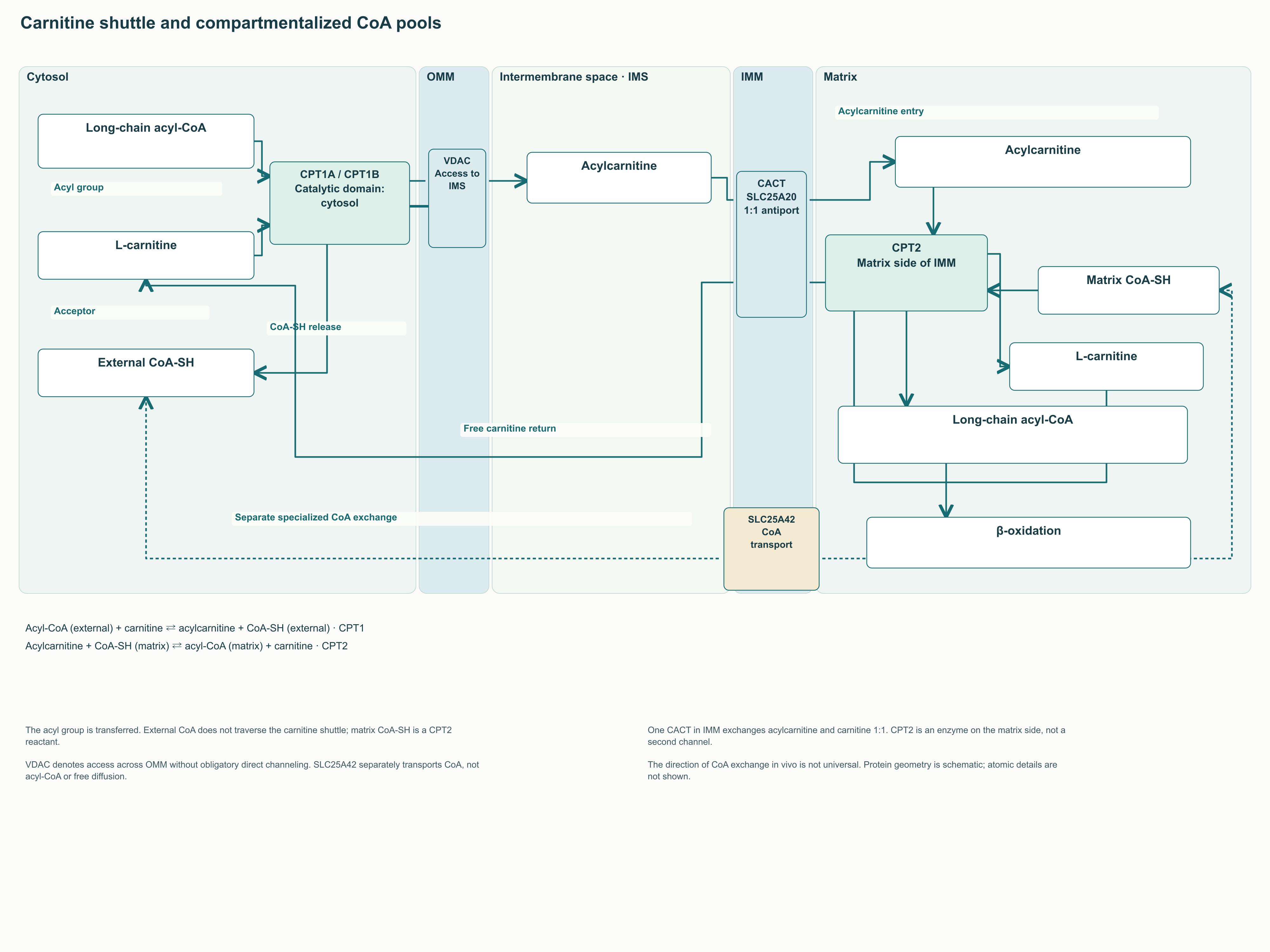
The carnitine shuttle comprises CPT1, carnitine–acylcarnitine translocase, CACT, and CPT2. It transfers the acyl group while maintaining the distinction between extramitochondrial and matrix pools of coenzyme A; the existence of the CoA transporter SLC25A42 does not eliminate the need for this route for long-chain acyl-CoA. Shuttle regulation depends on the CPT1 isoform, the local malonyl-CoA concentration, the membrane environment, AMPK, acetyl-CoA carboxylases and transcriptional programmes. However, faster substrate entry is useful only when subsequent reactions, the respiratory chain and ATP demand can accommodate that flux.
This monograph examines transport architecture, catalytic chemistry, tissue differences, energy balance and the consequences of impaired fatty acid oxidation. Inherited defects, carnitine supplements, exercise studies and the microbial pathway producing TMAO are discussed separately. A biochemical mechanism, an animal experiment and a clinical effect in humans carry different evidential weight, and that distinction is maintained throughout the text. The 106-ATP calculation is presented as a model using specified P/O ratios, while experimental interventions targeting CPT1 and the microbiota are described within the limits of their applicability.
1. Why long-chain fatty acids need a shuttle
1.1. Two membranes and two functionally compartmentalised CoA pools
The outer and inner mitochondrial membranes perform different tasks. The outer membrane contains VDAC channels through which many small metabolites exchange between the cytosol and the intermembrane space. The inner membrane controls transfer much more strictly: the movement of most polar substances depends on specific transport proteins. This maintains the electrochemical proton gradient and the conditions required for oxidative phosphorylation.
Low passive permeability of the inner membrane does not mean that CoA, ions or other polar compounds cannot cross it at all. In 2009, Fiermonte and colleagues expressed human SLC25A42, reconstituted it in liposomes and demonstrated the exchange-mediated transport of CoA and several adenine nucleotides. This provides direct experimental grounds for distinguishing an absence of free diffusion from the presence of specialised transport. A claim that mitochondria are completely isolated from external CoA contradicts these findings.
CoA-SH is a coenzyme with a reactive thiol group to which acyl residues attach. Its simplified structural sequence can be written as adenosine 3′-phosphate 5′-diphosphate — pantothenate — cysteamine-SH. Pantothenate itself already contains the β-alanine moiety, so adding it separately again would duplicate part of the molecule. Phosphate groups make CoA polar and charged, while the thioester bond enables activated carbon groups to be transferred between reactions.
The cytosolic and matrix CoA pools are functionally compartmentalised, but they are not two permanently sealed reservoirs. Extramitochondrial CoA participates in fatty acid activation and lipid assembly, whereas matrix CoA is needed for pyruvate dehydrogenase, the tricarboxylic acid cycle and β-oxidation. Their composition is determined by transport, synthesis, degradation and distribution between free and acylated forms. Total CoA content is therefore not equivalent to the availability of free CoA-SH for an individual reaction.
The situation differs for long-chain acyl-CoA: the canonical route delivering its acyl group into the matrix uses the carnitine shuttle. CoA transport through SLC25A42 is not palmitoyl-CoA transport and does not replace CPT1, CACT and CPT2. On the outer side, the acyl group is removed from one CoA molecule; in the matrix, it is attached to another. This separation explains both the need for the shuttle and the independent regulation of CoA in different compartments.
Cardiolipin supports the organisation of the inner membrane and the function of many mitochondrial proteins, but its presence alone does not explain the barrier's entire selectivity. Likewise, the negative matrix potential matters for bioenergetics without making the inner membrane an universally impassable wall. Proton leak, ion transport and the activity of specialised carriers coexist with maintenance of the gradient. This functional picture is sufficient for understanding CPT1; fixed cardiolipin percentages, pore sizes and potential values without a specified tissue or set of conditions would create false precision.
1.2. Fatty acid activation: energy is spent before β-oxidation begins
Fatty acids reach cells from food, lipoproteins and adipose tissue. Their uptake and intracellular distribution involve CD36, FATP-family proteins and other participants, as well as the physical properties of the membrane itself. Acyl-CoA synthetases then form activated derivatives that may be directed towards oxidation, triacylglycerol synthesis or phospholipid construction. Increased fatty acid delivery therefore does not yet determine which pathway will receive its carbon.
The activation reaction is written as R–COOH + ATP + CoA-SH → R–CO–S–CoA + AMP + PPᵢ. Here R denotes the hydrocarbon portion of the fatty acid, and PPᵢ is inorganic pyrophosphate. Subsequent hydrolysis, PPᵢ + H₂O → 2 Pᵢ, favours the directionality of the overall process. Because ATP is converted to AMP, the energy cost is two ATP equivalents, although the initial reaction uses one ATP molecule.
Long-chain acyl-CoA synthetases differ in tissue expression, localisation and substrate preferences. ACSL1 is especially important for oxidative fatty acid utilisation, ACSL3 is associated with lipid droplets, and ACSL4 participates in the incorporation of polyunsaturated chains into phospholipids. These associations describe predominant functions in particular cellular systems, rather than the exclusive fate of every acyl-CoA molecule produced. Substrate availability and interactions with neighbouring enzymes can alter flux distribution.
Complexes containing CPT1A, ACSL1 and VDAC have been detected in outer-membrane preparations. These findings support spatial coupling between activation and acyl-group transfer, but do not establish that every molecule is always passed directly from one active site to another. It is important to distinguish an observed protein association from quantitatively demonstrated substrate channelling. For practitioners, this means that enzyme localisation may matter as much as its total expression.
1.3. CPT1, CACT and CPT2: one continuous route
At physiological pH, carnitine contains a positively charged trimethylammonium group and a negatively charged carboxylate. Its simplified formula is ⁻OOC–CH₂–CH(OH)–CH₂–N⁺(CH₃)₃. This zwitterionic structure is compatible with solubility in an aqueous environment, and the hydroxyl group at C3 accepts the acyl group. Acylation changes the chemical cargo while preserving carnitine as a recyclable shuttle component.
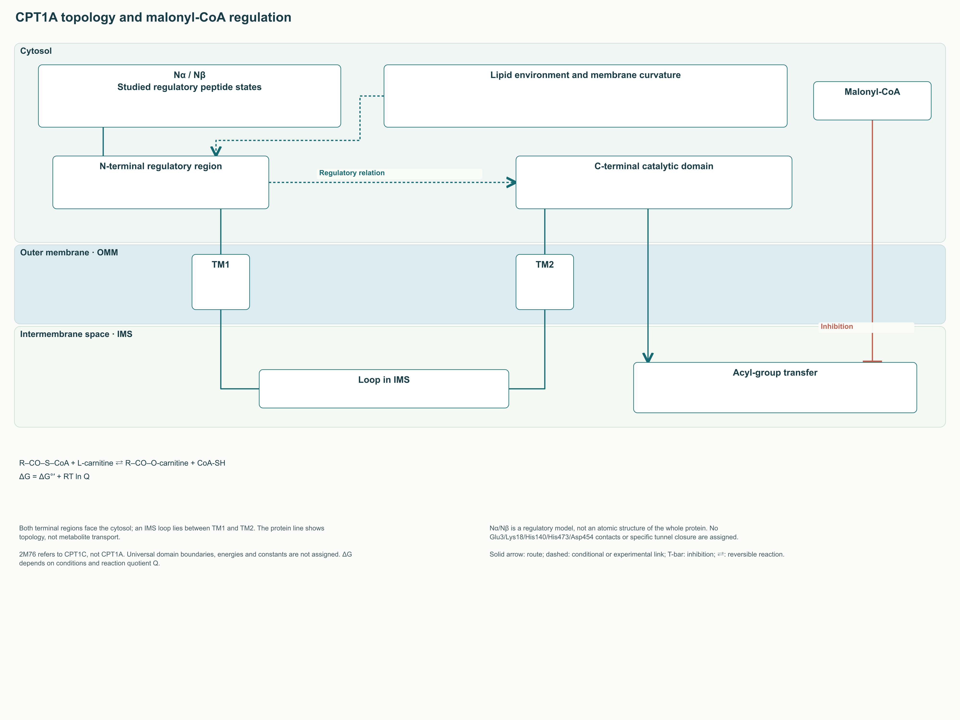
CPT1A and CPT1B are embedded in the outer mitochondrial membrane. In an experimental study of rat liver CPT1 topology, protein accessibility to proteases and antibodies demonstrated two transmembrane segments, cytosol-facing N and C termini, and a loop in the intermembrane space. This arrangement is consistent with enzyme access to external acyl-CoA and carnitine. It does not require the catalytic site to be situated inside the matrix.
The shuttle's first reaction is external acyl-CoA + L-carnitine ⇄ acylcarnitine + external CoA-SH. It transfers the acyl group to the oxygen of carnitine's hydroxyl group. The released CoA remains in the external pool, while acylcarnitine can reach the inner membrane. Carnitine acts here as a group carrier rather than a consumable energy source.
CACT, encoded by SLC25A20, transports acylcarnitine across the inner membrane mainly in exchange for free carnitine. In the physiological diagram, it is one transport protein with oppositely directed substrate fluxes, rather than two CACT proteins arranged in series. Work with the recombinant carrier in liposomes directly demonstrated carnitine exchange and transport of its acyl derivatives. Alternating access explains transport without a continuously open channel running through the membrane.
On the matrix side, CPT2 performs the reverse transfer: acylcarnitine + matrix CoA-SH ⇄ matrix acyl-CoA + L-carnitine. The resulting acyl-CoA is available to β-oxidation enzymes, and carnitine returns through CACT. Overall, the shuttle delivers the acyl group between CoA pools and regenerates its carrier. Directionality in a living cell is maintained by substrate availability and subsequent acyl-CoA utilisation, rather than by the irreversibility of each transferase reaction.
Free short-chain and some medium-chain fatty acids can use routes that depend less on CPT1. The boundary between a medium and a long chain is not identical in every tissue or condition, particularly for C12. Very long chains are often shortened first in peroxisomes, after which the products can enter mitochondrial metabolism. The carnitine shuttle is therefore a central route for long chains, but not the only transport system involved in all lipid catabolism.
2. Three CPT1-family proteins and different tissue tasks
2.1. CPT1A and CPT1B: a similar reaction, different kinetics
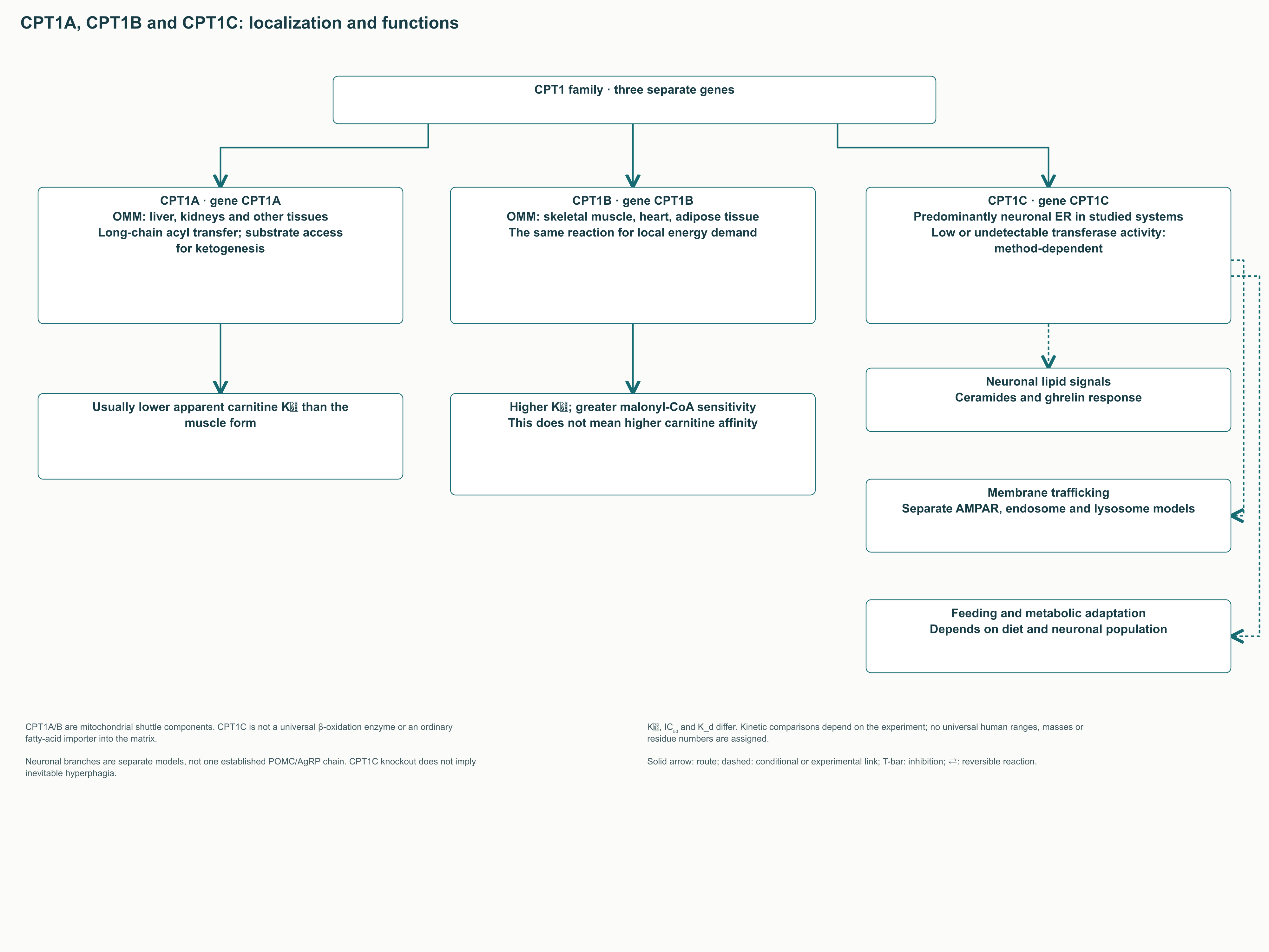
The terms “liver” and “muscle” isoform are useful provided they are not interpreted literally. CPT1A occurs in the liver and several other tissues; CPT1B is characteristic of skeletal muscle, the heart and some adipose cells. In the liver, fatty acid oxidation supplies energy for metabolism during fasting and acetyl-CoA for ketogenesis. In contracting muscle and the myocardium, it primarily helps meet local ATP demand.
CPT1A itself does not synthesise ketone bodies. It regulates long-chain substrate entry, whereas the formation of acetoacetate and β-hydroxybutyrate requires subsequent β-oxidation and ketogenesis enzymes. In the heart, the contribution of fatty acids to energy supply changes with workload, substrate availability and disease. Neither a fixed percentage of “energy from fat” nor a universal maximum CPT1 activity therefore describes every physiological state.
In a classic comparison of isolated mitochondria from several tissues, the apparent Kₘ for carnitine varied by approximately twentyfold. It was about 35 µmol/L in adult rat liver and human fetal liver, and substantially higher in some muscle and cardiac preparations. Muscle CPT1 was also more strongly inhibited by malonyl-CoA. In that comparison, the muscle form had a higher carnitine Kₘ, so its greater malonyl-CoA sensitivity cannot be explained by greater affinity for carnitine.
Kₘ, IC₅₀ and K_d should not be read as interchangeable characteristics. Kₘ describes how reaction velocity depends on substrate concentration, IC₅₀ is the inhibitor concentration that halves measured activity, and K_d describes equilibrium binding. All three depend on the measurement system, but in different ways. For a membrane enzyme, carnitine concentration, acyl-CoA binding to albumin, membrane preparation and the organism's species are especially important.
Property
CPT1A
CPT1B
CPT1C
Gene
CPT1A
CPT1B
CPT1C
Typical localisation
Outer mitochondrial membrane
Outer mitochondrial membrane
Predominantly neuronal ER in the systems studied
Characteristic tissues
Liver, kidney and other tissues
Skeletal muscle, heart, adipose tissue
Nervous system; also studied in tumour cells
Main established function
Formation of long-chain acylcarnitines
The same transport reaction in oxidative tissues
Regulation of neuronal lipid processes and membrane trafficking in experimental models
Malonyl-CoA
Inhibits; sensitivity depends on conditions
Usually greater sensitivity in comparable experiments
Binding and regulatory effects cannot be equated with CPT1A/B kinetics
Carnitine kinetics
Lower Kₘ in the classic tissue comparison
Higher Kₘ in several muscle preparations
Low or undetectable transferase activity depending on the method
Clinical interpretation
CPT1A deficiency impairs fasting tolerance
Energy metabolism in muscle and heart
No universal therapeutic rule to “activate” or “inhibit” it
The table compares functional classes rather than prescribing reference constants for every human tissue. Isoform names and the direction of kinetic differences are supported by comparative experiments, while CPT1C characteristics come from separate cellular studies. Exact values from one preparation cannot be inserted into calculations of flux in another tissue without further justification. This is particularly important when attempting to explain a supplement's effects through “saturating CPT1 with carnitine”.
2.2. CPT1C: linking energy signals to neuronal function
CPT1C differs from the two mitochondrial isoforms in more than tissue expression. Cellular localisation studies have predominantly detected it in the endoplasmic reticulum of neurons. Some early studies did not detect standard carnitine palmitoyltransferase activity, whereas Sierra and colleagues found weak activity in microsomal preparations. In their experimental systems, catalytic efficiency was lower than for CPT1A, and involvement in ordinary mitochondrial β-oxidation was not demonstrated.
This variability does not justify describing CPT1C either as a completely inactive protein or as a third equivalent gateway in the carnitine shuttle. The measurement method, cellular fraction and proposed substrate influence observed activity. Malonyl-CoA binding supports a sensory-function hypothesis without identifying a single downstream signal. CPT1C activity should be described in the context of the specific experimental system.
A mouse model involving ghrelin administration demonstrated a parallel signalling branch through CPT1C and changes in hypothalamic ceramides. Ghrelin's appetite-stimulating action was attenuated in animals lacking CPT1C, and central ceramide administration could restore part of the response. This connects neuronal lipid metabolism with the regulation of feeding behaviour. It does not establish that increasing or decreasing CPT1C activity in humans would produce a predictable therapeutic effect.
Other experiments link CPT1C to the delivery of AMPA receptors to the cell surface. GluA1 models examined changes in palmitoylation and receptor transport out of the ER, but this is a separate line of evidence that cannot simply be appended to a “satiety–POMC-neuron excitation” chain. The protein's participation in membrane trafficking has also been studied through late endosomes, lysosomes and interaction with protrudin. These studies broaden the functional picture while demonstrating why one universal CPT1C mechanism is insufficient.
The CPT1C-knockout phenotype also depends on diet and the neuronal population studied. In an early study, mice on a standard diet ate less and weighed less, but became more prone to fat accumulation on a high-fat diet. The original formula “knockout causes hyperphagia” is therefore incorrect as a general rule. New conditional models, including deletion of CPT1C from SF1 neurons, refine tissue-specific causality rather than removing the need to consider context.
3. Catalysis and malonyl-CoA: what is known about switching flux
3.1. Acyl-group transfer and the limits of atomic detail
CPT1's chemical task can be expressed by the general reaction R–CO–S–CoA + L-carnitine ⇄ R–CO–O-carnitine + CoA-SH. A thioester becomes an oxygen ester of carnitine, while the carbon chain is preserved. General catalytic models propose that activation of the hydroxyl group facilitates its attack on the carbonyl carbon of acyl-CoA. Bond rearrangement through a tetrahedral state ends with the release of free CoA-SH and acylcarnitine.
For the carnitine acyltransferase family, structural studies and mutagenesis support the participation of a conserved histidine and other residues in the catalytic environment. However, a homologous protein's structure, a sequence-based model and a directly solved structure of the specific complex represent different levels of evidence. A CPT1 model based on carnitine acetyltransferase was used to select residues for subsequent mutagenesis. Its results support the participation of individual positions, but do not establish the entire transition-state geometry or a universal contact network between substrates and the protein.
This monograph retains the molecular logic of transfer without using unsupported atomic geometry. Residue numbering requires identification of the protein, species and original study, and a mutation-induced loss of activity does not by itself prove a particular hydrogen bond. Similarly, reversibility does not mean that the reaction's standard free energy is precisely zero. Direction depends on chemical potentials, reactant ratios and their consumption by neighbouring processes.
The relationship between concentrations and thermodynamics is described by ΔG = ΔG°′ + RT ln Q. For the transfer reaction, Q = [acylcarnitine][CoA-SH] / ([acyl-CoA][carnitine]), using an appropriate approximation of activities. Here R is the gas constant, T is absolute temperature, and ΔG°′ is the standard transformed free-energy change. The expression shows why flux can change even with an unchanged amount of enzyme; it does not assign an unsupported numerical ΔG°′ or equilibrium constant.
3.2. McGarry's discovery: synthesis and oxidation share a signal
In 1977, McGarry, Mannaerts and Foster showed that malonyl-CoA inhibited ketone-body formation from long-chain oleic acid in rat liver preparations, but not from octanoate. They associated the inhibitory site with the first carnitine acyltransferase reaction. Subsequent work localised inhibition to CPT1. The actual experimental sequence matters: these results were not a clinical trial demonstrating that eating completely “switches off fat” in humans.
Malonyl-CoA is formed by the carboxylation of acetyl-CoA. The reaction acetyl-CoA + HCO₃⁻ + ATP → malonyl-CoA + ADP + Pᵢ combines the carbon source, bicarbonate and an energy cost; acetyl-CoA carboxylase, ACC, catalyses it. During lipogenesis, malonyl groups progressively lengthen the chain assembled by fatty acid synthase, FASN. At the same time, malonyl-CoA reduces long-chain substrates' access to mitochondrial oxidation.
For palmitate synthesis, FASN uses one initial acetyl group, seven malonyl groups and 14 NADPH. Seven ATP are spent beforehand to form seven malonyl-CoA molecules through ACC, so attributing that expenditure to FASN a second time is incorrect. This sequence helps explain why simultaneous intense synthesis and breakdown of the same chain would be energetically costly. Malonyl-CoA regulation reduces this conflict without imposing an absolute prohibition on lipogenesis and oxidation coexisting throughout a cell.
After food intake, insulin and nutrient signals generally support lipogenic processes and may increase CPT1 inhibition. During fasting and exercise, fatty acid delivery, ACC activity and energy status change in ways that favour greater fat utilisation. These tendencies should not be described as two completely mutually exclusive positions of an electrical switch. Different tissues simultaneously oxidise different substrate mixtures, while local flux depends on many constraints.
3.3. The regulatory domain and membrane influences
CPT1A's N-terminal region affects malonyl-CoA sensitivity through interaction with the rest of the protein and its membrane environment. An NMR study of the regulatory peptide identified Nα and Nβ states; whole-protein mutagenesis supported a model linking them differently to inhibition. This is an important result concerning conformational regulation, but not an atomic photograph of a closed carnitine tunnel. The authors explicitly distinguished experimental peptide data from the modelled orientation of domains in the complete enzyme.
When comparing structures, it is essential to identify the protein actually studied. Entry 2M76 corresponds to the regulatory domain of human CPT1C, whereas 2LE3 is discussed for CPT1A's Nβ state. The distinction matters because the two proteins do not have interchangeable functions. The allostery figure therefore shows established topology and a regulatory relationship without assigning unsupported atomic contacts.
CPT1A sensitivity to malonyl-CoA is indeed related to the lipid properties and curvature of the outer membrane. This does not imply a simple rule that any rise in cholesterol, mitochondrial fragmentation or fusion always changes oxidation in the same direction. Across an entire organelle, contact sites, protein distribution, respiratory function and substrate availability change simultaneously. Fragmentation and fusion should therefore be considered together with organelle status, avoiding a universal direct relationship between mitochondrial shape and CPT1 activity.
Cardiolipin oxidation should be kept distinct from hypotheses concerning CPT1 oligomers. Membrane-lipid damage can impair mitochondrial function, but a specific claim about the breakdown of CPT1 trimers and hexamers requires its own experimental support. For some downstream β-oxidation enzymes, the link to cardiolipin has been studied directly: acylation of particular VLCAD lysines altered membrane binding, which SIRT3 and SIRT5 could restore. This example supports the importance of the membrane environment without substituting evidence about another protein for CPT1 data.
4. Rapid and long-term regulation of oxidation
4.1. AMPK: energy metabolism becomes a signal
AMPK is a complex comprising a catalytic α subunit and regulatory β and γ subunits. Their different isoforms form combinations with differing tissue expression and regulation. A rise in AMP and ADP relative to ATP promotes complex activation and preservation of activation-loop phosphorylation. AMPK does not measure only one AMP concentration: it integrates nucleotide signals with upstream kinase activity and other cellular conditions.
An important activation site is Thr172 in the α subunit. Its phosphorylation is associated with LKB1, while calcium signalling can involve CaMKK2. AMP can allosterically activate the complex, whereas AMP and ADP help protect activating phosphorylation from removal. This arrangement allows responses both to energy expenditure and to signals accompanying contraction and changes in cellular activity.
The overall state of the adenylate pool is described by energy charge, EC = ([ATP] + ½[ADP]) / ([ATP] + [ADP] + [AMP]). The expression describes the proportion of available high-energy states of adenine nucleotides; it is not a direct equation for AMPK activity. If the total pool remains constant, conversion of ATP into ADP and AMP lowers EC. Local intracellular concentrations and exchange kinetics may differ from averages measured in a tissue homogenate.
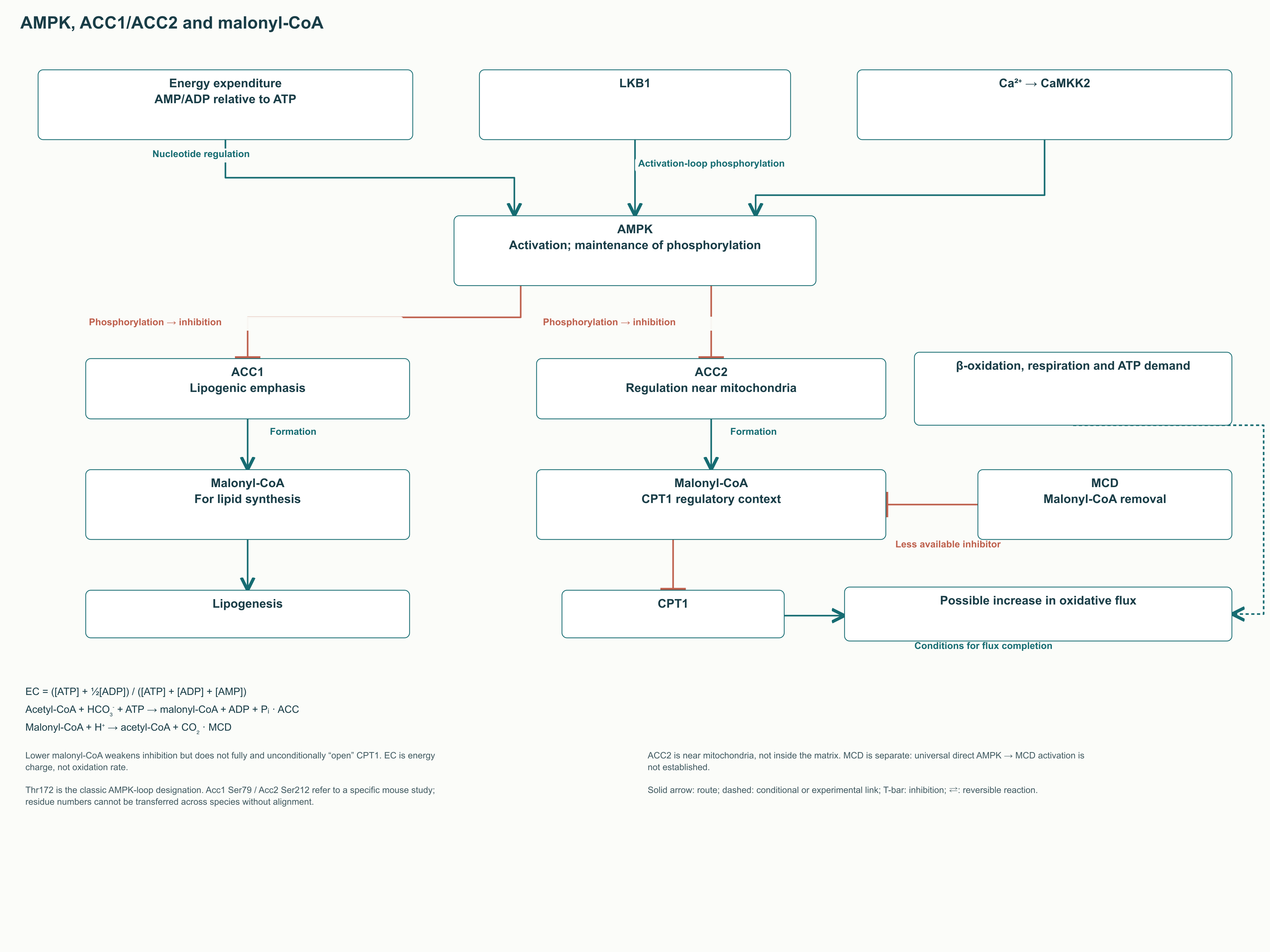
One key output of AMPK is ACC inhibition through phosphorylation. Reduced malonyl-CoA formation decreases lipogenic flux and may weaken CPT1 inhibition. Fullerton and colleagues studied mice in which key ACC1 and ACC2 phosphorylation sites were replaced with alanine, demonstrating disturbances in lipid metabolism and altered responses to metformin. These findings establish a causal role for the AMPK–ACC axis without implying complete CPT1 opening every time the axis is activated.
Phosphorylation-site numbers must be accompanied by the species and sequence used. The cited mouse model concerned ACC1 Ser79 and ACC2 Ser212; site numbers in another species must be determined from its corresponding sequence. For a conceptual figure, “ACC1/ACC2 phosphorylation” and “reduced malonyl-CoA” are sufficient labels. A detailed residue map is appropriate only when discussing a specific primary study.
SIRT1 connects acetylation regulation to this axis in particular experimental systems. In work by Lan and colleagues, changes in LKB1 acetylation accompanied changes in its localisation, interaction with STRAD and AMPK activity. This explains another way of coordinating energy and post-translational signals. It does not establish an identical direction of SIRT1 action in every tissue and cellular state.
4.2. ACC1 and ACC2: different emphases, the same chemical reaction
ACC1 and ACC2 catalyse the same acetyl-CoA carboxylation but differ in organisation and tissue roles. ACC1 is particularly important for supplying fatty acid synthesis with malonyl groups. ACC2 is associated with oxidation regulation near mitochondria and has N-terminal features affecting localisation. This division is useful as a working model without implying an absence of functional overlap between the isoforms.
In Abu-Elheiga and colleagues' 2001 study, deletion of ACC2 in mice was accompanied by increased fatty acid oxidation and reduced fat accumulation. This was a genetic animal experiment, not evidence that pharmacologically stimulating oxidation is safe in patients. Deleting a protein throughout development, briefly suppressing its activity and changing the diet are different interventions. The conclusion that “ACC2 inhibition treats obesity” therefore cannot follow from one model's phenotype alone.
Malonyl-CoA levels are determined by more than the amount of active ACC. Acetyl-CoA availability, allosteric signals, consumption of malonyl groups in synthesis and their removal by a decarboxylase all matter. A whole-tissue concentration need not match the concentration near the outer membrane. Comparisons among liver, muscle and neuronal preparations require particular caution because the metabolite serves different processes in each.
4.3. MCD and post-translational acylation
Malonyl-CoA decarboxylase, MCD, encoded by MLYCD, reduces the malonyl-CoA pool through malonyl-CoA + H⁺ → acetyl-CoA + CO₂. It can thereby relieve part of CPT1 inhibition and return carbon to acetyl-CoA. Its distribution among compartments matters because external malonyl-CoA concentration and matrix processes are not the same measurement. Increasing MCD activity does not guarantee the same outcome in every tissue.
The literature has discussed whether AMPK can directly phosphorylate and activate MCD. However, Sambandam and colleagues did not observe direct phosphorylation in H9c2 cells; activity changes were linked to protein distribution and expression. The parallel “AMPK turns ACC off and immediately turns MCD on” should therefore remain conditional, rather than a canonical direct arrow. The figure shows AMPK acting directly on ACC and presents MCD as an independent inhibitor-removal route.
Malonyl-CoA is also associated with covalent malonylation of protein lysines. Adding a malonyl group changes charge and may affect a particular protein region, but the direction of the functional effect depends on the enzyme and position. This does not imply that every malonylation event invariably inhibits all energy metabolism. Nor should the entire matrix malonyl-CoA pool be described as exclusively toxic: compartmental acyl derivatives have physiological functions of their own.
Du and colleagues demonstrated NAD⁺-dependent demalonylase and desuccinylase activities of SIRT5. In another experimental line, SIRT4 deacetylated and inhibited MCD, accompanying changes in mouse lipid metabolism. Deacetylation therefore does not always mean activation, and different sirtuins do not perform one universal “clean-up” function. These findings retain the subject of post-translational regulation while removing the simplified notion of one switch controlling every acylated enzyme.
4.4. PPAR, PGC-1α and adaptation of the enzyme machinery
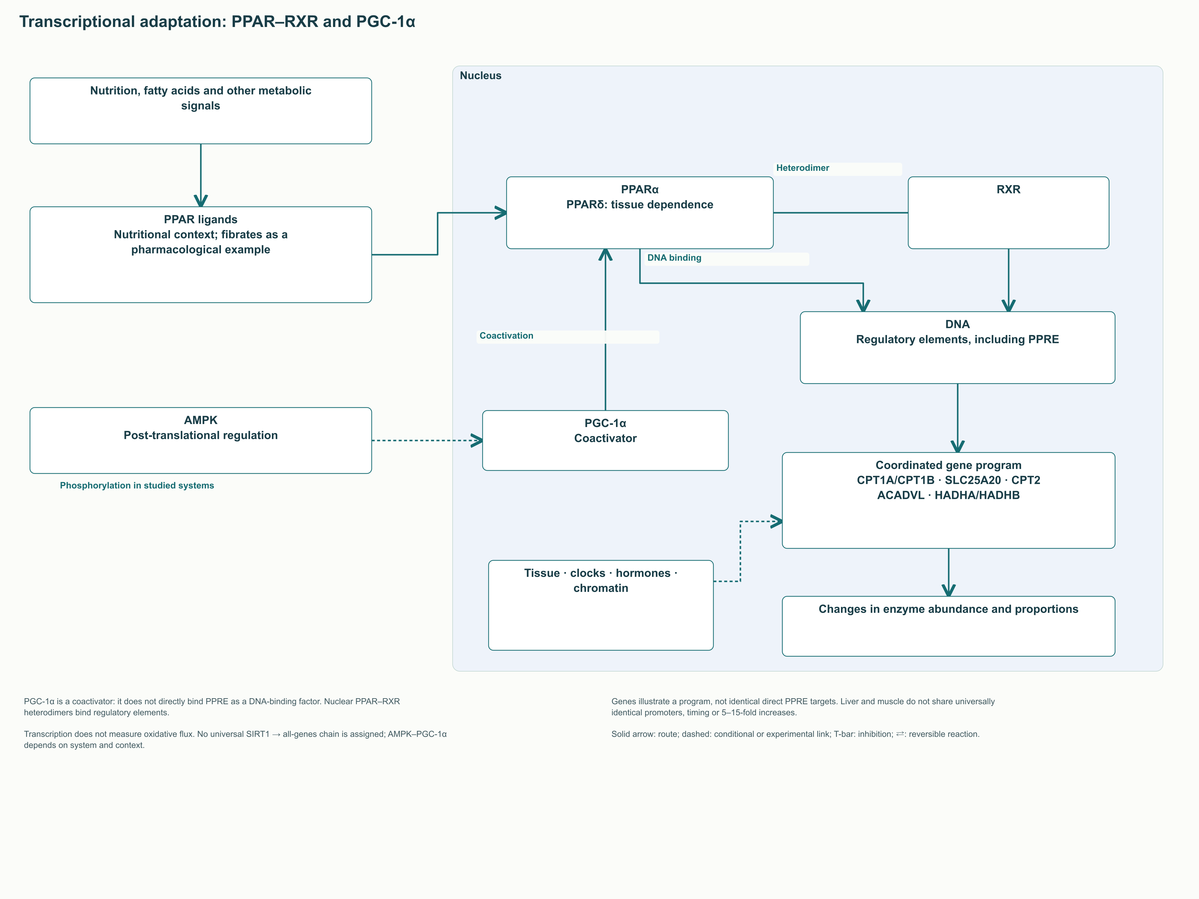
Rapid regulation changes the activity of existing proteins, whereas transcriptional adaptation reorganises their quantities and proportions. PPARα is important in the liver; PPARδ and other factors also participate in the muscle oxidative programme. PPARs interact with RXR and recognise DNA regulatory elements, but the responding gene set depends on tissue. A free fatty acid may act here both as fuel and as part of the signalling context.
In experiments by Kersten and colleagues, mice lacking PPARα adapted poorly to fasting: they accumulated liver lipids and had disturbed ketone and glucose metabolism. These results demonstrate the importance of a coordinated oxidation programme, rather than CPT1A expression alone. Such a programme includes transport enzymes, β-oxidation enzymes and related metabolic pathways. A change in one transcript cannot be assumed to reproduce adaptation of an entire tissue.
PGC-1α serves as a coactivator interacting with several transcription factors, including PPAR and ERR. It is not a fatty acid receptor and does not bind PPRE as an independent DNA-recognising factor. Jäger and colleagues demonstrated direct AMPK phosphorylation of PGC-1α in experimental muscle systems and dependence of part of the gene programme on this coactivator. Together with deacetylation, recruitment of chromatin complexes and altered expression, this links the energy sensor to long-term adaptation.
SIRT3 provides an example of mitochondrial post-translational regulation. Hirschey and colleagues examined altered LCAD acetylation and fatty acid oxidation in mice lacking SIRT3. However, LCAD differs in importance between species, so a mouse model cannot be converted without adjustment into a description of the leading human enzyme for every long chain. Sirtuin–oxidation relationships should be explained through a specific protein and model rather than a promise of generally “boosting mitochondria”.
Circadian clocks, diet, hormones and corepressors also influence metabolic programmes. A claim that CPT1A invariably peaks “at the beginning of wakefulness” requires data for the species, tissue and lighting schedule concerned. Likewise, the fold increase in expression during fibrate exposure depends on the drug, dose and experimental context. These directions help explain adaptation in practice without determining supplement timing or a predictable clinical response.
5. What happens after an acyl group enters the matrix
5.1. The four β-oxidation reactions
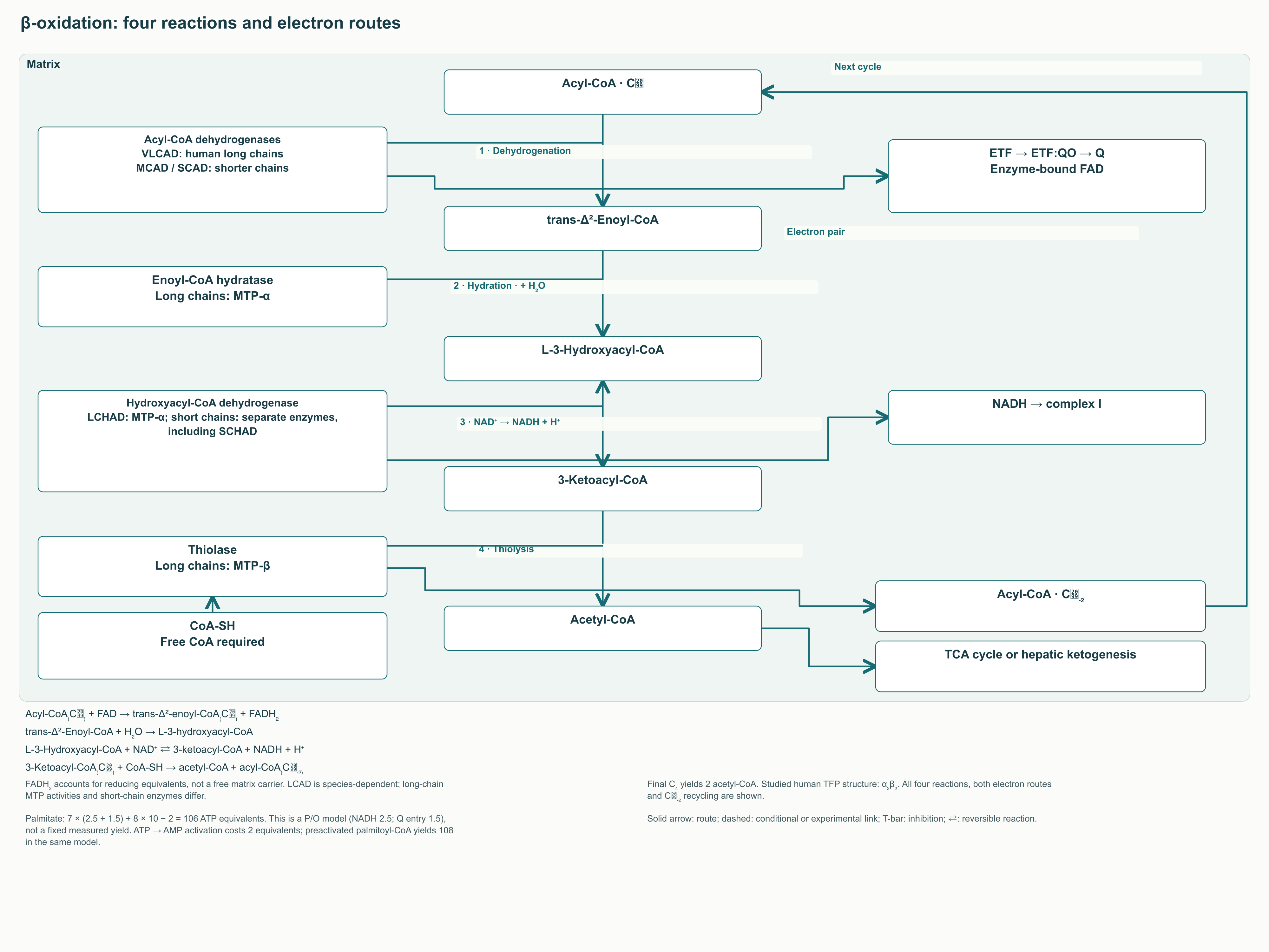
β-Oxidation progressively modifies the region near the acyl chain's carboxyl end. The name refers to the β carbon, the third atom in conventional biochemical numbering starting with the carbonyl carbon. One cycle comprises dehydrogenation, hydration, a second dehydrogenation and thiolysis. The chain usually shortens by two carbon atoms, producing acetyl-CoA and releasing electrons for the respiratory chain.
The first reaction is acyl-CoA₍Cₙ₎ + FAD → trans-Δ²-enoyl-CoA₍Cₙ₎ + FADH₂. An acyl-CoA dehydrogenase performs it, with FAD bound to the enzyme as a cofactor. FADH₂ in the textbook balance accounts for reducing equivalents, not a free molecule floating towards the respiratory chain. Electrons pass through ETF and ETF:Q oxidoreductase into the ubiquinone pool.
The second reaction is trans-Δ²-enoyl-CoA + H₂O → L-3-hydroxyacyl-CoA. Hydratase adds water across the double bond with a defined stereochemistry, supplying the next enzyme with the appropriate L isomer. The third step is L-3-hydroxyacyl-CoA + NAD⁺ ⇄ 3-ketoacyl-CoA + NADH + H⁺. The resulting NADH donates electrons through complex I, so the two reducing steps enter the respiratory chain through different routes.
The fourth reaction is 3-ketoacyl-CoA₍Cₙ₎ + CoA-SH → acetyl-CoA + acyl-CoA₍Cₙ₋₂₎. Thiolysis uses free CoA-SH, and typical thiolase chemistry includes a covalent acyl-enzyme intermediate involving a catalytic cysteine. The shortened chain can enter another cycle, while acetyl-CoA enters the tricarboxylic acid cycle or other pathways, including hepatic ketogenesis. In the final cycle, a four-carbon chain yields two acetyl-CoA molecules.
Step
Conversion
Catalytic system
Link to energy metabolism
1
Acyl-CoA → trans-Δ²-enoyl-CoA
Acyl-CoA dehydrogenases
Electrons pass through ETF and ETF:QO to Q
2
Enoyl-CoA → L-3-hydroxyacyl-CoA
Enoyl-CoA hydratase
Water addition
3
Hydroxyacyl-CoA → 3-ketoacyl-CoA
Hydroxyacyl-CoA dehydrogenase
NADH for complex I
4
Ketoacyl-CoA → acetyl-CoA + shortened acyl-CoA
3-Ketoacyl-CoA thiolase
Free CoA-SH consumption and release of a two-carbon fragment
5.2. Chain length, trifunctional protein and reaction coupling
Dehydrogenase families have overlapping substrate ranges. In humans, VLCAD is especially important for initial long-chain oxidation, while MCAD and SCAD process shorter products. Carbon-chain ranges provide orientation without imposing a rigid boundary beyond which an enzyme does not work at all. LCAD should not be portrayed as a universally equivalent major participant in human long-chain β-oxidation solely by analogy with rodents.
The three subsequent long-chain activities are combined in mitochondrial trifunctional protein, MTP or TFP. The HADHA subunit contains hydratase and long-chain hydroxyacyl-CoA dehydrogenase activities, while HADHB contains thiolase activity. Spatial organisation facilitates transfer of intermediates near the membrane. As the chain shortens, individual soluble matrix enzymes become more important.
The original text unconditionally described MTP as an α₄β₄ heterooctamer. However, structural studies of the human protein, including Xia and colleagues' work, describe an α₂β₂ functional unit and discuss other oligomeric states. A reaction diagram should therefore show the functions of the α and β subunits without imposing one unsupported assembly. This preserves the coupling mechanism and makes the description compatible with modern primary structural findings.
Oxygen is not directly included in any of the four reactions of a standard cycle. Nevertheless, regeneration of NAD⁺ and the oxidised flavin cofactor depends on electron removal by the respiratory chain. Without sufficient electron transfer to oxygen, reducing equivalents accumulate and constrain further flux. Substrate presence in the matrix therefore cannot bypass limitations in oxygen supply and oxidative phosphorylation.
5.3. Unsaturated, odd-chain and very-long-chain fatty acids
In unsaturated fatty acids, existing double bonds are not always suitable for the next hydratase reaction. Enoyl-CoA isomerase converts some intermediate configurations into the trans-Δ² form. Certain polyunsaturated substrates additionally require NADPH-dependent 2,4-dienoyl-CoA reductase. These bypass reactions alter reducing-equivalent and energy balances, making a uniform correction of “minus 1.5–2.5 ATP for every double bond” incorrect.
For example, bypassing the first dehydrogenase step removes the corresponding electron transfer through ETF. If reductase is needed, the cost of reduction using NADPH is added. Calculating the total for a particular fatty acid requires knowing double-bond positions and the sequence of intermediate reactions. The palmitate calculation cannot simply be copied to oleate or linoleate with identical coefficients for each step.
With an odd-chain substrate, the final carbon residue is propionyl-CoA. Its subsequent sequence is propionyl-CoA + HCO₃⁻ + ATP → D-methylmalonyl-CoA + ADP + Pᵢ, followed by D-methylmalonyl-CoA ⇄ L-methylmalonyl-CoA → succinyl-CoA. The first step requires biotin-dependent propionyl-CoA carboxylase; the last requires methylmalonyl-CoA mutase with adenosylcobalamin, a vitamin B₁₂ form. Conversion of the propionyl residue therefore depends on a different enzyme and cofactor set from ordinary acetyl-CoA release.
Succinyl-CoA replenishes tricarboxylic acid cycle intermediates, participating in anaplerosis. Entry into this pathway does not by itself establish that a small dietary quantity of odd-chain fatty acids will correct an oxaloacetate deficit or improve insulin sensitivity. Overall flux depends on the availability of other intermediates and their removal into biosynthetic pathways. A biochemical possibility cannot establish a therapeutic indication for dairy fat or a specialised supplement.
Peroxisomal oxidation of very long chains has a different energy organisation. At its initial step, electrons are transferred to oxygen to form H₂O₂ rather than entering the mitochondrial ETF route. Shortened products can subsequently be used by mitochondria, including after conversion into carnitine derivatives. This context also matters for CROT: carnitine octanoyltransferase is predominantly associated with peroxisomes, so placing it in the matrix as a universal CPT2 partner in the original text was incorrect.
5.4. Why MTP defects can affect pregnancy
LCHAD deficiency or deficiencies of other MTP activities impair completion of long-chain β-oxidation. Patients may develop hypoketotic hypoglycaemia, cardiomyopathy and other manifestations of energy deficiency. A separate clinical association concerns a mother carrying a fetus with impaired LCHAD function. In Ibdah and colleagues' primary study, fetal fatty acid oxidation defects were associated with severe maternal liver disease during pregnancy.
Acute fatty liver of pregnancy and HELLP syndrome should not be regarded as inevitable outcomes of any fetal HADHA defect. Not all such obstetric cases are explained by MTP, and not every pregnancy with an affected fetus has the same outcome. Accumulation and exchange of hydroxylated lipid metabolites are considered part of the mechanism, but the entire pathology cannot be reduced to one acylcarnitine. In clinical practice, recognising an emergency and subsequently arranging specialised family evaluation are more important than attempting to prescribe a universal “mitochondrial” protocol.
6. Energy balance: from palmitate to the calculated 106 ATP
6.1. Stoichiometry of seven cycles
Palmitate contains 16 carbon atoms and no double bonds. Complete conversion of its acyl chain into eight acetyl-CoA molecules requires seven β-oxidation cycles: the final C4 cleavage produces two C2 fragments at once. Generally, for a saturated even-chain Cₙ substrate, the number of cycles is n/2 − 1 and the number of acetyl-CoA molecules is n/2. These relationships describe carbon balance rather than reaction velocity in a particular tissue.
The accounting total for palmitoyl-CoA is palmitoyl-CoA + 7 CoA-SH + 7 FAD + 7 NAD⁺ + 7 H₂O → 8 acetyl-CoA + 7 FADH₂ + 7 NADH + 7 H⁺. As in the description of one cycle, FAD/FADH₂ represents exchange of reducing equivalents through bound cofactors. Acetyl-CoA subsequently undergoes oxidation in the tricarboxylic acid cycle, producing additional reduced carriers. CPT1 and carnitine transport are not separate ATP sources in this balance.
6.2. P/O is a calculation assumption, not a biological constant
The P/O ratio relates ATP synthesis to electron transfer to oxygen; in traditional notation, O denotes an oxygen atom, or half an O₂ molecule. Textbook calculations often assume 2.5 ATP equivalents per NADH and 1.5 for electrons entering the Q pool while bypassing complex I. Seven NADH then yield 17.5 ATP, and seven flavin reducing equivalents yield 10.5. β-Oxidation contributes a total of 28 ATP equivalents.
One acetyl-CoA in the tricarboxylic acid cycle produces 3 NADH, one flavin reducing equivalent and one GTP. With the selected P/O ratios, this is 3 × 2.5 + 1 × 1.5 + 1 = 10 ATP equivalents. Eight acetyl-CoA molecules yield 80, and two equivalents spent activating free palmitate are subtracted from the overall total. The resulting expression is ATP₍net₎ = 7 × (2.5 + 1.5) + 8 × 10 − 2 = 106.
Calculation component
Quantity
Assumed cost or yield
Result
NADH from β-oxidation
7
2.5 ATP per NADH
17.5
Flavin reducing equivalents from β-oxidation
7
1.5 ATP per electron pair delivered to Q
10.5
Acetyl-CoA completely oxidised in the TCA cycle
8
10 ATP equivalents per acetyl-CoA
80
Free-palmitate activation
1 ATP → AMP reaction
−2 ATP equivalents
−2
Total
Complete oxidation of one palmitate molecule
P/O = 2.5/1.5
106 ATP equivalents
A useful general expression under the same assumptions is ATP₍net,Cₙ₎ = (n/2 − 1) × 4 + (n/2) × 10 − 2 = 7n − 6. It applies to a free saturated even-chain fatty acid undergoing complete mitochondrial processing with the assumed textbook coupling. If counting begins with already activated palmitoyl-CoA, activation cost is not subtracted within that particular pathway segment, giving 108. If some acetyl-CoA is diverted into ketone bodies or synthesis, the condition of complete oxidation within one cell is not met.
ATP-synthase stoichiometry studies, including the c₈-ring structure in Watt and colleagues' work, show why an integer yield cannot be treated as an invariant property of a living mitochondrion. Coupling, proton leak, phosphate transport and nucleotide transport change the actual relationship between expenditure and synthesis. Textbook coefficients help compare pathways but do not measure actual ATP production in a person after fat intake. The expression “palmitate generates exactly 106 ATP molecules” must therefore include the calculation conditions.
6.3. Free energy and efficiency
Model thermodynamic efficiency can be expressed as η = N₍ATP₎ × ΔG₍ATP synthesis₎ / |ΔG₍substrate oxidation₎|. Both free energies must refer to comparable states and conditions. Standard transformed energy and the actual intracellular energy of ATP synthesis are not interchangeable. Calculation requires reactant concentrations or activities, temperature and defined system boundaries.
Mixing standard oxidation energy with an approximate intracellular ATP cost in one calculation does not produce a universally measured efficiency. Arithmetic division alone does not determine the efficiency of a living tissue. It is therefore more useful to establish conditions and calculation boundaries before interpreting the estimate. The important principle for practitioners is that a substantial part of the energy is stored, another part dissipates, and these proportions change with coupling status.
7. Incomplete oxidation, CoA and metabolic flexibility
7.1. When incoming flux exceeds downstream capacity
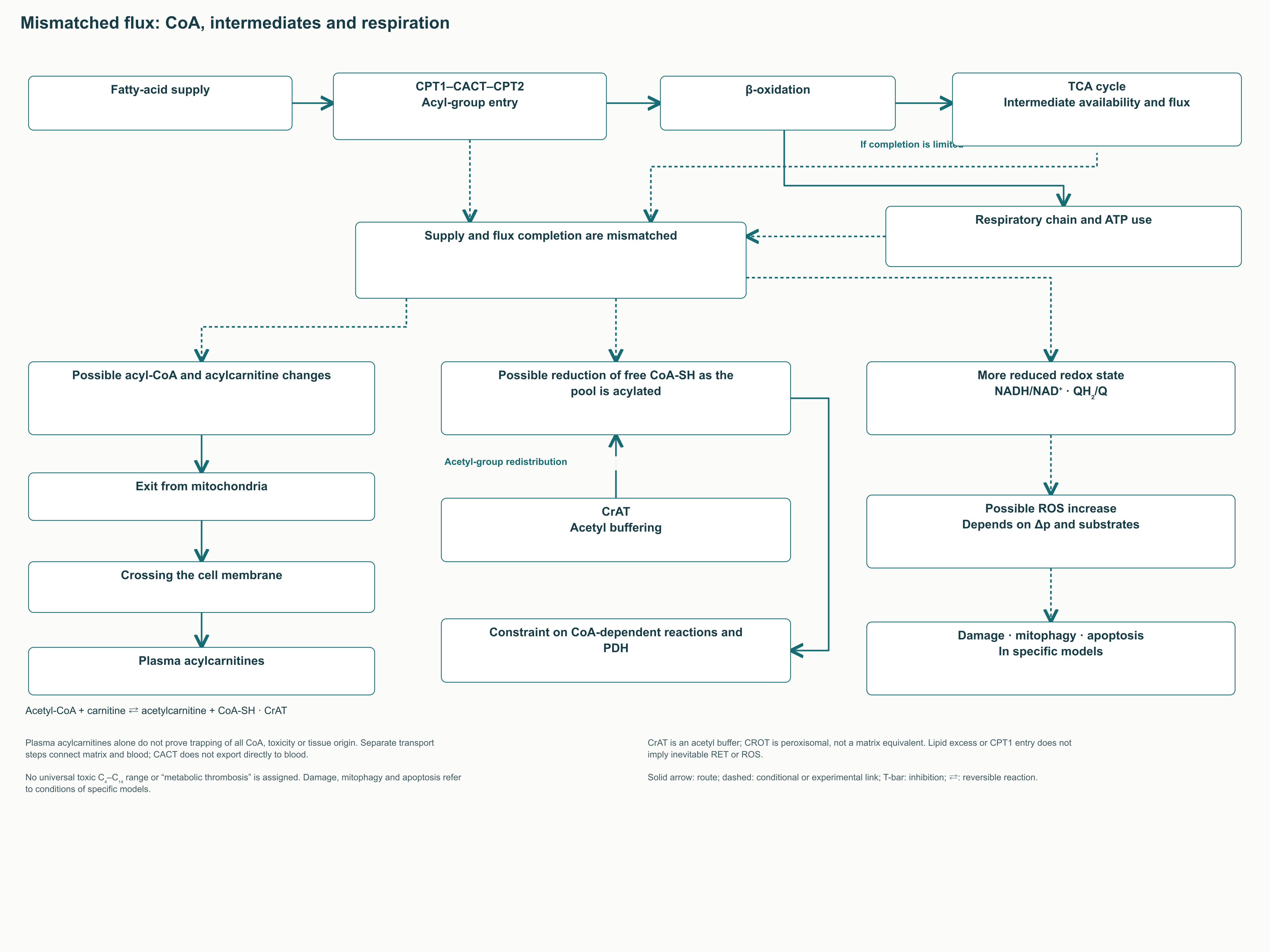
Increasing delivery of long-chain acyl groups into the matrix is only the first stage of their utilisation. β-Oxidation, the tricarboxylic acid cycle, the respiratory chain and ATP consumption must work in coordination. If substrate arrives faster than intermediates are oxidised, their concentrations change even when individual enzymes retain or increase activity. “More fat oxidation” therefore requires clarification: it may mean initial reactions, complete oxidation to CO₂ or increased electron flux.
In 2008, Koves and colleagues investigated metabolic overload in muscle models and animals. They associated insulin resistance with increased β-oxidation flux, altered switching to carbohydrate and changes in tricarboxylic acid cycle intermediates. In some experimental conditions, limiting fatty acid entry into mitochondria improved the metabolic response. This is a substantial objection to the idea that every increase in CPT1 must improve insulin sensitivity.
The experimental association does not imply that incomplete β-oxidation explains every form of diabetes. Acylcarnitine profiles differ among tissues, feeding states, exercise conditions and diseases. Sometimes elevated levels reflect rapid turnover; sometimes they indicate a downstream constraint or transport disorder. An acylcarnitine is not universal evidence of toxicity, and its concentration cannot replace measurement of metabolic flux.
An increased NADH/NAD⁺ ratio reduces oxidised-cofactor availability for several reactions. QH₂/Q status is also related to the capacity to accept new electrons through ETF and other respiratory-chain inputs. If ATP demand is insufficient or respiration is constrained, flux may slow even when CPT1 delivers substrate. These ratios explain feedback without establishing the same allosteric action on every TCA-cycle enzyme.
7.2. “CoA trapping” and acetylcarnitine's role
Free CoA-SH is consumed when acyl derivatives form and recovered when they are subsequently used. If a substantial portion of the pool remains acylated, available CoA may become insufficient for other reactions. “CoA trapping” describes that limitation, but establishing it requires information about free and bound fractions. Elevated concentrations of several plasma acylcarnitines do not by themselves show that matrix CoA is depleted.
Short-chain carnitine acetyltransferase, CrAT, helps redistribute acetyl groups through acetyl-CoA + carnitine ⇄ acetylcarnitine + CoA-SH. This is a separate carnitine-metabolism function that cannot be attributed solely to CPT1. Muoio and colleagues found that muscle deletion of CrAT in mice impaired glucose tolerance and substrate switching; the work also included human-cell and participant investigations. The results supported a model of acetyl-group buffering and limitation of their inhibitory influence on the pyruvate dehydrogenase complex.
Acetylcarnitine, long-chain acylcarnitines and medium-chain products have different origins and functions. CPT2 can catalyse reversible long-chain transfer, CrAT acts predominantly on short groups, and CROT is associated with peroxisomal metabolism. Furthermore, a metabolite leaving a mitochondrion, crossing the cell membrane and appearing in plasma involves separate transport stages. An arrow stating “CACT ejects acylcarnitines directly into blood” therefore conceals necessary intermediate processes.
Oxidative stress, reverse electron transfer and mitochondrial permeability transition pore opening are possible with particular combinations of a reduced Q pool, membrane potential and cellular damage. They are not inevitable continuations of every acylcarnitine elevation. Apoptosis, mitophagy and uncoupling also have separate mechanisms and conditions. In the overload figure, these events appear as possible consequences rather than an inevitable linear chain.
7.3. RER, respiratory quotient and fuel switching
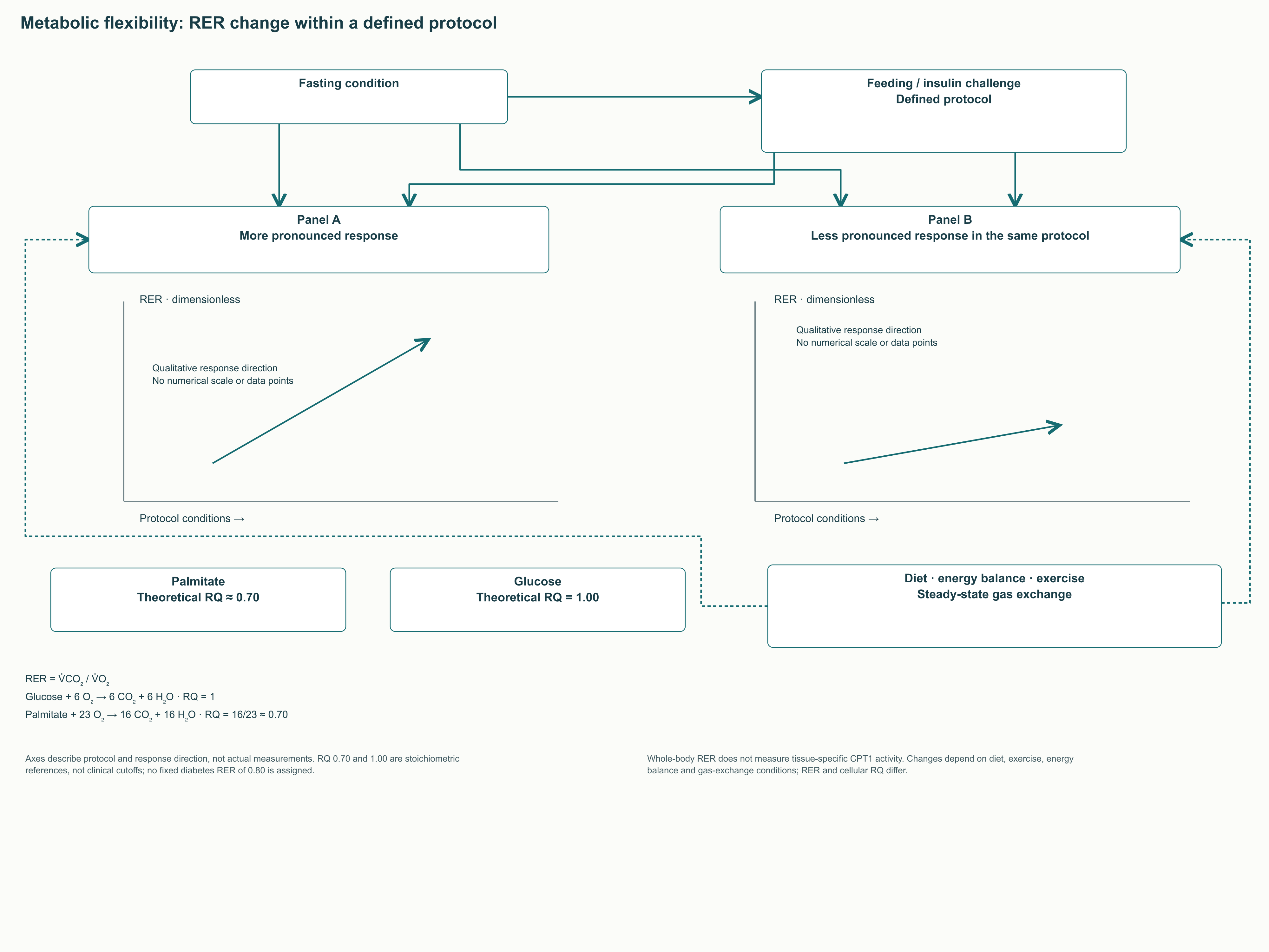
Metabolic flexibility is the ability to change substrate utilisation when moving from fasting to feeding, during insulin exposure and during exercise. One investigative method is indirect calorimetry, measuring O₂ consumption and CO₂ production. The ratio of respiratory fluxes is RER = V̇CO₂/V̇O₂. At the chemical-reaction level, the ratio of CO₂ formed to O₂ consumed is the respiratory quotient, RQ; under steady conditions the two may be close, but they are not completely identical.
For glucose, C₆H₁₂O₆ + 6 O₂ → 6 CO₂ + 6 H₂O, so RQ = 6/6 = 1. For palmitate, C₁₆H₃₂O₂ + 23 O₂ → 16 CO₂ + 16 H₂O, giving RQ = 16/23 ≈ 0.70. With mixed-substrate utilisation, the value generally falls between these textbook extremes. Protein oxidation, ketone bodies, lipogenesis and changes in CO₂ stores complicate interpretation.
After a meal or during insulin exposure, a greater proportion of carbohydrate oxidation may accompany an increased RER. Impaired metabolic switching may produce a smaller change, but diabetes has no universal “fixed RER of 0.80–0.83”. Baseline diet, energy balance, fitness and measurement conditions substantially affect results. The figure therefore compares response directions without invented clinical thresholds or imitation of actual measured curves.
During intense exercise, RER can exceed one because of acid buffering and increased CO₂ elimination. This cannot be interpreted literally as the body oxidising “more than 100% carbohydrate”. Likewise, an isolated fasting measurement neither diagnoses CPT1 deficiency nor determines a need for carnitine. Practitioners gain more from comparing standardised responses under the same protocol than from judging metabolism by one number.
7.4. The Randle cycle and reversible PDH regulation
Fatty acid oxidation changes acetyl-CoA/CoA-SH and NADH/NAD⁺ ratios, influencing pyruvate utilisation. The pyruvate dehydrogenase complex, PDH, converts pyruvate to acetyl-CoA and thereby links carbohydrate metabolism to the tricarboxylic acid cycle. PDKs phosphorylate the complex and reduce activity, while pyruvate dehydrogenase phosphatases remove those phosphate groups. Phosphorylation and dephosphorylation allow reversible changes in PDH activity between feeding and exercise states.
PDK4 is one isoform whose expression responds to fasting and hormonal signals. FoxO1, KLF15 and other regulators may contribute to reorganisation of this programme, but their contribution varies by tissue and state. Insulin signalling through Akt changes FoxO1 activity, linking glucose availability to gene regulation. Different PDKs use PDH phosphorylation sites differently, so an identical PDK4 action on all three sites cannot be assumed without primary evidence.
The Randle cycle is useful as a substrate-competition model without replacing the entire pathophysiology of insulin resistance. Limited pyruvate oxidation, reduced cellular glucose entry and impaired insulin signalling are related but distinguishable events. Not all glucose is “dumped into lactate” when PDH activity falls: it may enter glycogen and other pathways. This distinction matters when interpreting carnitine-buffering mechanisms and proposed therapeutic effects.
8. Lipotoxicity: which lipids and which signalling pathways
8.1. Diacylglycerols, ceramides and localisation
A fatty acid may become an energy substrate, storage lipid, membrane component or signalling molecule. Triacylglycerol in a lipid droplet is not equivalent to diacylglycerol near a signalling protein. For DAG, stereochemistry, acyl composition and accessibility to protein-kinase C1 domains matter. Total “muscle fat” content therefore does not automatically describe lipotoxic load.
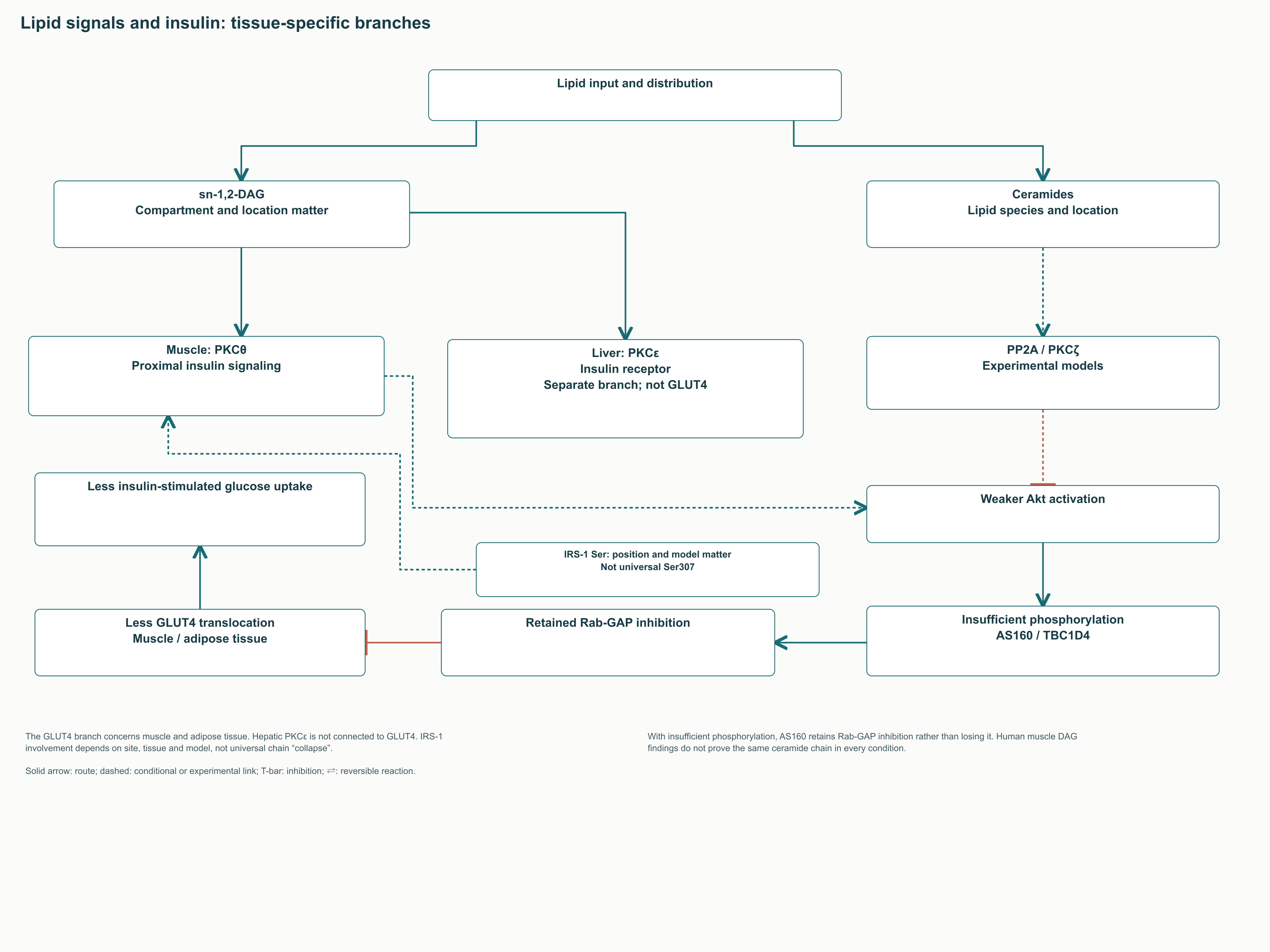
In Szendroedi and colleagues' study, sequential human muscle biopsies during lipid infusion tracked changes in DAG, PKCθ activation and insulin signalling. Associations with IRS1 Ser1101 phosphorylation and a weaker subsequent response were observed. In the same experimental system, changes in ceramides and acylcarnitines explaining insulin resistance were not detected. These results demonstrate why all lipid metabolites cannot unconditionally be combined into one causal chain.
A different pathway has been studied experimentally in the liver: DAG–PKCε and altered insulin-receptor activity. Petersen and colleagues identified human-receptor Thr1160 as a PKCε substrate and investigated the corresponding substitution in mice. Liver and muscle mechanisms therefore have different specific intervention points. Nor does the liver use the muscle GLUT4 route as its principal mechanism of insulin-dependent glucose uptake.
In muscle, Akt is linked to phosphorylation of TBC1D4, known as AS160. This reduces its inhibitory GAP action on particular Rab proteins and facilitates movement of GLUT4-containing vesicles to the membrane. The sequence may be disrupted when insulin signalling weakens. “AS160 remains inactive” inverted the meaning: the retained inhibitory function of the unphosphorylated protein may obstruct the normal response.
8.2. Serine phosphorylation does not always mean harm
IRS1 has many phosphorylation sites, whose actions depend on position, timing and cellular context. In experiments by Copps and colleagues, replacement of mouse Irs1 Ser307, corresponding to human Ser312, increased insulin resistance. This directly contradicts the rule that any phosphate on that serine must destroy insulin signalling. Ser307 therefore cannot be drawn as a universal pathological target without identifying the species and specific experiment.
Ceramides can influence Akt through PP2A and atypical PKCs and can participate in membrane stress and cell death. However, individual ceramide species have different functions, and an experimental role does not always correspond to a change in total content in a human biopsy. Palmitate does supply substrate for sphingolipid synthesis, but the specific outcome depends on lipid composition and cellular processing capacity. A causal conclusion requires intervention, measurement of the relevant fraction and an outcome at a specified system level.
ER stress through PERK and IRE1, proinflammatory signalling and mitochondrial injury may complement these pathways. Their presence cannot predict an identical 70–80% loss of glucose uptake or inevitable heart failure. Those fixed values in the original text lacked sufficient support linked to a specific experiment. Clinical interpretation benefits more from treating lipotoxicity as a contextual network of mechanisms than as a single cascade from palmitate to diabetes.
9. Inherited defects and the Arctic CPT1A variant
9.1. CPT1A deficiency: hypoglycaemia with insufficient ketogenesis
With marked CPT1A deficiency, the liver uses long-chain fatty acids less effectively during fasting. Energy requirements continue after glycogen is exhausted, but ketone-body production and support of glucose metabolism become insufficient. A characteristic clinical signal is hypoketotic hypoglycaemia: low glucose with an inappropriately small ketone rise. Lethargy, seizures, liver involvement and hyperammonaemia may occur, particularly during infection or reduced food intake.
An increase in free carnitine relative to long-chain acylcarnitines is associated with restriction of the CPT1 reaction itself. Screening may use the C₀/(C₁₆ + C₁₈) ratio, but the specific fractions and cut-offs depend on the laboratory protocol. “Above 50–100” cannot be presented as a universal diagnostic criterion for every sample. Confirmation rests on combined biochemical and molecular genetic evidence rather than one ratio.
Preserved muscle energy metabolism is explained by the presence of another isoform, but does not make hepatic decompensation safe. The practical management principle is to prevent prolonged fasting and maintain an individual illness-management plan. Specialists may consider medium-chain fats because some components depend less on CPT1. Diet, permissible fasting intervals and other measures must nevertheless be determined for the individual patient; this monograph does not replace that plan.
9.2. CPT2 and CACT: the block lies downstream of CPT1
CPT2 deficiency may present as severe neonatal or infantile forms, or as predominantly muscular disease. The myopathic form typically causes recurrent episodes of muscle pain, weakness, rhabdomyolysis and myoglobinuria, sometimes with normal health between attacks. Triggers include prolonged exercise, infection, fasting or cold. Age at presentation and severity depend on genotype and circumstances, so “adult form” does not exclude earlier onset.
Severe early forms involve the liver, heart and other organs. Kidney and brain involvement may occur in the most severe neonatal cases, but the whole spectrum cannot be described through one obligatory set of congenital malformations. The recognised p.Ser113Leu variant is associated with the myopathic phenotype but does not explain every case. Clinical form cannot reliably be determined from one common substitution without the complete genetic and clinical context.
CACT deficiency is associated with SLC25A20 and impairs transfer of already formed acylcarnitine across the inner membrane. Severe cases may likewise involve hypoglycaemia, hyperammonaemia, cardiomyopathy and arrhythmias. Long-chain acylcarnitine profiles can overlap with CPT2 deficiency, so a biochemical result does not always identify the site of the block. Genetic confirmation and specialised interpretation are particularly important for distinguishing these conditions.
Disorder
Site of restriction
Characteristic diagnostic logic
Practical relevance
CPT1A deficiency
Long-chain acylcarnitine formation
Relatively abundant free carnitine and few long-chain derivatives; CPT1A confirmation
Acylcarnitine profile may overlap with CACT; CPT2 confirmation
Early systemic forms or recurrent rhabdomyolysis
Primary carnitine deficiency
Cellular uptake and renal carnitine retention
Low free carnitine; SLC22A5 confirmation
A different defect requiring replacement therapy
This table illustrates defect topology and is not a diagnostic algorithm based on one number. Some abnormalities may be less pronounced outside an attack, and a dried-blood-spot sample does not always exclude myopathic CPT2 deficiency. The combination of a clinical trigger, biochemical profile and genetic findings matters more than a change in carnitine alone. Explaining recurrent rhabdomyolysis as an ordinary “lack of a supplement” is particularly dangerous.
9.3. p.Pro479Leu: common does not mean harmless
The CPT1A c.1436C>T, p.Pro479Leu variant occurs frequently in some Arctic populations. In Rajakumar and colleagues' study of 1111 Greenland Inuit, L479 allele frequency was about 0.73, and genotype was associated with particular lipid measures. This describes a specific sample and association, not the entire Arctic or proven protection from cardiovascular events. Frequencies in other groups and regions should not automatically be replaced with one universal value.
Biochemically, reduced residual activity and weaker malonyl-CoA sensitivity are discussed. These can act in opposing directions: less inhibition does not automatically compensate for lower catalytic capacity. A proposed adaptation to a traditional diet therefore remains a hypothesis about selection. It does not establish continuously high ketogenesis or clinical advantage in every carrier.
Gillingham and colleagues studied the response to medically supervised fasting in five children homozygous for this variant. Long-chain acylcarnitine and ketone production was attenuated, and two tests were stopped because of hypoglycaemic symptoms. The findings demonstrate a physiologically meaningful restriction of fasting tolerance despite the variant's high prevalence. They do not support the idea of unconditionally increased ketogenesis in carriers.
Population associations with childhood mortality require consideration of infection, nutrition, access to care and other factors. They cannot be explained solely by transition to a high-carbohydrate diet. The clinical task is to prevent dangerous episodes and interpret genotype correctly, rather than prescribe an ethnically defined diet. Selection history and individual medical risk answer different questions.
10. Carnitine in the body: transport, forms and supplements
10.1. OCTN2 should not be confused with CACT
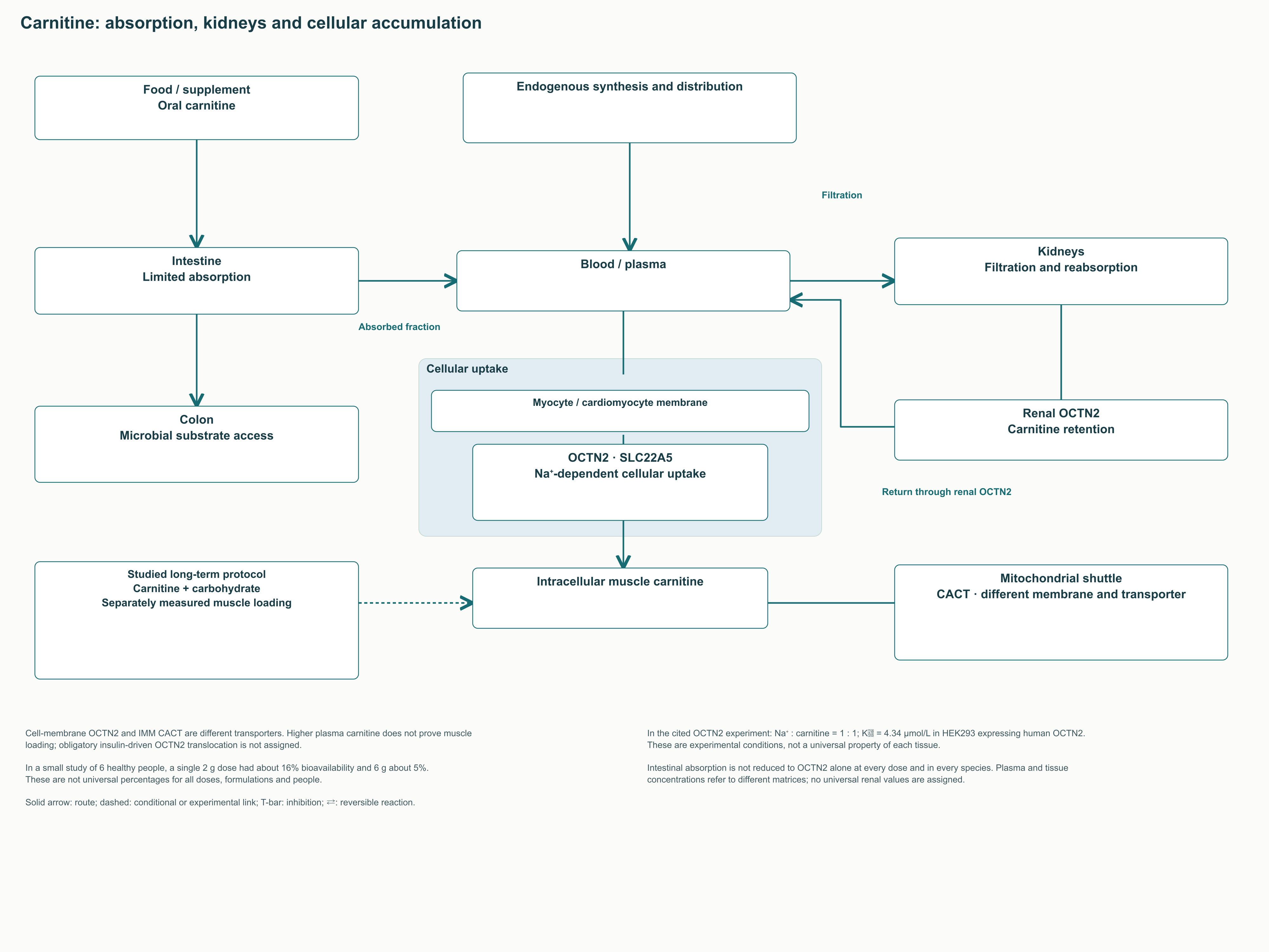
OCTN2, encoded by SLC22A5, is located in the cell membrane and enables high-affinity sodium-dependent carnitine uptake. CACT is located in the inner mitochondrial membrane and exchanges carnitine with acylcarnitine within the shuttle. These are two different barriers and transport mechanisms. Increased blood carnitine does not automatically show that myocyte carnitine has increased or mitochondrial flux has risen.
Tamai and colleagues measured a Kₘ of about 4.34 µmol/L when expressing human OCTN2 in HEK293 cells. This characterises a particular cellular experiment rather than a clinical carnitine target. Transport studies describe sodium–carnitine symport at 1:1; the cited functional data do not support two Na⁺ ions per carnitine. Transporter function is associated with the sodium gradient but is not direct insulin-mediated substrate transfer across the mitochondrial membrane.
Most tissue carnitine is concentrated in muscle, while the plasma pool is much smaller. Renal reabsorption helps conserve carnitine, and its balance also includes dietary intake and endogenous synthesis. At high oral doses, absorption and retention become constraints, so the effect is not proportional to the amount ingested. Correct comparisons require matching units and measurement matrices: millimoles per kilogram of tissue cannot simply be divided by micromoles per litre of plasma without additional conditions.
In a small pharmacokinetic study by Harper and colleagues involving six healthy participants, a single 2-g dose had approximately 16% bioavailability, whereas a 6-g dose had about 5%. These figures illustrate limited assimilation of large doses but are not universal values for every salt, meal or patient. Carnitine from ordinary food and a pharmacological bolus dose also have different kinetics. Poor muscle loading therefore cannot reliably be overcome simply by increasing a single dose.
10.2. Primary systemic carnitine deficiency
Marked loss of OCTN2 function impairs tissue uptake and renal retention of carnitine. Primary carnitine deficiency may present with hypoketotic hypoglycaemia, cardiomyopathy and muscle weakness, but its spectrum also includes milder and asymptomatic variants. This inherited disease differs from an ordinary diet-related change in plasma carnitine. A very low level requires investigation, including differentiation of a primary disorder from secondary changes in other metabolic diseases.
Levocarnitine replacement for confirmed primary deficiency has a different purpose from use by a healthy person for sport or weight control. It replenishes substrate in defective homeostasis and involves dose adjustment and biochemical monitoring. Introducing a universal “extremely high dose” and attributing its action solely to passive diffusion would be incorrect. Genetic diagnosis, severity, tolerance and monitoring determine the individual regimen.
Secondary deficiency may occur with some acquired conditions, medications and other inherited disorders. A low test result does not establish that carnitine explains fatigue or impaired exercise tolerance. Carnitine should also not automatically be prescribed for every β-oxidation defect: block sites and the risk of long-chain derivative accumulation differ. The key practical step is establishing the metabolic cause rather than choosing a “stronger” supplement form.
10.3. L-carnitine, tartrate, fumarate and ALCAR
L-carnitine is the physiologically used stereoisomer; tartrate and fumarate are its salt forms, while acetyl-L-carnitine is an acyl derivative. This matters when calculating carnitine quantity: salt mass is not free-substance mass. In anhydrous L-carnitine L-tartrate with the corresponding stoichiometry, carnitine makes up about 68%, but the particular product label should identify which quantity is stated. A specific salt does not by itself establish superior exercise or cardiovascular outcomes.
Fumarate is a tricarboxylic acid cycle intermediate, but this does not establish cardiac synergy for every carnitine–fumarate salt. Likewise, faster dissolution does not equal better muscle loading. Claiming a form advantage requires direct comparisons using equal active-substance doses and clinically meaningful outcomes. Such comparative findings are specifically needed to declare tartrate preferable for exercise performance.
Acetyl-L-carnitine, ALCAR, participates in acetyl-group metabolism and is studied in the nervous system. Its transport and conversion cannot be reduced to “crosses the barrier better — becomes acetylcholine — protects synapses”. Tissue availability, metabolic effects and symptom improvement are separate questions. In particular, an acetyl group's involvement in metabolism does not necessarily increase acetylcholine synthesis to a clinically useful extent.
In Thal and colleagues' multicentre year-long study, ALCAR did not slow deterioration in the main outcomes across the overall Alzheimer disease group. A subsequent early-onset disease trial also failed to demonstrate the expected overall benefit in principal outcomes. “Neuroprotectant” should therefore not serve as ready-made evidence for treating cognitive impairment. Individual diseases and study findings should be discussed separately rather than combined into a universal morning ALCAR course.
11. Clinical studies: what outcome was actually measured
11.1. Glycemia, body weight, and body composition
Carnitine's mechanistic role makes it a plausible candidate for metabolic research, but the clinical effect must be assessed separately from its transport function. Studies differ in participants' diagnoses, baseline diet, duration, the form of the substance, and concomitant treatment. The pooled result applies to this set of studies and does not imply the same response in a healthy athlete and a patient with diabetes. It is particularly important to distinguish a change in a laboratory measurement from prevention of disease complications.
The meta-analysis by Zamani and colleagues, published in 2023, pooled 41 randomized studies involving 2,900 participants. The mean difference versus control was −3.22 mg/dL for fasting glucose, −0.27 percentage points for HbA1c, and −0.73 for HOMA-IR. Heterogeneity was high: I² was 88.6%, 90.1%, and 98.2%, respectively. A statistically significant average effect therefore cannot be presented as a consistent individual reduction in blood glucose or a proven treatment for insulin resistance.
A percentage point and a relative percentage mean different things here. A change in HbA1c from 8.00% to 7.73% is −0.27 percentage points, whereas the relative change is −0.27 / 8.00 × 100% ≈ −3.4%. HOMA-IR is calculated from fasting insulin and glucose concentrations; it does not directly measure flux through CPT1. Even simultaneous improvement in these measurements does not identify which of the possible metabolic branches produced the result.
For body weight, Talenezhad and colleagues pooled 37 randomized studies involving 2,292 participants. The mean difference in weight was −1.21 kg, with a 95% confidence interval from −1.73 to −0.68 kg, and the difference in BMI was −0.24 kg/m². Fat mass was assessed in kilograms, whereas no significant overall effect was found on body fat percentage or waist circumference. In the analysis of high-quality studies, evidence of an effect on body weight remained, limiting stronger promises about changes in body composition.
Question
What the selected analysis showed
Remaining limitation
Does glycemia change?
A small average reduction in several measurements across a heterogeneous set of studies
High heterogeneity, different baseline conditions, and no automatic conclusion about clinical complications
Does body weight change?
A modest average difference in weight and BMI
The same response in all participants and superiority over sustained dietary changes have not been established
Does body fat percentage change?
The meta-analysis of 37 studies did not establish a significant overall effect
Fat mass in kilograms and body fat percentage are not interchangeable
Can an optimal dose be identified from meta-regression?
A statistical association in the available studies can be described
It does not replace a direct comparison of doses or individualized prescribing
This allows carnitine to be discussed as an adjunct under investigation in particular clinical situations, while keeping the magnitude of the observed effect in perspective. It does not make the supplement mandatory for excess body weight or justify replacing prescribed treatment. If a particular outcome is of interest, it should be specified in advance: weight, exercise tolerance, HbA1c, or the frequency of events. Choosing a substance without a defined goal makes even a positive analytical result difficult to interpret.
11.2. Cardiovascular outcomes and CEDIM-2
Ischemia changes oxygen delivery and the balance between carbohydrate and fat oxidation. Carnitine and other metabolic interventions have therefore been studied after myocardial infarction, but physiological plausibility is not equivalent to a proven reduction in mortality. The timing of treatment initiation, route of administration, background reperfusion therapy, and primary endpoint all matter. Studies from different eras should not be combined into a promise of universal contemporary secondary prevention.
The 2013 meta-analysis by DiNicolantonio and colleagues included 13 controlled studies with 3,629 participants. For all-cause mortality, the odds ratio was 0.73 with a 95% confidence interval of 0.54–0.99, whereas the risk ratio was 0.78 with an interval of 0.60–1.00. The authors also found signals of fewer ventricular arrhythmias and less angina, but did not find a convincing overall reduction in heart failure or recurrent myocardial infarction. Saying that the “risk of death falls by 27%” without identifying the odds ratio confuses different statistical measures and creates a stronger impression of certainty.
The randomized CEDIM-2 trial enrolled 2,330 patients with anterior ST-elevation myocardial infarction. The study ended with fewer participants than planned, and the primary composite endpoint of death or heart failure at six months occurred in 9.2% versus 10.5%, p = 0.27. A secondary assessment of early mortality showed a positive signal, but this does not turn a negative primary result into established overall efficacy. It is a useful example of why a study's conclusion is determined by the prespecified endpoint rather than its most favorable comparison.
Result of the post-infarction meta-analysis
Effect measure
Interpretation
All-cause mortality
OR 0.73; 95% CI 0.54–0.99; RR 0.78; 95% CI 0.60–1.00
A borderline result that depends on the effect measure
Ventricular arrhythmias
RR 0.35; 95% CI 0.21–0.58
A signal in a historical set of controlled studies
Angina
RR 0.60; 95% CI 0.50–0.72
Not equivalent to a reduction in mortality or recurrent myocardial infarction
Heart failure
RR 0.85; 95% CI 0.67–1.09
A statistically convincing overall effect was not established
Recurrent myocardial infarction
RR 0.78; 95% CI 0.41–1.48
An uncertain estimate with a wide interval
11.3. Steatotic liver disease
Interest in carnitine for steatotic liver disease concerns the balance between fatty acid delivery, synthesis, storage, and oxidation. In a randomized study by Malaguarnera and colleagues, carnitine was studied alongside a dietary intervention for 24 weeks in patients with nonalcoholic steatohepatitis. The authors assessed biochemical and histological measurements, so the work can be discussed as a clinical experiment. Its result, however, does not establish carnitine as standard treatment for all forms of what is now termed metabolic dysfunction-associated steatotic liver disease.
Increasing fatty acid entry into mitochondria does not guarantee resolution of inflammation or fibrosis. Mismatched flux can lead to intermediate accumulation and increased oxidative stress, whereas insufficient entry can lead to lipid accumulation. The clinical question is therefore broader than CPT1 activity: it includes liver status, body weight, diabetes, diet, and drug treatment. The promise that “a CPT1 activator eliminates steatosis” omits these limitations and substitutes a single enzyme arrow for a clinical outcome.
12. Carnitine, exercise, and the integration of metabolic signals
12.1. Muscle loading requires time and a specific protocol
Wall and colleagues studied prolonged carnitine intake together with carbohydrates in a small group of healthy men. For 24 weeks, participants in the intervention group received a drink containing 2 g of L-carnitine L-tartrate and 80 g of carbohydrate twice daily; controls received carbohydrate. This design tested the combination under specific conditions, including a substantial carbohydrate intake. It differs considerably from taking a single capsule immediately before exercise or taking a supplement while fasting.
The study found an approximately 21% increase in muscle carnitine content and changes in fuel use at different exercise intensities. At the lower intensity, glycogen use decreased, whereas at the higher intensity, pyruvate dehydrogenase complex activity and lactate accumulation changed; there was also a positive signal for work performed. However, the small sample and prolonged combined protocol limit extrapolation to other populations and regimens. The study establishes that the muscle pool can be altered, but does not demonstrate a universal acute ergogenic effect of carnitine.
Glycogen sparing under one set of conditions does not mean that carbohydrate oxidation should be suppressed as much as possible under every set of conditions. At high intensity, the ability to use glucose and glycogen rapidly becomes part of exercise performance. In addition, increased acetylcarnitine can reflect buffering of acetyl groups rather than exclusively faster fatty acid import. Carnitine's role during exercise is therefore better explained through metabolic flexibility and free CoA availability than by the single term “fat burner.”
Studies of L-carnitine L-tartrate have also assessed recovery after resistance exercise. In the study by Volek and colleagues, changes concerned markers of damage and recovery following an experimental stress protocol. These measurements are not equivalent to established hypertrophy, increased maximal strength, or injury prevention. An isolated positive observation concerning recovery does not establish that tartrate is superior to every other form when the amount of carnitine is equal.
In a subsequent study by Stephens and colleagues, 12 men received a carbohydrate drink with or without carnitine for 12 weeks. In the carnitine group, the muscle pool and energy expenditure during low-intensity exercise increased, while the increase in fat mass observed in controls did not occur. Maximum CPT1 activity measured in the laboratory did not change significantly. The result distinguishes metabolic flux from maximum enzyme activity, and prevention of weight gain during a carbohydrate protocol should not be renamed proven weight loss on a usual diet.
12.2. FatMax is a measured characteristic, not a universal heart-rate zone
FatMax refers to the intensity at which the maximum absolute rate of fat oxidation is reached in a particular test. It depends on training status, the exercise protocol, previous food intake, and the conditions of gas-exchange measurement. The proportion of energy supplied by fat and the amount of fat oxidized in grams per minute are different quantities. During very light exercise, the proportion from fat may be large even though absolute fuel expenditure remains low.
In the study by Achten and Jeukendrup involving 55 trained men, the mean maximum rate was 0.52 ± 0.15 g/min at 62.5 ± 9.8% of VO₂max. The variation within this relatively homogeneous group already shows why fixed values of “60–65% for everyone” and “0.6–0.8 g/min” are not universal norms. Test reproducibility was assessed separately and was also limited. A mean value helps explain the typical study profile but does not prescribe a person's heart rate without individual measurement.
Even accurate identification of FatMax does not guarantee the greatest loss of fat mass over weeks or months. Weight change depends on sustained energy balance, compensation through appetite, and activity during the rest of the day. Exercise develops different capacities, and the choice of intensity should consider these alongside recovery and tolerance. A substantial carbohydrate contribution during intense exercise can accompany useful adaptation, so trying always to oxidize as much fat as possible narrows the training objective.
12.3. What remains of the “metabolic stack” idea
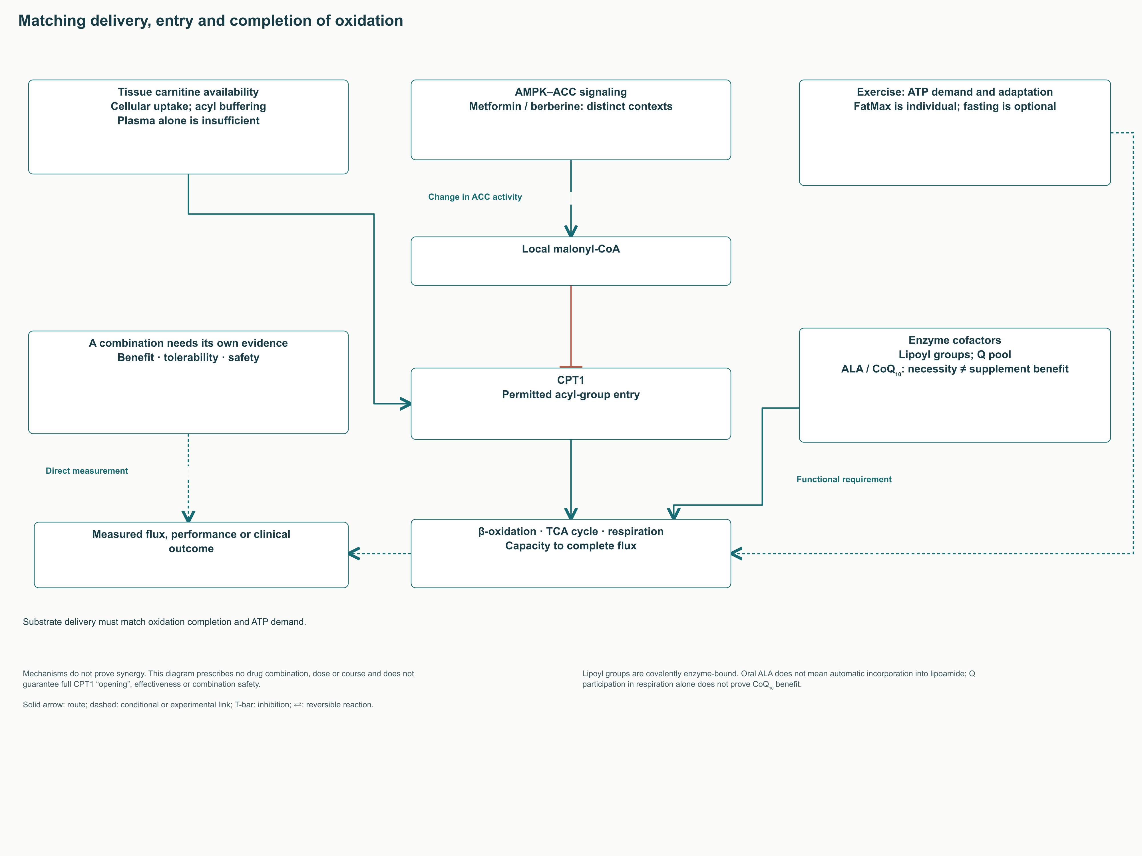
The idea of influencing metabolism through a combination usually brings together AMPK activation, lower malonyl-CoA, added carnitine, and aerobic exercise. These represent four different levels: a signal to change enzyme activity, release of inhibition, availability of a transported substrate, and energy demand. Coordinating them is biologically plausible, but the sum of plausible mechanisms is not yet a tested combination. If downstream oxidation or ATP demand is limited, additional substrate delivery may not improve the outcome.
Metformin has been studied in the AMPK–ACC context, but its clinical actions cannot be reduced to directly switching on CPT1. Berberine and other compounds that influence energy metabolism in experimental systems likewise cannot be combined with it into a ready-made regimen for self-administration. A combined intervention requires separate evidence on doses, interactions, tolerance, and the selected outcome. Neither synergy nor the safety of the entire combination can be calculated from the mechanism of a single component.
Cofactors of subsequent reactions were also included in the combination idea: lipoyl groups are required by some dehydrogenase complexes, while ubiquinone accepts electrons in the respiratory system. However, enzyme-bound lipoamide is a covalently attached cofactor, and oral α-lipoic acid does not automatically replenish every such site. Additional coenzyme Q10 likewise cannot be considered a universal way to remove a respiratory-chain limitation. The biochemical necessity of these molecules must be distinguished from the established benefit of adding them when no cause of deficiency has been identified.
Taking carnitine while fasting does not reproduce the prolonged muscle-loading protocol with carbohydrate. On the contrary, changing the study conditions changes the proposed mechanism of carnitine delivery to muscle. For a person with impaired fatty acid oxidation, fasted exercise may carry a very different risk from that faced by a healthy participant in a physiological experiment. The integration figure therefore shows levels of regulation and their limitations, preserving the substantive idea without turning it into a drug prescription.
Level of intervention
Biological meaning
Condition for a beneficial effect
What cannot be concluded in advance
Energy signal and AMPK
A change in the activity of ACC and other targets
An intact regulatory system and an appropriate context
That every AMPK activator lowers body weight
Malonyl-CoA
Regulation of CPT1 inhibition
Relevance of the local pool to the tissue concerned
That the lowest concentration is always optimal
Carnitine
Acyl-group transfer and buffering of the acetyl pool
Intracellular availability and intact transport pathways
That increasing the plasma level loads muscle
Exercise
ATP demand and long-term adaptation
Appropriate intensity and recovery
That maximum fat oxidation equals maximum benefit
Subsequent oxidation and respiration
Completion of flux and regeneration of coenzymes
Oxygen delivery, enzyme capacity, and ATP utilization
That accelerating CPT1 removes every downstream limitation
13. The microbiota, carnitine, and TMAO
13.1. Several microbial pathways lead to one metabolite
Not all ingested carnitine reaches human tissues. The fraction available to intestinal microorganisms may contribute to the formation of trimethylamine, TMA, which after absorption is oxidized predominantly by hepatic FMO3 to trimethylamine N-oxide, TMAO. The magnitude of the response depends on microbial composition and enzymes, substrate availability, liver function, and renal elimination. The same dose of carnitine therefore need not produce the same TMAO concentration.
Oxygen-dependent systems, including CntA/B, have been described for the direct conversion of carnitine. An anaerobic sequence also exists and is particularly relevant to conditions in the large intestine: γ-butyrobetaine forms first, and other microorganisms can then convert it to TMA. Rajakovich and colleagues identified a pathway in Emergencia timonensis and the associated bbu gene cluster. This is multistep enzymatic chemistry that cannot be reduced to an unsupported reaction involving simple dehydration of γ-butyrobetaine.
A simplified representation of the flux is carnitine → γ-butyrobetaine → TMA → TMAO. It shows the direction of metabolism of the carbon skeleton and trimethylammonium group, but is not a balanced equation for a single reaction. For the hepatic step, the following equation can be written: TMA + NADPH + H⁺ + O₂ → TMAO + NADP⁺ + H₂O. Here NADPH supplies reducing equivalents, and oxygen is required for the monooxygenase reaction, which should be distinguished from the anaerobic microbial branch.
13.2. Human associations and causality in experiments
Koeth and colleagues combined studies of carnitine conversion in humans with animal experiments. The microbiota's response differed between participants with different dietary habits, while animal models allowed the pathway's influence on atherosclerosis to be investigated. However, a habitual dietary category does not guarantee the absence of the relevant microorganisms or complete cessation of TMAO formation. Nor can the effect of a selected diet in mice automatically be extrapolated to myocardial infarction frequency in a supplement user.
In human cohorts, higher TMAO was associated with greater cardiovascular risk. Its level also depends on renal function, diet, and other characteristics that are themselves associated with prognosis. Platelet and animal experiments support the possibility of biological effects, including increased platelet reactivity, but do not determine a safe carnitine dose for every patient. Association, experimental mechanism, and the outcome of a prolonged clinical intervention must remain separate layers of inference.
Animal models have also been used to discuss changes in macrophage lipid-uptake receptors, including CD36, foam-cell formation, and reverse cholesterol transport. These branches help frame mechanistic questions about cholesterol movement between tissues. A change in receptor or enzyme expression does not mean that an entire pathway is completely switched off in humans. They should therefore be shown as experimentally studied processes, while vascular associations and platelet effects remain separate.
This literature does not establish a universal clinical threshold of “6–10 μmol/L” above which a person should stop taking carnitine. Values have different distributions in different samples, and a single measurement does not describe the full risk. It is particularly inappropriate to ignore renal clearance while trying to explain a high value solely by the amount of red meat or capsules consumed. The metabolite's significance should be discussed in its research and clinical context, retaining these dependencies.
13.3. DMB is an experimental tool, not dietary advice
Wang and colleagues used 3,3-dimethyl-1-butanol, DMB, to investigate nonlethal suppression of microbial TMA production. The work involved enzyme and microbial systems as well as mouse models of atherosclerosis. It demonstrated the principle of intervening in microbiota metabolism without destroying the entire community. However, research efficacy in these systems does not establish safety or therapeutic benefit in patients.
Specificity is particularly important: in that study, DMB did not inhibit the CntA/B system. The authors observed effects on other enzyme systems and on TMA formation under particular microbial conditions. It therefore cannot be depicted as a universal competitive blocker of every carnitine-conversion pathway or labeled with an unsupported “80–90% suppression.” The diagram identifies effects on experimentally sensitive systems, and the anaerobic branch is not assumed to be automatically blocked.
Finding a compound in foods does not mean that an effective and safe dose can be obtained through ordinary consumption. It also does not turn garlic, oil, wine, or resveratrol into proven prevention of TMAO-associated events. Such claims require direct studies of the relevant product, dose, and clinical outcome. Without them, listing foods alongside DMB creates the appearance of a ready-made solution that the data do not provide.
13.4. How to discuss the benefits and limitations of carnitine
Situation
What can be said on the basis of the evidence
What does not become a recommendation
Confirmed primary carnitine deficiency
Replacement therapy has a defined medical purpose
Stopping necessary treatment solely because of mechanistic concerns about TMAO
A healthy person interested in weight or sports
Effects depend on the goal and protocol; a universal advantage has not been established
Mandatory continuous use or a standard course
Renal dysfunction or high cardiovascular risk
Clinical context, elimination, and the reason for prescribing matter
Independently calculating risk from a single TMAO threshold
The idea of combining carnitine with DMB or a plant product
This is a research hypothesis requiring separate investigation
Taking DMB, garlic, or resveratrol as a proven “antidote”
Choosing ALCAR instead of L-carnitine
The forms undergo different transformations and have different studied indications
Guaranteed elimination of microbial TMAO formation
Short alternating courses
A change in regimen requires evidence concerning the desired outcome
An automatically safe regimen of “6–8 weeks with a break”
The balance of benefit and uncertainty is determined by the indication rather than by a single general assessment of the molecule. Treating an established deficiency and attempting to improve performance in a healthy person offer different expected benefits. A successful effect on a laboratory measurement does not replace a clinical outcome, and an experimental risk does not provide a ready-made rule for stopping treatment. Practical discussion begins with the reason for prescribing, the available evidence, and the outcome that is actually needed.
14. CPT1 as a pharmacological target
14.1. Why tumors use fatty acid oxidation
Fatty acid oxidation can help tumor cells survive nutrient restriction and other forms of metabolic stress. In particular models, CPT1A or related pathways contribute to energy maintenance and adaptation, but this dependence is not the same across tumors. Zaugg and colleagues studied CPT1C and demonstrated an association with cell survival and tumor growth under stressful conditions. The result extends CPT1C's functions beyond neurons while retaining the distinction between an experimental dependence and a clinically established target.
Increased protein expression does not prove that inhibiting the protein will kill the tumor. The reaction or regulatory function that limits survival must be identified and distinguished from effects on normal tissues. Genetic deletion, suppression of expression, and the action of a small-molecule compound can produce different results. For CPT1C in particular, an antitumor effect cannot be attributed in advance to blocking the conventional reaction that imports long-chain fatty acids into mitochondria.
14.2. Etomoxir and the problem of off-target effects
Etomoxir has been widely used as an experimental CPT1 inhibitor. Its use requires attention to concentration, exposure time, the cellular model, and CoA status. Divakaruni and colleagues showed that some effects in macrophages at high concentrations were explained by disruption of CoA homeostasis and could not be reduced to blocking fatty acid oxidation. Cell death or a change in phenotype after treatment is therefore not, by itself, evidence of CPT1 dependence.
This limitation has methodological importance for the entire literature on metabolic targets. An inference supported by a combination of genetic intervention, flux measurement, and pharmacological intervention under sufficiently selective conditions is more convincing. Even this combination does not yet establish the therapeutic window or benefit to patients. Etomoxir should not be turned from a research tool into advice for weight management or cancer treatment.
14.3. Different metabolic drugs cannot be grouped under one mechanism
Teglicar, or ST1326, was developed as a compound acting on CPT1A in experimental studies. Perhexiline is discussed among antianginal agents capable of altering myocardial metabolism, while its use is linked to drug exposure and toxicity. A randomized study before coronary surgery did not find the expected myocardial protection, further limiting a simple model of its action through CPT1. These directions demonstrate interest in controlling substrate metabolism, but each compound requires its own evidence on the target, dose-dependent effects, and clinical outcome.
Ranolazine should be considered separately. In primary electrophysiological studies, its effects were associated with ionic currents, including the late sodium current. Calling it a direct CPT1 inhibitor on the basis of the general idea of a “metabolic antianginal drug” is incorrect. A physiological outcome, pharmacological class, and direct molecular target are not interchangeable concepts.
Approach
Research or clinical context
Main limitation of interpretation
Etomoxir
Experimental intervention in CPT1 and fatty acid oxidation
At high concentrations, effects on CoA and other processes are substantial
Teglicar / ST1326
Studies of intervention in CPT1A
Experimental selectivity does not establish broad clinical use
Perhexiline
Metabolic antianginal therapy in particular clinical contexts
Evidence is needed concerning exposure, toxicity, and the specific indication
Ranolazine
Antianginal and electrophysiological effects
The late Na⁺ current cannot be replaced with a label saying “direct CPT1 inhibition”
Proposed CPT1 activators
A search for ways to increase oxidative flux
Increased import must be coordinated with subsequent oxidation and ATP demand
14.4. Activation also requires coordinated flux
In steatosis or insulin resistance, the opposite strategy is attractive: increase oxidation and reduce lipid accumulation. However, cells face several limitations, and CPT1 is not always the main one. Changes in fatty acid delivery, mitochondrial mass, the tricarboxylic acid cycle, and respiration can alter the effect of the same intervention. The therapeutic objective is therefore to restore coordinated metabolism rather than maintain maximum activity of the entry enzyme at all times.
15. A practical reading of the monograph
15.1. For clinical and laboratory interpretation
When the question concerns hypoglycemia, inadequate ketogenesis, or recurrent rhabdomyolysis, the shuttle diagram helps establish the appropriate diagnostic logic. Carnitine deficiency must be distinguished from deficiencies of CPT1A, CACT, CPT2, and the enzymes of subsequent β-oxidation. The same complaint does not identify a single blockage point, and an increase or decrease in one acylcarnitine does not replace a complete profile and clinical history. The state at sampling, the relationship to fasting or exercise, and genetic confirmation are particularly important.
When studying insulin resistance, it is useful to distinguish excess substrate delivery, incomplete oxidation, and impaired lipid signaling. Acylcarnitines can provide information about flux status, but are not a universal diagnosis of “mitochondrial overload.” RER describes whole-body gas exchange under the conditions of measurement and does not measure CPT1 activity in a particular tissue. This separation prevents attempts to derive a diagnosis and treatment from a single attractive diagram.
15.2. For nutrition, training, and assessment of research
When discussing supplements, the first question concerns the person's goal and baseline condition. Correcting a confirmed deficiency, prolonged muscle loading, and seeking a small average effect on body weight are different objectives. Each requires an appropriate study design and suitable endpoint. A molecule's ability to participate in fat transport does not prove that additional intake will improve any of these objectives.
When reading a new study, it is helpful to identify the compartment, measured flux, model, and outcome in sequence. Next, establish whether causality is supported by independent methods and how closely the conditions match the proposed application. A liposome result establishes transport capacity, a mouse result establishes an effect within a particular physiological model, and a randomized clinical trial establishes the effect of a specific intervention on a selected outcome. Keeping this sequence makes it possible to use detailed biochemistry without premature clinical conclusions.
CPT1 occupies an important position between lipid availability and mitochondrial utilization. However, the carnitine shuttle operates together with malonyl-CoA regulation, matrix CoA, β-oxidation enzymes, and the respiratory system. Normal function is defined by the ability to change fluxes to match the tissue's task. It is coordination and adaptability, rather than maximum fat oxidation at every moment, that connect the molecular mechanisms to the practical significance of this system.
FAQ
Why is the carnitine shuttle needed if a CoA carrier exists?
SLC25A42 transports CoA itself, whereas the shuttle delivers a long-chain acyl group. CPT1 removes it from external CoA and transfers it to carnitine, while CPT2 attaches it to matrix CoA. These are different transport tasks.
Do CPT1A, CPT1B, and CPT1C perform the same reaction?
CPT1A and CPT1B participate in the canonical mitochondrial shuttle. CPT1C is studied predominantly in the neuronal ER, and its weak or undetectable catalytic activity depends on the model. Neuronal signaling functions cannot be turned into a universal clinical mechanism.
Does increasing CPT1 activity always improve fat utilization?
Substrate entry must match the capacity of β-oxidation, the tricarboxylic acid cycle, respiration, and ATP utilization. When flux is mismatched, intermediates can accumulate. Greater acyl-group entry therefore does not guarantee more complete oxidation or improved insulin sensitivity.
Why is palmitate oxidation said to yield 106 ATP?
This is a calculation using P/O = 2.5 for NADH and 1.5 for flavin-derived reducing equivalents. Complete oxidation yields 28 + 80 ATP equivalents, from which 2 are subtracted for activation. Actual coupling in living tissue varies.
Does plasma carnitine indicate muscle loading?
Plasma and intramuscular pools differ. Cellular uptake depends on OCTN2, and a prolonged muscle-loading protocol cannot be replaced by a single increase in blood concentration. Feeding conditions, duration, and direct measurement of the tissue pool matter in studies.
Is carnitine proven to be a universal weight-loss agent?
A meta-analysis found a modest average difference in body weight in particular studies. This does not imply the same individual effect or an established reduction in body fat percentage. The indication, baseline condition, and selected outcome determine its practical significance.
Can FatMax be identified using a general formula based on the percentage of VO₂max?
A group mean is not an individual result. FatMax depends on test conditions and shows substantial variation between individuals. Maximum fat oxidation during a single test also does not determine fat loss over months.
Does the CPT1A P479L variant mean continuous ketogenesis?
No: reduced activity and reduced sensitivity to malonyl-CoA can coexist. A study of homozygous children found a limited ketogenic response to fasting. Population prevalence and a selection hypothesis do not eliminate individual medical risk.
Can DMB or garlic be taken to block TMAO?
DMB studies were conducted in experimental systems and do not establish a patient treatment regimen. DMB did not inhibit CntA/B in the primary study. Foods cannot be considered a proven “antidote” simply because they contain a particular compound.
Should carnitine be stopped whenever TMAO increases?
A single TMAO value does not determine the benefits and risks of treatment. Renal function, diet, the reason for prescribing, and clinical context matter. Correcting confirmed primary deficiency offers a different expected benefit from taking an optional sports supplement.
What does a high acylcarnitine level indicate?
It may reflect metabolic intensity, a limitation at a particular step, or an inherited transport disorder. Interpretation requires the complete profile, the state at measurement, and the clinical circumstances. One measurement does not establish universal mitochondrial toxicity.
Is ranolazine a direct CPT1 inhibitor?
Its electrophysiological effects are linked to ionic currents, including the late sodium current. The general designation of a metabolic antianginal drug does not identify its direct target. CPT1 therefore cannot be used as a universal explanation of its action.