Acetyl-L-carnitine: brain acetyl metabolism, mitochondria, and clinical limits
An in-depth account of ALCAR transport, acetyl exchange, acetylcholine and chromatin, mitochondrial function, and human studies of neuropathy, cognitive impairment, and depression. Studied doses are distinguished from prescriptions and cellular mechanisms from clinical outcomes.
Acetyl-L-carnitine, or ALCAR, connects several areas of biochemistry that are usually discussed separately: acetyl-group metabolism, mitochondrial function, acetylcholine synthesis, and the regulation of gene activity. It is a naturally occurring short-chain acylcarnitine formed in the body from L-carnitine and acetyl-CoA. Its ability to carry an acetyl group makes it an interesting research tool, but the biochemical function of a molecule and the benefits of taking additional amounts are different questions. Answering the second requires human studies involving a specific disease, formulation, dose, and clinical outcome.
Human data on ALCAR concern peripheral neuropathy, depressive symptoms, and cognitive impairment, but the findings are heterogeneous and sometimes contradict an appealing mechanistic picture. The experience of preventing neuropathy during taxane treatment is particularly instructive: a substance considered a neuroprotectant worsened symptoms in a large randomized trial. Professional discussion should therefore move from chemistry and cellular compartments to individual clinical questions. This progression makes it possible to understand where a mechanism is established, where it remains hypothetical, and exactly what was measured in patients.
What the acetyl group changes
L-carnitine contains a negatively charged carboxylate group and a positively charged quaternary ammonium group. Such a molecule is called a zwitterion: its net charge may be zero even though local charges remain. In ALCAR, the hydroxyl group of carnitine is esterified with an acetyl group containing two carbon atoms. Acetylation does not eliminate the charged groups or turn the compound into a fat-soluble molecule that passes through membranes without restriction.
The abbreviated structure of L-carnitine is written as (CH₃)₃N⁺–CH₂–CH(OH)–CH₂–COO⁻, and that of ALCAR as (CH₃)₃N⁺–CH₂–CH(O–CO–CH₃)–CH₂–COO⁻. These formulas show both the retained charges and the point at which the acetyl group is attached. The designation L refers to the stereochemistry of the carnitine backbone, not to the number of acetyl groups. Clinical studies examine the L-form specifically; their results cannot automatically be transferred to a mixture of stereoisomers.
Property
L-carnitine
Acetyl-L-carnitine
Molecular mass of the inner salt
161.20 g/mol
203.24 g/mol
Presence of an acetyl group
None
One group attached by an ester bond
Main role in the reactions discussed
Acceptance and transport of acyl groups
Exchange and transport of acetyl groups
Transport in a blood–brain barrier model
OCTN2 is involved
OCTN2 is involved
Mass of ALCAR hydrochloride
Not applicable to this comparison
239.70 g/mol
The difference between these compounds matters, but it does not prove that a supplement preferentially targets the cortex or hippocampus. Distribution is determined by transporters, metabolism, blood flow, and tissue condition, not simply by the presence of an acetyl group. A calculated octanol/water partition coefficient likewise cannot replace measurement of transport across a living barrier. The comparison “ordinary carnitine stays in the blood, whereas the acetylated form freely enters the brain” therefore oversimplifies the experimental findings.
How the molecule crosses the blood–brain barrier
The blood–brain barrier consists of the endothelium of brain capillaries, with tight intercellular junctions and specialized transport systems. For a hydrophilic molecule, the presence of a transporter may matter more than passive partitioning into the lipid phase. OCTN2, encoded by SLC22A5, participates in sodium-dependent transport of carnitine and ALCAR. The energy for this secondary active transport comes from the Na⁺ electrochemical gradient maintained by Na⁺/K⁺-ATPase; OCTN2 itself does not hydrolyze ATP with each transport event.
The schematic expression ALCARoutside + Na⁺outside → ALCARinside + Na⁺inside describes the direction of coupled uptake. It does not prove identical stoichiometry and kinetics at every brain membrane, nor does it describe the entire journey from blood to neuron. Between the luminal surface of an endothelial cell and the brain's extracellular fluid there is another, abluminal membrane. A separate step of entry into nervous-system cells is then required, so the rate of accumulation in the endothelium cannot be treated as the rate of delivery to a presynaptic terminal.
Kido and colleagues used rat brain perfusion, endothelial cultures, and the RBEC1 brain endothelial cell model. In that model, estimated Kₘ values were 33.1 ± 11.4 μM for L-carnitine and 31.3 ± 11.6 μM for ALCAR; disruption of OCTN2 was associated with reduced uptake. Kₘ describes the concentration at which the rate of a particular transport system reaches half its maximum. These values belong to the experimental system and do not establish a universally high rate of ALCAR transport across the human barrier.
Transport studies support the involvement of OCTN2, but do not justify constructing a complete human route featuring obligatory receptor-mediated transcytosis, free abluminal diffusion, or a leading role for OCTN1. The possibility that another transporter participates in a particular tissue does not establish its function at the blood–brain barrier. The practical implication is that an increase in plasma concentration, the appearance of the substance in cerebrospinal fluid, and an increase in intracellular acetyl-CoA are successive but independent questions. Measuring one does not allow the others to be reconstructed.
The reversible CrAT reaction and acetyl-group availability
Carnitine acetyltransferase, abbreviated CrAT, catalyzes the reaction acetyl-CoA + L-carnitine ⇌ ALCAR + CoA-SH. CoA-SH denotes free coenzyme A with an available thiol group; it is required for many metabolic reactions. In the direction that forms ALCAR, an acetyl group is transferred from acetyl-CoA to carnitine, releasing free CoA. In the reverse direction, ALCAR can provide acetyl for the formation of acetyl-CoA if the enzyme and CoA are available in the relevant compartment.
The reaction does not require direct participation of ATP, but this does not mean that the entire process of supplying a cell with acetyl is energetically free. The ester bond in ALCAR is not identical to the thioester bond in acetyl-CoA, and membrane transport and gradient maintenance are linked to the cell's overall energy metabolism. The direction of a reversible reaction depends on substrate and product concentrations, not on the promotional description “energy donor.” In one situation the carnitine pool accepts excess acetyl; in another, acetylcarnitine transfers it to available CoA.
The inner mitochondrial membrane restricts the free movement of acetyl-CoA, so cells use transportable intermediates. One well-studied route begins with citrate: following its exit through the citrate carrier, ATP-citrate lyase, ACLY, carries out the reaction citrate + CoA-SH + ATP → acetyl-CoA + oxaloacetate + ADP + Pᵢ. Another source outside mitochondria is acetate, which ACSS2 activates through acetate + CoA-SH + ATP → acetyl-CoA + AMP + PPᵢ. The importance of each route depends on cell type, nutrient supply, and metabolic state.
Experiments in cells lacking ACLY and ACSS2 showed that acetyl-equivalent transport through acetylcarnitine can sustain lipogenesis and histone acetylation. However, these results come from cellular models, including tumor cell lines, and do not establish a single pathway in the human neuron. CrAT is discussed primarily in relation to mitochondria and peroxisomes; the enzyme that returns acetyl to an extramitochondrial pool in a particular system requires separate identification. A universal “nuclear CrAT” that immediately converts a swallowed capsule into chromatin-associated acetyl-CoA remains an unproven construct.
From an acetyl group to acetylcholine
Acetylcholine is formed from choline and acetyl-CoA in the choline acetyltransferase, or ChAT, reaction: acetyl-CoA + choline → acetylcholine + CoA-SH. Choline enters a cholinergic terminal through mechanisms including the high-affinity transporter CHT1. The completed transmitter is loaded into synaptic vesicles by VAChT and released through calcium-dependent exocytosis. After interacting with nicotinic or muscarinic receptors, acetylcholine is broken down by acetylcholinesterase, and choline can be reused.
ALCAR is not choline and does not replace it in this reaction. It can transfer an acetyl group through enzymatic formation of acetyl-CoA, but the result depends on CoA availability, enzyme localization, and preservation of the cholinergic cell. If a terminal has died or ChAT expression is reduced, adding one precursor does not restore the entire transmission apparatus. Likewise, increased transmitter synthesis does not guarantee better memory, because cognition depends on many neuronal networks.
In experiments by White and Scates, the acetyl group of ALCAR was used to synthesize acetylcholine in an enzyme system and preparations of rat brain synaptosomes. Earlier studies of rat brain slices also showed incorporation of label from acetylcarnitine into acetylcholine alongside other carbon sources. This confirms that acetyl transfer is possible, but does not establish a constant contribution from this route in humans. These studies do not support the claim that 30% of newly formed acetylcholine in the human cortex necessarily comes from ingested ALCAR.
For professionals, the distinction between tracing a label and demonstrating a functional effect is especially important. A radioactive or stable-isotope label shows where an atom came from, but does not tell us whether total synthesis flux increased or synaptic transmission improved. Even a change in flux must then be compared with behavior and clinical function. The cholinergic mechanism therefore explains research interest in ALCAR, whereas decisions about treating cognitive impairment must rest on human trial results.
Chromatin acetylation and the mGlu2 receptor
Histones help package DNA, and modifications of their amino-acid residues participate in regulating chromatin accessibility. During lysine acetylation, its positively charged amino group becomes acetylated; schematically, Lys–NH₃⁺ becomes Lys–NH–CO–CH₃. Acetyltransferase enzymes obtain the acetyl group from acetyl-CoA. Enzymes of the p300/CBP family and other acetyltransferases act selectively, so the availability of substrate does not mean that every gene is switched on at once.
Studies of ACLY and ACSS2 demonstrate a link between cellular metabolism and histone acetylation. In neuronal models, ACSS2 was detected on chromatin, and its suppression in the mouse hippocampus was associated with impaired spatial memory. This helps explain why a cell needs local sources of acetyl-CoA, but the ACSS2 study itself does not prove the efficacy of an ALCAR supplement. A change in an epigenetic mark must be considered in the context of the specific locus, cell type, and experimental intervention.
In the study by Nasca and colleagues in FSL rats and mice exposed to chronic stress, ALCAR produced antidepressant-like behavioral changes after approximately three days. The researchers linked the effect to increased mGlu2 expression, NF-κB p65 acetylation, and an increase in the H3K27ac mark at the Grm2 promoter. The relevant residue is specifically lysine 27 of histone H3, not H3K9ac. Separate experiments in neural progenitor cells examined p65 acetylation and differentiation at ALCAR concentrations of hundreds of micromoles, which cannot be converted into an oral patient dose.
mGlu2 is a metabotropic glutamate receptor encoded by GRM2 in humans. It couples to the Gᵢ/Gₒ family and participates in regulating adenylyl cyclase, cAMP, and presynaptic transmitter release, including calcium-dependent processes. This feedback can limit glutamate release at particular synapses, but it is not a universal switch that turns excitation off. Depression likewise cannot be reduced to obligatory glutamate excess, a uniform loss of CA3 spines, or a single state of the GRM2 promoter.
Translating these findings to humans requires showing more than animal behavior: a clinical effect, its timing, and its connection to the proposed target must be demonstrated. In human studies, low plasma ALCAR concentrations were associated with certain characteristics of major depressive disorder, but an observational design does not establish causality. Plasma is not a direct measurement of nuclear acetyl-CoA or neuronal H3K27ac, and there is no diagnostic threshold for “epigenetic ALCAR deficiency” in routine practice. A rapid effect in an animal model therefore cannot be promised to a person as relief of depression within 48–72 hours.
Two carnitine processes inside the mitochondrion
Long-chain fatty acids and acetyl groups use related but different carnitine processes. CPT1 on the outer mitochondrial membrane transfers a long-chain acyl group from CoA to carnitine. The translocase CACT, encoded by SLC25A20, carries acylcarnitine across the inner membrane in exchange for free carnitine. CPT2 on the matrix side of the inner membrane returns the acyl group to CoA, after which acyl-CoA can enter β-oxidation.
The short acetyl group in ALCAR does not make it a direct replacement for a long-chain CPT1 substrate. CrAT is discussed principally as a participant in short acyl-group exchange, whereas CPT1–CACT–CPT2 provides transport at a different scale. Malonyl-CoA regulates the entry of long-chain substrates through CPT1, linking oxidation to the overall state of lipid metabolism. Additional ALCAR should therefore not be assumed to increase β-oxidation in every brain cell.
Neurons and astrocytes differ in substrate preferences and in their handling of fatty acids. Experimental work by Ioannou and colleagues showed transfer of potentially toxic fatty acids from active neurons to astrocytes, their accumulation in lipid droplets, and subsequent processing. This explains a protective role for intercellular lipid exchange, but does not make long-chain fats the universally predominant fuel for neurons. Glucose, lactate, and other substrates retain central roles in nervous-tissue energy metabolism.
The CPT1C isoform deserves separate treatment because it is localized mainly in the neuronal endoplasmic reticulum and is not equivalent to the ordinary mitochondrial “gateway” for fat oxidation. In mouse experiments, its absence was associated with changes in ceramide content, dendritic-spine maturation, and spatial learning. These findings reveal a connection between lipid metabolism and neuronal function, but do not prove that oral ALCAR stimulates CPT1C. Still less do they automatically explain worsening neuropathy during chemotherapy.
Free CoA and regulation of pyruvate oxidation
The pyruvate dehydrogenase complex, PDH, links glycolysis with mitochondrial carbon oxidation: pyruvate + CoA-SH + NAD⁺ → acetyl-CoA + CO₂ + NADH + H⁺. If acetyl-group input exceeds subsequent utilization, the acetyl-CoA/CoA-SH and NADH/NAD⁺ ratios change. These ratios participate in feedback that limits pyruvate oxidation. PDK kinases phosphorylate and inhibit PDH, whereas PDP phosphatases participate in its reactivation.
CrAT allows part of the acetyl pool to be temporarily transferred into acetylcarnitine, releasing CoA-SH. This buffering means redistribution of chemical groups, not destruction of excess energy. Reversibility permits acetyl to be returned later, while acetylcarnitine export through transport systems connects compartments. A limitation follows: supplying ALCAR from outside does not necessarily shift the reaction toward CoA release, because it also adds one of the reaction's products.
Mice with muscle-specific deletion of CrAT showed impaired metabolic flexibility—the ability to switch between predominantly using fats and carbohydrates. In a small crossover study of 11 volunteers with impaired glucose tolerance, 2 g of L-carnitine daily for 36 days improved measures of metabolic flexibility and acetylcarnitine formation in muscle. Whole-body insulin sensitivity did not improve. These are human data on L-carnitine and muscle tissue, not evidence that acetyl-L-carnitine restores brain energy metabolism.
The sequence “excess acetyl-CoA → PDK → PDH inhibition” should therefore be understood as regulated metabolic feedback. It does not mean that every high acetyl-CoA level causes clinical lactic acidosis or that an ALCAR capsule immediately resolves it. Lactic acidosis requires assessment of oxygen delivery, circulation, medications, and other causes; a biochemical diagram cannot replace that assessment. Brain research requires direct evidence on substrate flux and tissue function rather than extrapolation from a muscle model.
Cardiolipin, the respiratory chain, and cytochrome c release
Cardiolipin is a characteristic lipid of the inner mitochondrial membrane with four acyl chains. It participates in the organization of membrane proteins, crista curvature, and interactions among respiratory-chain components. Its proportion and chain composition depend on the tissue and the state of the organelle, so the linoleic-acid-rich cardiolipin of the heart should not be treated as the universal composition in the brain. Cristae are formed by a complex system of lipids and proteins, rather than simply by negatively charged regions of one phospholipid.
Cardiolipin supports the organization of respiratory-chain complexes, including supercomplexes containing complexes I, III₂, and IV. Electrons pass through several carriers, and complex activity generates the proton electrochemical gradient used by ATP synthase. Δψₘ denotes the electrical component of this gradient; a higher potential does not always mean a healthier mitochondrion. When reduced carriers accumulate, high polarization can coexist with increased production of reactive oxygen species.
Cardiolipin oxidation changes its interaction with cytochrome c and other proteins. However, detachment of cytochrome c from the inner membrane and its release into the cytosol are different events: release requires altered permeability of the outer membrane. MOMP denotes this outer-membrane permeabilization, whereas mPTP refers to the permeability transition of the inner membrane. These processes can interact, but mPTP should not be drawn as the sole door through which cytochrome c directly reaches the cytosol.
In the classic study by Paradies, changes in cardiolipin and the phosphate carrier were examined in heart mitochondria from old rats. ALCAR improved the relevant measures in that model, but the study did not demonstrate direct activation of tafazzin, restoration of all lipids in the human brain, or prevention of dementia. Complex I should likewise not be described as an obligatory direct source of hydroxyl radicals: superoxide generation and subsequent chemical transformations are separate stages. Clinical neuroprotection must be established independently of preservation of a membrane measure in an animal.
Mitochondrial biogenesis and dynamics
Mitochondrial biogenesis requires coordinated activity of the nuclear and mitochondrial genomes. The coactivator PGC-1α participates in regulating nuclear transcriptional programs together with NRF1 and GABP, also called nuclear respiratory factor 2. This name must not be confused with Nrf2, or NFE2L2, a regulator of the antioxidant response. TFAM is imported into mitochondria and participates in packaging, maintaining, and expressing mitochondrial DNA.
The mitochondrial genome encodes 13 polypeptides of the respiratory system, whereas most necessary proteins are encoded in the nucleus. Complex II consists entirely of nuclear-encoded subunits; an increase in mitochondrial DNA therefore does not equal a proportional increase in every complex from I to V. In energy regulation, AMPK can phosphorylate PGC-1α, whereas SIRT1 can deacetylate it. These reactions modify the protein in different ways and do not establish an obligatory sequence of action for every dose of ALCAR.
In work by Nicassio and colleagues in 28-month-old rats, one to two months of ALCAR were associated with changes in PGC-1α/β, NRF1, TFAM, and several brain mitochondrial measures. An important detail is that the age-related decline concerned PGC-1β, whereas baseline PGC-1α was unchanged. DRP1 and FIS1 did not change, while the age-related increases in MFN2 and the long form of OPA1 moved toward young-animal values. Many effects largely disappeared one month after withdrawal, so the study does not show irreversible rejuvenation of nervous tissue.
MFN1/2 participate in outer-membrane fusion, OPA1 in the organization and fusion of the inner membrane, and DRP1 in fission. Fission is necessary not only during damage, but also for organelle distribution and quality control; an excessively branched network is not a universal treatment goal. A mitochondrion in a long axon must also be delivered to the appropriate region and sustain local ATP demand. Biogenesis, fusion, fission, transport, and removal of damaged organelles should therefore be considered together, rather than as a linear program of “more mitochondria means better memory.”
Diabetic neuropathy: pain, sensation, and nerve structure
Diabetic distal symmetric polyneuropathy develops against a background of combined metabolic and vascular disturbances. Increased flux through the polyol pathway, formation of advanced glycation end products, oxidative stress, and changes in lipid metabolism can damage axons, Schwann cells, and nerve microcirculation. Long axons are particularly vulnerable, so symptoms often begin in the feet and spread proximally. Thin unmyelinated C fibers and thin myelinated Aδ fibers relate to pain and temperature sensation, whereas large fibers relate to vibration and other functions.
Measured outcomes reflect different aspects of the disease. A pain score reports a subjective symptom, vibration perception a sensory function, nerve-conduction testing mainly large-fiber function, and biopsy the structure of a selected nerve. Less pain therefore does not prove regeneration, and more regenerating clusters do not guarantee normalized conduction or prevention of foot ulcers. This principle is especially important when reading ALCAR studies, in which positive and negative findings coexist within the same publication.
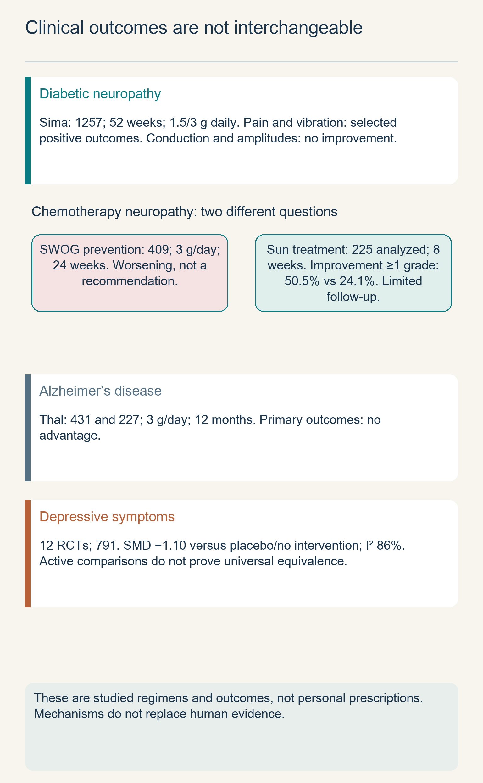
Sima and colleagues analyzed two placebo-controlled studies lasting 52 weeks, with 1,257 participants in the intention-to-treat analysis. They studied 500 or 1,000 mg of ALCAR three times daily, equivalent to 1.5 or 3 g per day. Positive findings concerned fiber numbers and regenerating clusters in biopsies and vibration perception; improvement in pain appeared in one study and in the combined analysis, particularly at 3 g daily. However, nerve-conduction velocities and response amplitudes did not improve—this negative finding must not be replaced with the general statement “the nerve recovered.”
A separate study by De Grandis and Minardi enrolled 333 patients: 1 g was given intramuscularly for the first ten days, followed by 2 g orally for another 355 days. The authors reported improvements in several neurophysiological measures, but this regimen differs from Sima's oral-only trials. In addition, the completeness of the available neurophysiological data limits confidence in the conclusion. Differences in formulation, administration route, population, and analysis help explain why the findings cannot be combined into one guaranteed effect.
The meta-analysis by Li included four randomized trials reported in three publications, with 523 participants. The average advantage for pain was 1.20 cm on a ten-centimeter visual analogue scale, with a 95% confidence interval of 0.68–1.72 cm. The estimate in the diabetic subgroup was 1.47 cm, whereas in the nondiabetic subgroup it was 0.71 cm with an interval that included no difference. This is an absolute difference on a scale, not a universal 20.2% reduction in pain; an average effect likewise does not mean that every patient responds identically.
Practical discussion of ALCAR does not replace diabetes management, foot examinations, protection of insensate skin, or investigation of other neuropathy causes. Diabetes does not exclude B12 deficiency, toxic exposure, or another nerve disease. AAN guidance on painful diabetic polyneuropathy considers drug classes with an established clinical role, including certain antidepressants and antiseizure medicines, and requires assessment of sleep and mood. ALCAR should not be presented as a proven substitute for such care or as a treatment already shown to alter the disease's natural history.
Chemotherapy: prevention and treatment have different results
Neuropathy after anticancer treatment involves different damaging mechanisms. Taxanes disrupt microtubule function and axonal transport, platinum drugs can damage sensory neurons in dorsal root ganglia, and mitochondrial changes participate in several models of toxicity. However, similarities between individual cellular injuries and diabetic neuropathy do not guarantee the same response to a supplement. The specific chemotherapy, the start of supplementation, and baseline symptom severity all matter.
In a small open-label study by Bianchi, 25 patients with neuropathy following paclitaxel or cisplatin received 1 g of ALCAR three times daily for eight weeks. The authors described improvements in sensory symptoms and certain neurophysiological measures. Without a control group, a drug effect cannot be separated from natural recovery, symptom fluctuation, and participant expectations. The study provided a reason for further investigation, but did not establish neuropathy prevention or a universal recovery protocol.
There is also a positive placebo-controlled study by Sun of treatment for established chemotherapy-induced neuropathy. The full analysis set contained 225 patients receiving 3 g of ALCAR daily or placebo for eight weeks. Improvement by at least one grade on the NCI-CTC scale occurred in 50.5% versus 24.1%; the difference was 26.4 percentage points, with a 95% interval of 14.1–38.5. However, follow-up was short, the analyzed groups differed from the original enrollment, and the publication describes some chemotherapy details ambiguously; the result does not establish long-term benefit for every cancer survivor.
A more concerning finding for prevention comes from SWOG S0715. Among 409 evaluable women receiving adjuvant taxane treatment for breast cancer, ALCAR at 3 g daily for 24 weeks was compared with placebo. There was no advantage at week 12, and by week 24 FACT-NTX neuropathy scores were worse in the ALCAR group. A decline of more than five points occurred in 38% versus 28%, an absolute difference of ten percentage points; the negative direction persisted during two-year follow-up.
This finding cannot be reduced to a ban on taking a capsule on the infusion day and permission to use it on every other day. ALCAR was taken as part of a months-long prevention regimen, and the worsening was not a brief event around taxane administration. Proposed explanations involving CPT1C or accelerated clearance of “protective factors” were not demonstrated mechanisms in the trial. Clinical harm remains important even when its molecular cause is unknown.
ASCO guidance explicitly does not support ALCAR for prevention of chemotherapy-induced neuropathy. For established painful neuropathy, duloxetine has the most adequate evidence among the pharmacological approaches considered, although its benefit is limited. Positive small ALCAR treatment studies should be discussed alongside their limitations and the negative prevention findings. A decision after chemotherapy likewise requires an oncology context rather than automatic prescribing “for regeneration.”
Cognitive decline and Alzheimer's disease
Alzheimer's disease involves changes in synapses, protein pathology, and energy metabolism, including impaired cholinergic transmission. These processes offer plausible targets for ALCAR, but none explains the disease in full. Mild cognitive impairment is also heterogeneous: a similar memory complaint can have different causes and different probabilities of progression. Findings from older studies using broad diagnostic categories therefore cannot be transferred to modern individual diagnosis without qualification.
The meta-analysis by Montgomery, Thal, and Amrein combined placebo-controlled studies of mild cognitive impairment and mild Alzheimer's disease, with doses of 1.5–3 g daily and assessments at three, six, or twelve months. The pooled effect size was approximately 0.20, and approximately 0.32 for global clinical impression. Such standardized estimates describe a difference relative to variability in measurements and do not equal a fixed increase in MMSE points. They suggest a small average effect rather than restoration of lost function or prevention of dementia.
Large individual trials provide an important limitation to this picture. In Thal's one-year study, 431 patients with mild or moderate Alzheimer's disease received 3 g of ALCAR daily or placebo; there was no advantage on the primary outcomes in the overall population. The suggestion of benefit in younger patients was then tested in a separate early-onset disease trial. Among 227 randomized participants aged 45–65, the same regimen showed no advantage in the intention-to-treat analysis of the primary ADAS-Cog and CDR outcomes.
The discrepancy between a small pooled effect and large negative trials requires discussion of study composition, different scales, and possible publication biases. A meta-analysis does not turn an investigational intervention into a proven disease-modifying treatment. It also does not establish slower brain atrophy or restoration of glucose metabolism on PET: such conclusions require separate studies with the corresponding endpoints. ALCAR should not be offered to a healthy person as an established means of preventing age-related memory loss.
Depressive symptoms and the limits of comparison with antidepressants
Cellular acetylation mechanisms and experimental regulation of mGlu2 make ALCAR interesting for depression research. However, a depressive symptom, dysthymia, and major depressive disorder are not interchangeable clinical categories. Severity, age, comorbidities, and previous treatment matter. An observed association between plasma ALCAR and depression likewise does not mean that a low concentration causes the illness or predicts response to supplementation.
Veronese's meta-analysis included 12 randomized studies with 791 participants. Comparisons with placebo or no intervention produced a standardized mean difference of −1.10, with a 95% interval of −1.65 to −0.56, but heterogeneity was high: I² = 86%. This means that findings differed substantially between studies, and the pooled estimate describes no single typical patient particularly well. In three comparisons with active treatment, the estimate was 0.06, with an interval of −0.22 to 0.34; it does not demonstrate universal equivalence to all antidepressants.
The distinction between no statistically significant difference and demonstrated noninferiority is particularly clear in the study by Zanardi and Smeraldi. In it, 204 patients with dysthymia received ALCAR 500 mg twice daily or amisulpride 50 mg daily for twelve weeks. The between-group difference did not reach statistical significance, but the prespecified noninferiority criterion with a two-point margin was not met. Changing the allowable margin after obtaining the findings does not replace successful testing of the original hypothesis.
ALCAR was well tolerated in several studies, but this does not mean that sexual, gastrointestinal, neurological, or other adverse effects are absent in every person. Questions about rapid onset require a human assessment of timing rather than a three-day finding in a rodent model. ALCAR should not be used to discontinue effective treatment independently or delay help for severe depression. Professionals can more appropriately discuss it as an investigated intervention with heterogeneous findings than as an established rapid epigenetic antidepressant.
Combinations with lipoic acid, CoQ10, and PQQ
Combining substances is often justified by their participation in different aspects of mitochondrial function. CoQ10 transfers electrons from complexes I and II to complex III, PQQ is studied in relation to cellular signaling pathways, and lipoic acid is associated with enzyme complexes and redox processes. However, compatible explanations do not establish proven synergy. Demonstrating synergy requires comparison of the combination with its separate components using clinical outcomes, rather than merely a diagram with converging arrows.
In Hagen's study, old rats received ALCAR in drinking water at a concentration of 1.5% and R-α-lipoic acid in feed at 0.5% for up to one month. Metabolic and oxidative measures were examined, including in isolated liver cells. The study did not use nicotinamide adenine dinucleotide as its second component, and did not determine a human ratio of “1,000 mg ALCAR to 200–300 mg lipoic acid.” Concentrations in an animal's water and feed cannot be mechanically converted into a ready-made human regimen.
In an earlier ALCAR study in old rats, improvements in certain mitochondrial and behavioral measures coexisted with increases in some indicators of oxidative burden. This emphasizes that increasing metabolism does not necessarily reduce every reactive oxygen species at the same time. PQQ experiments used mouse Hepa1-6 hepatoma cells, concentrations of 10–30 μM, and exposure for 24–48 hours, finding a connection with CREB and PGC-1α. A cellular PQQ concentration is not evidence for the efficacy of an oral “triple stack” for fatigue or brain rejuvenation.
The R-form of lipoic acid has biochemical significance, but that does not justify a mandatory combination of only this form with ALCAR in every clinical situation. Likewise, the involvement of Nrf2 and glutathione in the antioxidant response does not prove that an added component neutralizes every risk of another substance. Protocols involving PQQ, ubiquinol, SAMe, Alpha-GPC, uridine, bacopa, lion's mane, or phosphatidylserine require their own combination-specific evidence. In its absence, a diagram should show different mechanisms and the limits of their study, not a guaranteed chain of enhanced effects.
Absorption, formulation, and studied doses
The persistent view that ALCAR has high oral bioavailability needs reconsideration. A comparative study in 2025 examined doses of 0.5 and 1.5 g of ALCAR and L-carnitine in healthy volunteers, measuring metabolites for up to 48 hours. At 1.5 g, the incremental area under the acetylcarnitine curve after ALCAR was approximately 7.7 times smaller than the incremental carnitine area after L-carnitine. This measure cannot be converted into an absolute bioavailability percentage without an intravenous comparison; it also does not measure the substance's concentration in the brain.
An earlier pharmacokinetic study included 11 patients with Alzheimer's disease who first received 30 mg/kg intravenously for ten days, followed by 1.5 g orally for fifty days. The sequence of different administration routes and the small population limit extrapolation to an ordinary single supplement dose. Time to peak and half-life values depend on the regimen, formulation, and substance measured. A single 40–45% bioavailability value, peak time, or half-life therefore cannot be declared for every product and patient.
Hydrochloride differs in mass from the inner salt. If the package specifically states the mass of hydrochloride, the conversion is 1,000 mg × 203.24 / 239.70 ≈ 848 mg ALCAR. However, the wording “acetyl-L-carnitine, as hydrochloride” may already indicate the active-substance equivalent, in which case the dose should not be reduced a second time. The precise label wording and product specification matter, not only the salt's name.
Clinical question
Example of an actually studied regimen
Limitation on use
Diabetic neuropathy
1.5 or 3 g/day orally for 52 weeks; separately, 1 g intramuscularly for 10 days, then 2 g/day orally
Not every functional outcome improved; different routes cannot be treated as one protocol
Prevention of taxane neuropathy
3 g/day for 24 weeks
Symptoms worsened; this preventive approach is not recommended
Treatment of established chemotherapy-induced neuropathy
3 g/day for 8 weeks in individual studies
Limited evidence and short follow-up; not a universal prescription after cancer treatment
Alzheimer's disease
3 g/day for 12 months in large trials
Primary outcomes showed no advantage
Dysthymia
1 g/day for 12 weeks compared with amisulpride
Prespecified noninferiority was not demonstrated
“Rejuvenation” of a healthy brain
No established regimen
Animal and cellular findings do not define a human protocol
This table describes studies rather than issuing a prescription. The range of 1–3 g daily is not an established therapeutic window, because different diseases and regimens produce different findings. There is likewise no proven universal requirement to take ALCAR fasting, finish doses before 3 p.m., or undertake two courses per year. If an individual treatment trial is discussed, its objective, baseline symptom severity, and stopping criteria should be defined in advance; additional laboratory investigations should be selected for the clinical question rather than a promise to “check the mitochondria.”
The microbiota and TMAO production
Carnitine compounds can serve as substrates for intestinal microorganisms. One route of trimethylamine, or TMA, production involves the CntA/CntB system and requires oxygen. Another, important in the anaerobic intestinal environment, involves conversion of carnitine to γ-butyrobetaine and subsequent reactions catalyzed by enzymes of the Bbu system. These routes involve different bacteria and conditions, so the diagram cannot be reduced to one universal “carnitine lyase” shared by every member of the microbiota.
After TMA is absorbed, hepatic FMO3 catalyzes its oxidation: TMA + NADPH + H⁺ + O₂ → TMAO + NADP⁺ + H₂O. TMAO concentration depends not only on microbial production, but also on kidney function, diet, and other circumstances. L-carnitine studies have found associations with cardiovascular risk and mechanistic effects in animal models. However, an association with concentration in humans does not prove that a particular ALCAR dose necessarily causes a clinical vascular event.
The comparative study in 2025 did not confirm the proposed protection of the acetylated form against this metabolic route. The authors estimated that a substantial proportion of both compounds was metabolized to TMAO—approximately 90% under the study conditions—and observed TMAO concentrations around 50 μM. This is a finding from a particular pharmacokinetic study, not a constant percentage for everyone. The study did not assess myocardial infarction or stroke, but makes it unjustified to promise that ALCAR leaves the microbiota too little substrate for substantial TMAO formation.
The inhibitor of microbial TMA formation 3,3-dimethyl-1-butanol, or DMB, has been studied in experimental systems and mice. The existence of related dietary sources does not prove that olive oil, vinegar, or garlic neutralizes the consequences of ALCAR use. Taking it in courses is likewise not a tested way to eliminate TMAO risk. In practical terms, these findings call for discussion of the rationale for long-term use and kidney function, rather than banning ordinary foods or prescribing a home “antidote.”
Safety, thyroid function, and medication-related situations
Carnitine supplements can cause gastrointestinal symptoms, including nausea, cramps, and diarrhea, while formation of odorous metabolites can change body odor. The absence of an established upper intake level for carnitine does not mean that every dose is safe. Small trials are often insufficient to identify rare adverse events and do not replace evidence on long-term use in healthy people. For pregnancy, breastfeeding, children, and severe comorbidities, regimens from adult neuropathy trials cannot be adopted automatically.
In experimental work, L-carnitine restricted entry of thyroid hormones into cell nuclei. A six-month study of 50 women receiving a fixed TSH-suppressive dose of levothyroxine examined 2 or 4 g of L-carnitine daily and found reductions in several symptoms of iatrogenic hyperthyroidism. These are findings on L-carnitine, a defined population, and a particular medication-related situation. They do not prove that ALCAR effectively treats arrhythmia, blocks hormone binding to its receptor, or necessarily causes tissue hypothyroidism despite normal TSH.
For a person with thyroid disease or taking levothyroxine, additional ALCAR should be discussed with the treating clinician. There is no basis for requiring TSH, free T3, and free T4 in every healthy user simply because they intend to take the supplement. Investigation should be determined by symptoms and established disease, while changing thyroxine dosage requires clinical assessment. Fatigue after starting a supplement likewise does not confirm a specific endocrine mechanism without considering alternative causes.
Official information on levocarnitine describes reports of seizures in patients with and without epilepsy. These reports require attention to medical history, but levocarnitine and ALCAR are not identical, and they do not establish a specific seizure-threshold-lowering mechanism for ALCAR. One supplement should not simultaneously be explained through obligatory suppression of glutamate transmission and an invented obligatory activation of the same transmission. In unstable seizure disorders, any supplement decision should take treatment and the patient's condition into account.
Valproate can disturb carnitine metabolism and be associated with hyperammonemia, but toxicology guidance discusses levocarnitine—L-carnitine—not an interchangeable ALCAR capsule. Poisoning or pronounced symptoms require specialist care rather than self-correction of a “deficiency.” Official levocarnitine information also calls for attention to INR when combined with warfarin; applying a specific interaction to ALCAR requires caution. The medication list, kidney function, seizure history, and cancer treatment matter more than universal advice about the time of day.
Using these findings in professional practice
Assessment of ALCAR should begin with the question of which outcome needs to change. Pain, foot sensation, nerve-conduction velocity, memory, mood, and the ability to perform daily activities are not one “neuroprotection” scale. The diagnosis, causal factors, existing therapy, and the patient's correspondence to the studied population should then be clarified. This sequence prevents findings from a cellular model or one disease from being transferred to an entirely different clinical situation.
When reading the science, it is useful to distinguish four levels in sequence: the biochemical possibility of a reaction, a marker change in a model, a functional effect in an animal, and a clinical outcome in a human. The reversibility of acetyl exchange in ALCAR is readily explained, and experimental connections with acetylation exist, but clinical findings remain selective. In diabetic neuropathy certain outcomes improved, in taxane-neuropathy prevention harm was found, and large Alzheimer's disease trials showed no expected advantage. No general molecular diagram cancels these differences.
Routine measurement of plasma ALCAR cannot select a “brain dose” or determine the state of histones. Acylcarnitine analysis has specialized diagnostic uses, but is not a universal panel for assessing cognitive reserve. Likewise, subjective alertness after a capsule does not prove mitochondrial biogenesis, axon regeneration, or elimination of excitotoxicity. For patients and professionals, the value of knowledge lies in precisely understanding the observed result and its limits, rather than a promise to correct every part of metabolism simultaneously.
FAQ
How does ALCAR differ from L-carnitine?
ALCAR carries an acetyl group and participates in its reversible exchange through CrAT. Both compounds are linked to carnitine metabolism, but their clinical studies and formulations cannot automatically be considered interchangeable.
Has better memory been demonstrated in healthy people?
There is no established prevention of age-related memory loss. A small positive pooled estimate from older cognitive-impairment studies does not override negative primary outcomes in large one-year Alzheimer's disease trials.
Can ALCAR be taken during chemotherapy?
ALCAR is not recommended for prevention of taxane neuropathy: a large trial found worsening symptoms. Decisions after chemotherapy also require discussion in an oncology setting, because individual positive treatment studies do not establish a universal regimen.
What dose is established for the brain?
There is no universal brain dose. Trials used different regimens, such as 1.5–3 g/day for diabetic neuropathy or 3 g/day for Alzheimer's disease, with differing results; a studied dose is not a personal prescription.
Are lipoic acid, CoQ10, or PQQ necessary?
No mandatory combination has been established. Studies of separate mechanisms and experiments in old rats do not prove synergistic improvement of human memory, pain, or longevity through a ready-made stack.
Can a blood test measure ALCAR deficiency?
Plasma concentration does not measure nuclear acetyl-CoA or the state of brain histones. Acylcarnitine analysis has specialized diagnostic uses, but is not a universal way to select a supplement for memory or mood.