Melatonin: Mitochondria, Receptors, and Clinical Use — NutriFit
Melatonin: Mitochondria, Circadian Receptors, and Clinical Use
An in-depth account of melatonin synthesis, MT1/MT2 and mitochondrial signaling, redox chemistry, cell death, and clinical chronobiology. Human studies of sleep and glucose metabolism are distinguished from neuroprotection models; formulations, interactions, and the limits of laboratory measures are explained.
Melatonin is best known as a signal of biological night, but its biology does not end with the pineal gland or falling asleep. N-acetyl-5-methoxytryptamine is detected in different tissues, and in experimental systems it participates in chemical reactions with reactive oxygen and nitrogen species, changes cellular signaling, and affects mitochondrial function. These areas are connected, but they address different questions: where the molecule is produced, which target it interacts with, and whether taking it improves a particular outcome in humans. For professionals, it is especially important not to replace the last question with a convincing picture of the first two.
This article connects the pathway from tryptophan to melatonin with membrane architecture, receptor signaling, redox chemistry, and clinical chronobiology. Mitochondrial experiments are considered at their own scale, while sleep and glucose metabolism studies are described with their formulation, dose, population, and measurement method. This approach makes it possible to explain potential cellular protection without turning it into an unsupported treatment protocol for stroke, cancer, or “mitochondrial deficiency.” The practical value of melatonin depends on the indication, administration time, and formulation, rather than the number of molecular mechanisms listed.
An evolutionary hypothesis and the modern night signal
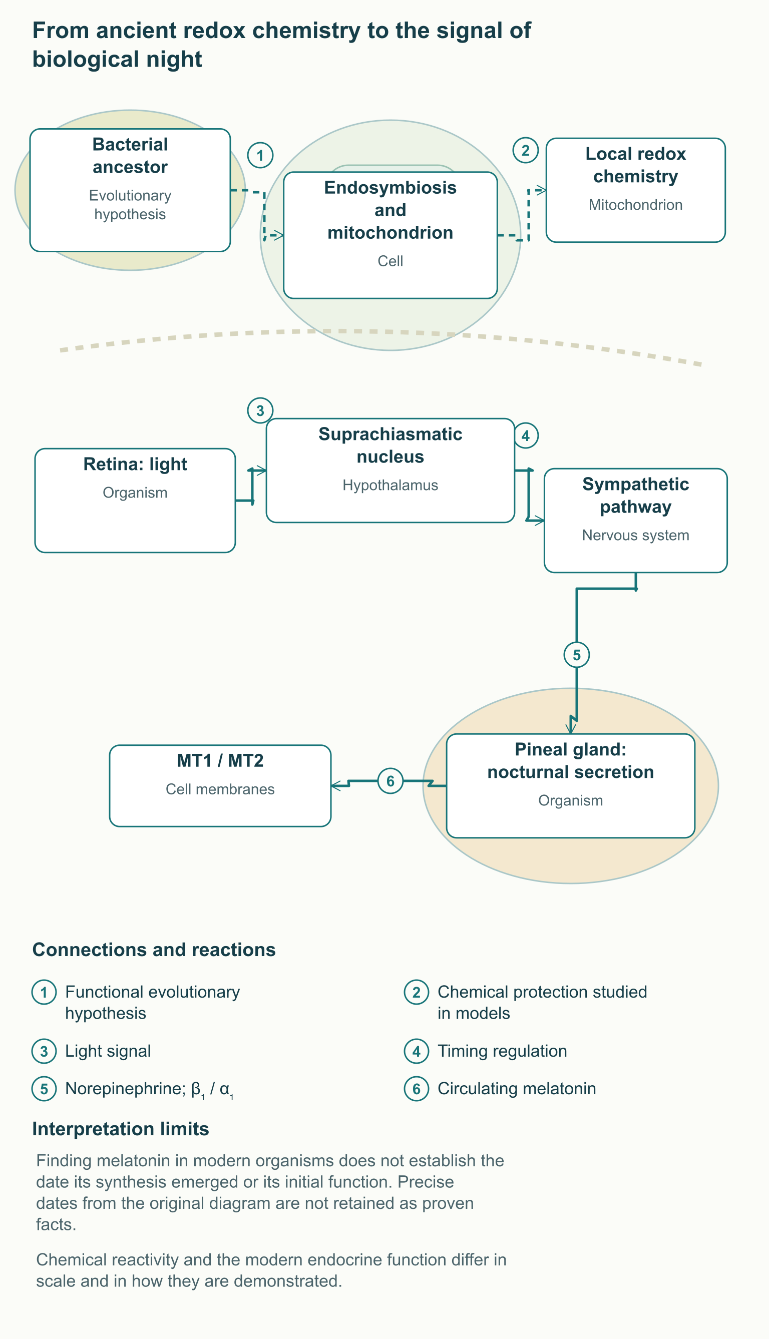
Mitochondria originated from a bacterial ancestor, and their respiratory chain retains a connection to that evolutionary history. Transferring electrons to oxygen supports energy production, but some electrons can be diverted into superoxide formation, so cells need systems that control oxidative reactions. The distribution of melatonin beyond vertebrates makes the hypothesis of an ancient chemical protective function plausible. However, detecting a substance in modern organisms does not by itself date the emergence of its synthesis or establish its first biological function.
For this reason, a story in which melatonin appeared exactly 2.5 billion years ago and acquired an endocrine function exactly 500 million years ago is too definite. Such a timeline would require independent evolutionary evidence, rather than transferring the age of changes in Earth's oxygen environment to a particular enzyme system. A more careful reconstruction connects the ancient redox chemistry of indole compounds with the subsequent specialization of signaling pathways in animals. The diagram presents these events as a hypothesis of functional development, without unsupported dates or a claim that all ancestors lacked any receptor targets.
In humans, pineal secretion creates a timing signal that helps the body distinguish biological day from night. Information about illumination passes from the retina to the suprachiasmatic nucleus of the hypothalamus, then reaches the pineal gland through intermediate neural structures and a sympathetic pathway. This chain involves the paraventricular region, spinal sympathetic neurons, and the superior cervical ganglion; norepinephrine acts on β₁- and α₁-adrenoceptors in pinealocytes. At night, this mechanism supports melatonin synthesis, whereas light during biological night can suppress secretion and change circadian phase.
Sleep also depends on accumulated sleep pressure, behavior, disease, and the environment, so the nocturnal hormonal signal is not equivalent to an off switch for consciousness. A person can remain awake despite high endogenous melatonin or feel sleepy when its level is low. This is why increasing the amount of the hormone does not always remove the cause of insomnia. Understanding the complaint requires distinguishing insufficient opportunity to sleep, difficulty sleeping on an appropriate schedule, and a mismatch between internal night and the socially desired time.
Circulating melatonin and local tissue systems
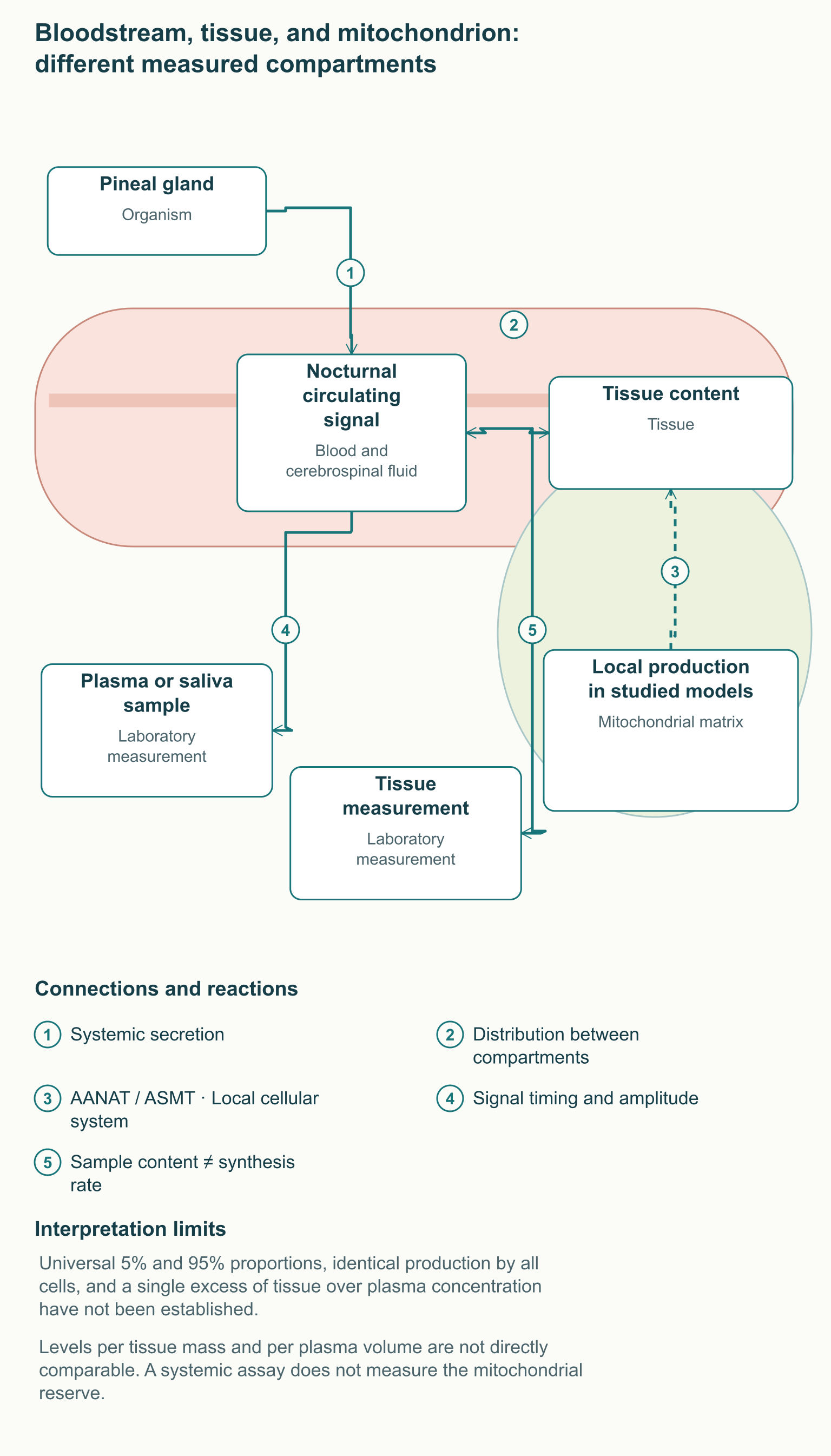
The pineal gland is the most important source of the pronounced nocturnal rhythm of circulating melatonin, but the substance and enzymes involved in its production have also been detected outside this gland. These tissues include the gastrointestinal tract and certain brain cell systems; specific findings depend on the species, tissue, and analytical method. A biopsy or homogenate study addresses the amount of substance in a sample, but does not necessarily establish its synthesis rate. Evidence for local production is strengthened by measurements of enzyme activity, metabolic conversion of a labeled substrate, and control of the purity of the isolated organelle.
In a study of the human hepatobiliary and gastrointestinal tract, melatonin was measured in mucosal biopsies, liver, bile, and blood samples from different vascular beds. This confirms that plasma is only one measurable compartment, but radioimmunological determination in tissue is not a direct measurement of mitochondrial synthesis. Content per tissue mass also cannot be compared with concentration per plasma volume without additional assumptions. The denominator, binding, distribution, and potential entry of circulating substance into the tissue all differ.
The idea of two entirely independent pools is useful as an initial approximation, but is too rigid for actual physiology. Circulating melatonin can enter tissues, while local production does not necessarily mean absolute isolation from the bloodstream. It is not established that every nucleated human cell continuously synthesizes an equally large mitochondrial pool. Still less do the available experiments allow the pineal gland to be assigned less than 5%, and mitochondria more than 95%, of total production in the human body.
Nighttime melatonin levels depend on age, individual rhythm amplitude, illumination, sampling time, and assay method. Even a convincingly high measurement in an individual tissue experiment does not establish a universal hundredfold or thousandfold excess over plasma levels. Removing the pineal gland in an animal model can help investigate its contribution to a particular measure, but does not provide a ready-made quantitative map of the entire human body. Clinically, blood or saliva measurements are used to assess the timing signal, rather than to count an intracellular antioxidant reserve.
Synthesis: from tryptophan to acetylation and methylation
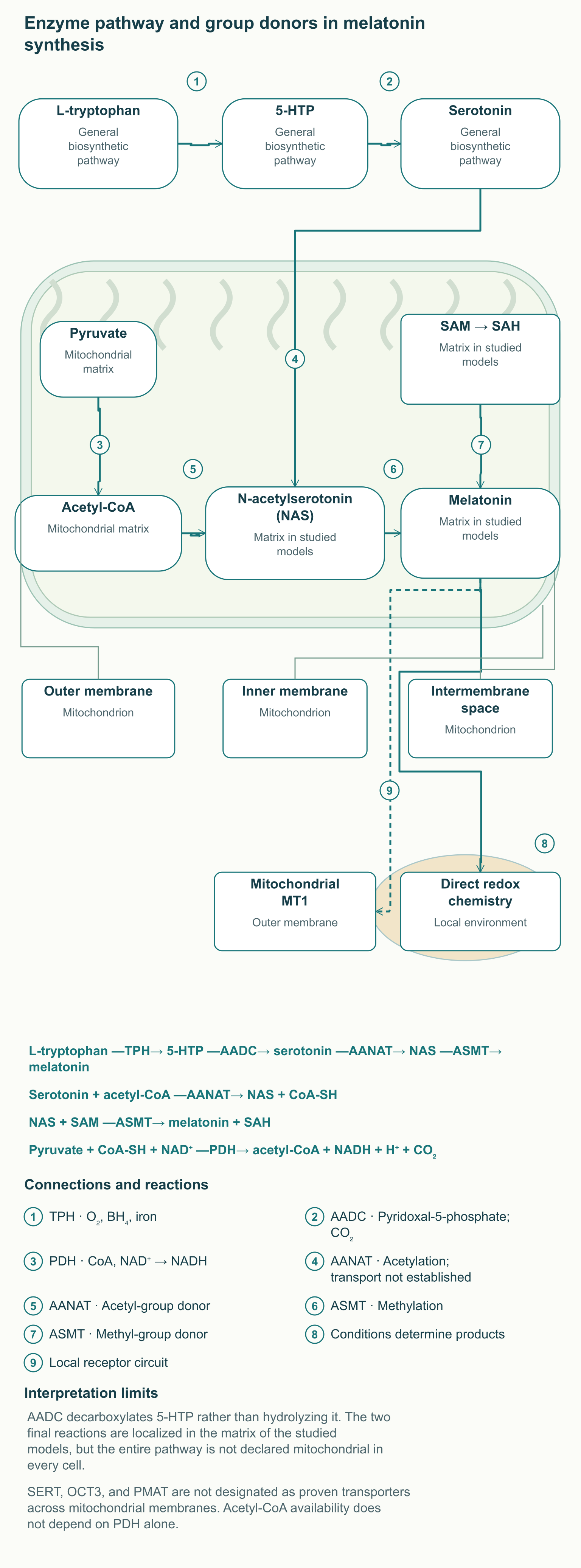
The classical melatonin synthesis pathway starts with L-tryptophan and includes four successive transformations. Tryptophan hydroxylase, TPH, forms 5-hydroxytryptophan, or 5-HTP, using oxygen and tetrahydrobiopterin, BH₄; its catalytic system depends on iron. Aromatic L-amino acid decarboxylase, AADC, then removes the carboxyl group, forming serotonin and CO₂. AADC uses pyridoxal-5-phosphate, the active form of vitamin B6, as a cofactor: this is decarboxylation, not hydrolysis.
The last two reactions use other chemical group donors. Arylalkylamine N-acetyltransferase, AANAT, transfers an acetyl group from acetyl-CoA to serotonin, producing N-acetylserotonin, or NAS, and free CoA. Acetylserotonin O-methyltransferase, ASMT, also known as HIOMT, transfers a methyl group from S-adenosylmethionine, SAM, to the hydroxyl group of NAS. The products are melatonin and S-adenosylhomocysteine, SAH, so acetylation and methylation require not only the enzymes but also the availability of their respective substrates.
The pathway can be read without specialized chemical notation: L-tryptophan —TPH→ 5-HTP —AADC→ serotonin —AANAT→ NAS —ASMT→ melatonin. Here, an arrow represents an enzyme reaction, not movement of the molecule between cellular compartments. In the pineal gland, AANAT is an important regulated step in nocturnal secretion, but the description “the only rate-limiting enzyme” should not automatically be transferred to every tissue or experimental setting. Flux through the entire pathway also depends on precursors, ASMT activity, and removal of products.
In the work by Suofu and colleagues, AANAT and ASMT were detected in the mitochondrial matrix in experimental systems involving mouse brain and neuronal cells. The study included isolated mitochondria and N2a cells, making it important for the local synthesis hypothesis, but it does not describe all human tissues. Finding the two final enzymes in the matrix does not prove that all four stages always take place there. Precursors may arrive from other cellular compartments, and their transport pathway requires separate evidence.
The serotonin transporter SERT, OCT3, and PMAT participate in monoamine transport in particular cellular systems, but this does not establish them as transporters across both mitochondrial membranes. Accordingly, the diagram does not draw a confirmed serotonin delivery route through them into the matrix. The distinction between a known reaction and an unidentified transport step is especially important for professionals; otherwise, the map appears complete only because an unsupported detail has been added. For the same reason, the participation of B6 or SAM in a reaction does not prove that additional supplementation improves human sleep in the absence of a relevant deficiency.
Acetyl-CoA, PDH, and energy metabolism
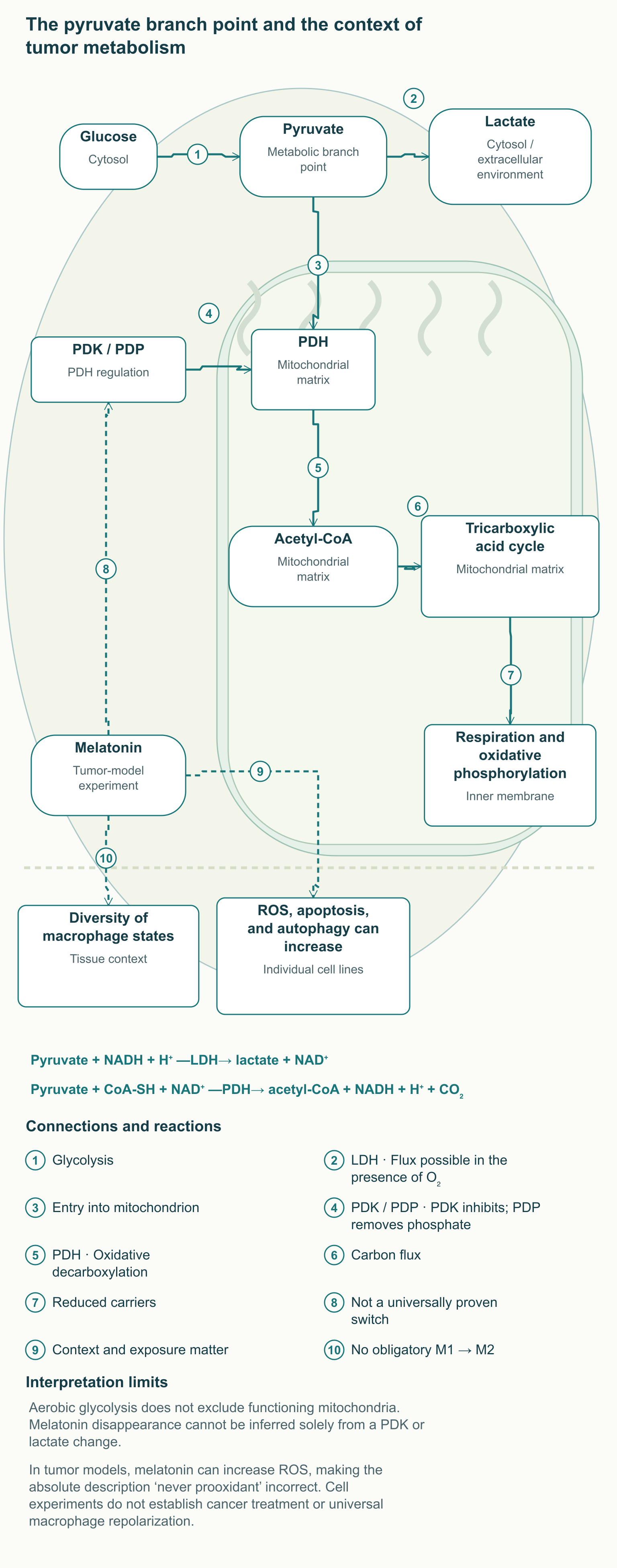
Acetyl-CoA links melatonin production to a broader network of cellular metabolism. The pyruvate dehydrogenase complex, PDH, catalyzes oxidative decarboxylation of pyruvate entering mitochondria from glycolysis. The reaction is written as follows: pyruvate + CoA-SH + NAD⁺ —PDH→ acetyl-CoA + NADH + H⁺ + CO₂. The acetyl group can then be used in the tricarboxylic acid cycle or in other reactions, including serotonin acetylation wherever the corresponding enzyme system is present.
PDH is a multicomponent complex, not a simple channel that opens with good sleep. Its activity depends on thiamine pyrophosphate, the lipoamide group, FAD, CoA, and NAD⁺, as well as the energy and redox state of the matrix. PDK kinases phosphorylate the complex and reduce its activity, whereas PDP phosphatases remove this inhibition. The NADH/NAD⁺ and acetyl-CoA/CoA ratios reflect how much the cell needs further pyruvate oxidation.
The substrate connection between PDH and AANAT does not mean that inhibiting PDH necessarily eliminates mitochondrial melatonin. Acetyl-CoA can be produced from fatty acids and other sources, and its availability depends on tissue and metabolic state. Even a reduction in a particular flux is not equivalent to zero substance content in the organelle. Claiming complete cessation of synthesis would require directly measuring synthesis under a defined intervention, rather than deriving the result solely from an attractive chain of arrows.
In practice, this connection helps explain why melatonin research overlaps with bioenergetics, but does not justify independently prescribing “PDH activators.” It also does not allow mitochondrial melatonin deficiency to be diagnosed from blood lactate or fatigue. These measures have many causes and lack the necessary specificity. When assessing a mechanism, it is important to establish whether the authors measured enzyme activity, metabolic flux, product concentration, or only expression of the corresponding gene.
MT1 and MT2: receptor signaling in the membrane
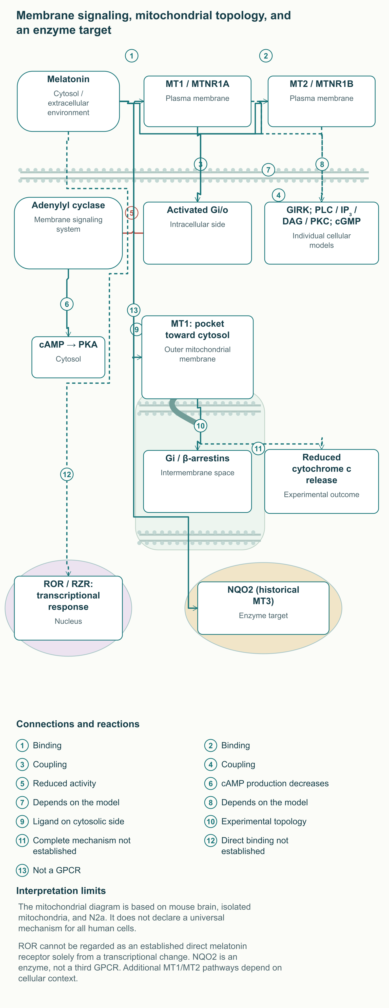
MT1 and MT2 are G protein-coupled receptors encoded by MTNR1A and MTNR1B. Their architecture includes seven transmembrane helices and a ligand-binding site; structural studies of the human receptors have revealed details of agonist recognition. On activation, a receptor changes its interaction with a G protein rather than simply holding a molecule near the membrane. Coupling to Gi/o can reduce adenylyl cyclase activity and formation of cyclic AMP, or cAMP, one of the intracellular signaling intermediates.
Reducing cAMP can decrease protein kinase A activation, but the final response is determined by the architecture of the particular cell. A G protein consists of α and βγ components; their signals can affect ion channels and other enzyme pathways. Neuronal systems have implicated GIRK potassium channels and changes in excitability, while individual models have implicated PLC, IP₃, DAG, PKC, and cGMP. These observations should not be assembled into a mandatory program performed identically by every MT1 or MT2 receptor in every organ.
Characteristic
MT1
MT2
Gene and chromosomal region
MTNR1A, 4q35.2
MTNR1B, 11q14.3
Target class
Seven-transmembrane GPCR
Seven-transmembrane GPCR
Well-supported coupling
Gi/o, reduced cAMP in the relevant systems
Gi, reduced cAMP in the relevant systems
Additional pathways
Ion channels and kinases; the result depends on the cell
PLC/PKC and other intermediates in individual models
Physiological context
Night signaling and neuronal regulation
Circadian regulation and metabolic responses
How to interpret affinity
A parameter of a particular ligand and method
A parameter of a particular ligand and method
The dissociation constant, Kd, describes binding under particular experimental conditions, so it cannot be presented as an invariant number for all tissues and all medicines. Structures obtained with ramelteon or other agonists do not mean that these compounds have exactly the same pharmacokinetics and clinical effects as melatonin. Likewise, the distinction between MT1 and MT2 cannot be reduced to “one induces sleep, the other shifts the clock.” The circadian system brings together different cells and signals, and the resulting phase response depends on exposure time.
Vascular effects also cannot be reduced to a universal opposition between MT1-mediated constriction and MT2-mediated dilation. The response depends on the vascular bed, tissue state, concentration, and other regulatory systems. This simplified diagram is particularly dangerous if it is used to infer a predictable blood pressure reduction in a particular patient. Clinically, blood pressure and how the patient feels are assessed directly, while a mechanistic description is used to frame a question rather than replace an observed outcome.
Mitochondrial MT1: precise orientation and experimental limits
Suofu's study proposed a mechanism of local melatonin signaling that the authors called automitocrine. In the studied systems, MT1 was located on the outer mitochondrial membrane, with its ligand-binding side facing the cytosol. Gi and β-arrestins were found on the intermembrane-space side. This is an unusual topology, and reversing its sides changes the meaning of the entire diagram: melatonin should not be shown binding to a pocket facing into the intermembrane space.
Locally produced melatonin can leave the matrix and interact with a target on the outer membrane, but its movement pathway cannot be considered fully described solely from enzyme localization. In the experiments, activating the receptor system was accompanied by reduced local cAMP and less cytochrome c release. Gi is activated as an inhibitory regulator of adenylyl cyclase: saying “melatonin inhibits Gi” reverses the causal connection. The authors did not conclusively establish the complete mechanism limiting cytochrome c release, so arrows linking Bax, PKA, and the pore should not be presented as a finished universal sequence.
The study's technical limitations also matter for interpretation. Alongside mouse brain tissue, N2a cells expressing recombinant MT1 were used, and the specificity of endogenous receptor detection posed a methodological difficulty. The authors did not directly demonstrate that melatonin of mitochondrial origin provides the same neuroprotection in vivo in humans. Thus, a local receptor system is a research explanation for a possible cellular effect, not a basis for calculating human daily production or selecting an oral dose to protect the brain.
ROR/RZR and NQO2: different levels of interaction
Nuclear receptors in the ROR family regulate transcription, but direct binding of melatonin to them should not be considered established on the basis of an old receptor diagram. A primary study in ovine pituitary tissue investigated RORα presence and specific nuclear binding of labeled melatonin; receptor RNA was present without detection of the corresponding nuclear binding. Changes in RORα activity were described in breast cancer cells exposed to melatonin, but a transcriptional effect can be indirect. The distinction between changing a factor's activity and physically binding to it is fundamental when choosing the term “receptor.”
Accordingly, regulation of COX-2, iNOS, NF-κB, or inflammatory genes is not drawn as a proven universal chain of “melatonin — ROR — inflammation suppression.” Observed changes in these pathways may depend on the cell model, time, and dose, as well as on other signaling targets. The expression “nuclear effect” describes the site of the eventual response, but not necessarily the site of initial molecular binding. This qualification preserves the substance of genomic regulation while removing an incorrectly assigned direct intermediary.
The historical MT3 site is associated with a different molecular target, the enzyme quinone reductase 2, NQO2. It was identified using protein isolation, molecular methods, and expression of the human enzyme in cells; the interaction was subsequently studied with kinetic and structural methods. NQO2 is not a third seven-transmembrane melatonin receptor and should not be placed alongside MT1 and MT2 as another GPCR. Binding to or inhibiting an enzyme also does not guarantee general “detoxification of all quinones”: the outcome depends on the substrate and redox reaction conditions.
Direct redox chemistry: reaction rate and cellular competition
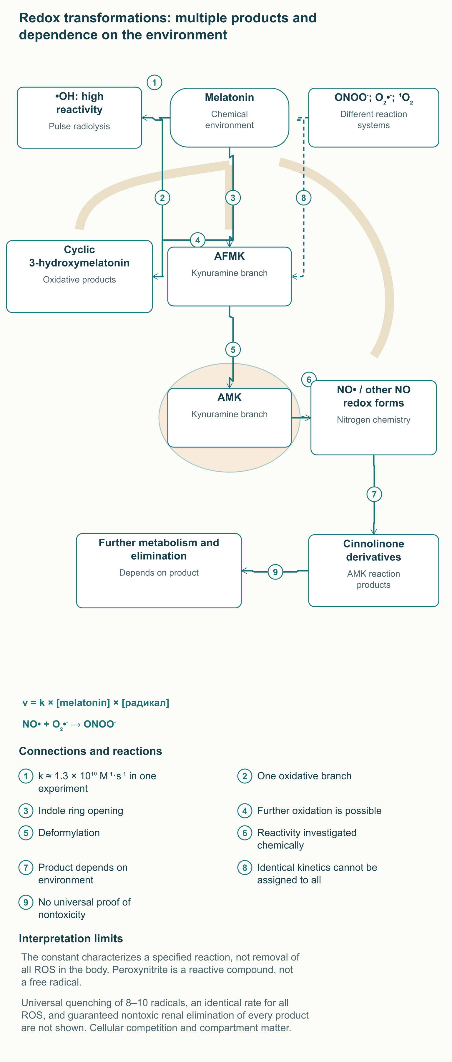
Melatonin can react with particular reactive oxygen and nitrogen species without a membrane receptor. In one pulse radiolysis study, the rate of reaction with the hydroxyl radical was characterized by a constant of approximately 1.3 × 10¹⁰ M⁻¹·s⁻¹, close to the diffusion limit. This value demonstrates high chemical reactivity under the specified experimental conditions. It does not mean that a small oral dose neutralizes every hydroxyl radical in all tissues of the body.
The rate of a second-order reaction depends not only on the constant k, but also on the concentrations of both reactants: v = k × [melatonin] × [radical]. In a cell, a radical simultaneously encounters proteins, lipids, nucleic acids, and other small molecules. The probability of reaction therefore depends on competition, distance from the site of formation, and substance availability in the relevant compartment. The hydroxyl radical is extremely short-lived, and preventing its formation is often more important than delivering a “trap” from the bloodstream after damage has begun.
Other reactive species cannot automatically be equated with the hydroxyl radical. Experiments using different generation and detection methods have produced different estimates of melatonin activity, including a substantially weaker reaction with superoxide. Peroxynitrite, ONOO⁻, is not itself a free radical, although it contributes to nitrosative damage and formation of secondary reactive products. A scientific diagram should distinguish chemical species rather than grouping them as “radicals” with one identical removal rate.
Some oxidative transformations open melatonin's indole ring to form AFMK, N¹-acetyl-N²-formyl-5-methoxykynuramine. Deformylation of AFMK can yield AMK, N¹-acetyl-5-methoxykynuramine, which also reacts with nitrogen-containing species. In primary chemical experiments, AMK reacted with different redox forms of NO, producing, among other compounds, a cinnolinone derivative. This is more precise than a single arrow leading to a “nontoxic cinnoline” supposedly ending every melatonin reaction.
The idea of an antioxidant cascade means that some transformation products may retain activity. But “one molecule reliably quenches eight to ten radicals” is not universal cellular or clinical stoichiometry. The pathway depends on the medium, oxidant, enzyme participation, and subsequent fate of the products, while absence of toxicity must be established separately. It is also incorrect to contrast melatonin with vitamins C and E as an absolutely safe molecule versus inevitably prooxidant antioxidants: the biological effects of all these substances depend on conditions.
Nrf2, antioxidant enzymes, and the glutathione cycle
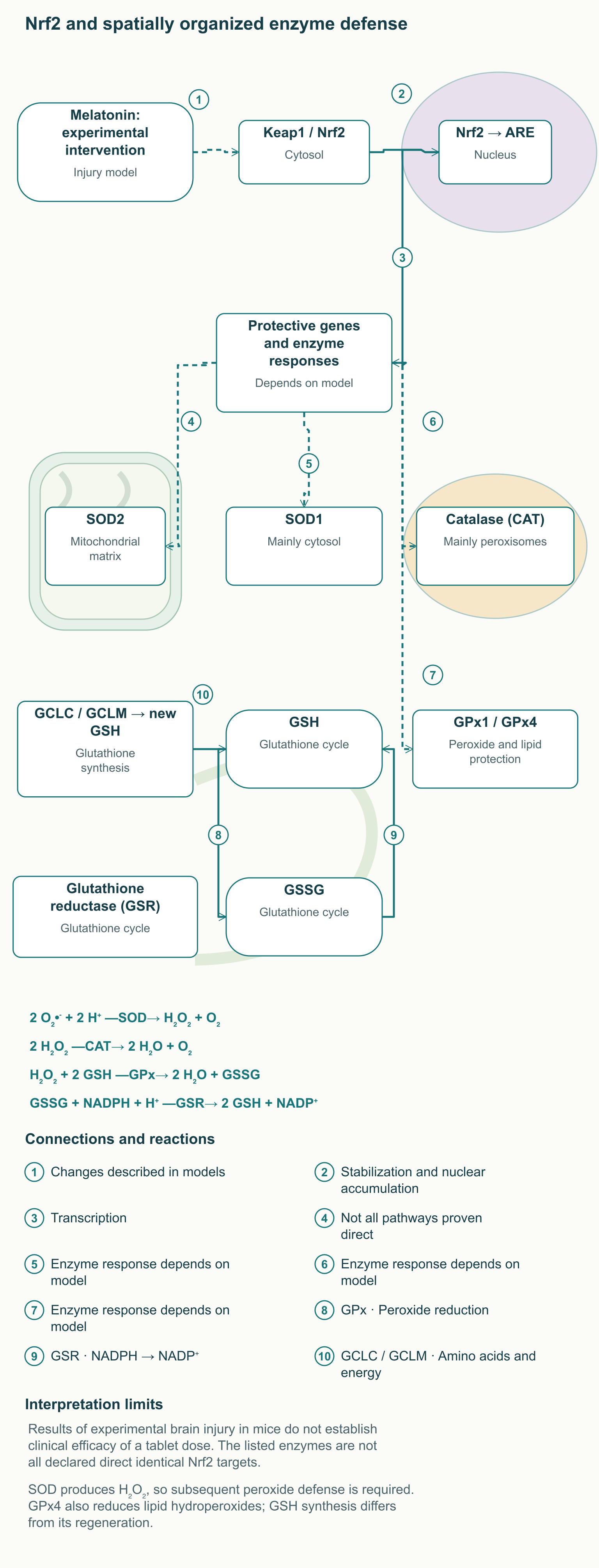
Receptor signaling and direct reaction with reactive species do not exhaust the possible effects of melatonin. Experimental models also investigate changes in protective enzyme expression and activity, including those mediated by Nrf2. Under basal conditions, the Keap1 system helps target Nrf2 for degradation; when redox signals change, the factor becomes more stable and can accumulate in the nucleus. There, Nrf2 works with other proteins to regulate genes containing the appropriate antioxidant response elements, AREs.
In experiments by Ding and colleagues on traumatic brain injury in mice, Nrf2-ARE was studied as a possible mediator of melatonin action. Changes were observed in nuclear Nrf2, protective measures, and enzyme responses; animals with disrupted Nrf2 helped clarify the pathway's involvement. This is experimental injury and experimental administration, not a clinical trial of an ordinary tablet to prevent brain damage. A change in Nrf2 also does not prove that melatonin always directly activates ERK, disengages Keap1, and increases every antioxidant enzyme to the same extent.
Enzyme defense is spatially organized. SOD2 acts mainly in the mitochondrial matrix, whereas SOD1 is located mainly in the cytosol and also in certain other compartments; catalase is predominantly associated with peroxisomes. Superoxide dismutase converts superoxide to H₂O₂: 2 O₂•⁻ + 2 H⁺ → H₂O₂ + O₂. This removes one reactive species while forming another, so the system needs subsequent control of hydrogen peroxide.
Catalase catalyzes 2 H₂O₂ → 2 H₂O + O₂, while glutathione peroxidases use reduced glutathione, GSH. For hydrogen peroxide, the reaction can be written as H₂O₂ + 2 GSH → 2 H₂O + GSSG, where GSSG is oxidized glutathione disulfide. Glutathione reductase regenerates reduced glutathione using NADPH: GSSG + NADPH + H⁺ → 2 GSH + NADP⁺. GPx4 is especially important for reducing lipid hydroperoxides, so its function cannot be confined to the same picture of free H₂O₂.
New GSH is produced in two enzyme stages, the first involving glutamate-cysteine ligase with catalytic GCLC and regulatory GCLM. Amino acid availability, energy, and reducing capacity determine cycle function together with protein expression. The diagram separates the established architecture of the defense system from experimental changes induced by melatonin. This makes it possible to understand potential enhancement of protection without presenting all the listed genes as equally proven direct Nrf2 targets in every tissue.
mPTP, the outer membrane, and two different cell death pathways
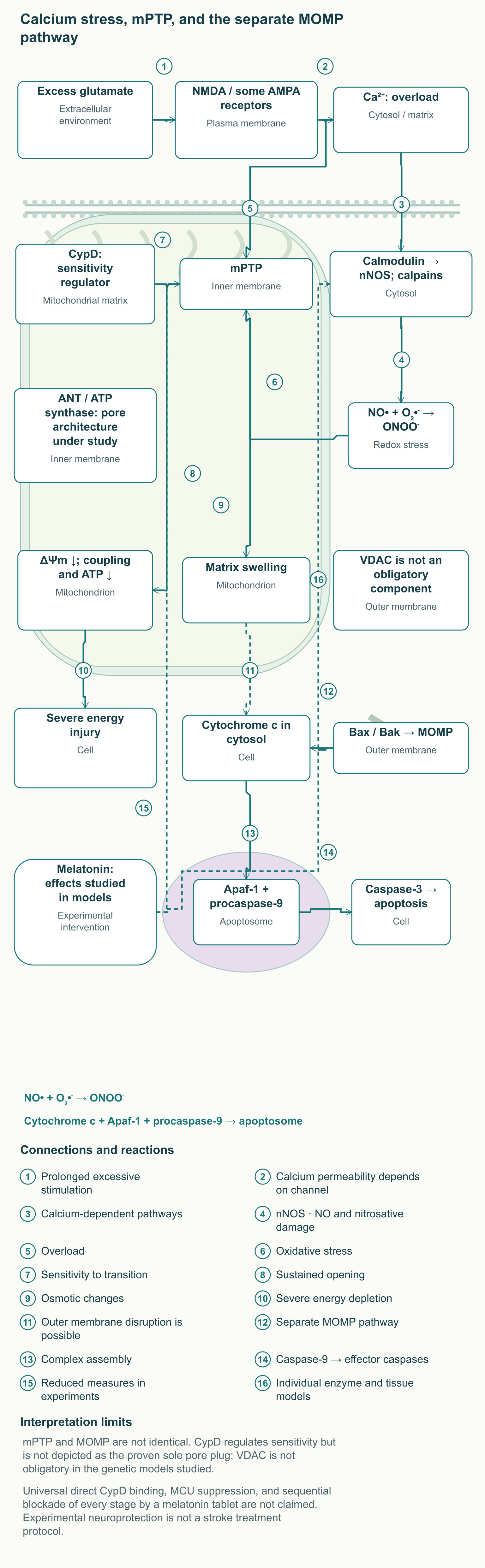
Mitochondrial permeability transition involves increased permeability of the inner membrane to small solutes. With sustained mPTP opening, the membrane potential, ΔΨm, weakens, coupling between respiration and ATP synthesis is disrupted, and water can cause matrix swelling. The transition is facilitated by calcium overload and oxidative stress, but is not a single linear reaction occurring in every injury. The consequences depend on duration, cellular energy status, and the ability to restore the membrane gradient. When the gradient is lost, ATP synthase can, under certain conditions, work in reverse, consuming ATP to move protons instead of producing ATP.
Cyclophilin D, or CypD, is a matrix regulator of sensitivity to permeability transition. Experiments in mice lacking Ppif showed mitochondrial resistance to particular calcium and oxidative challenges and protection in certain ischemia-reperfusion models. However, these changes did not prevent every form of apoptotic cell death. In particular, the results highlighted the distinction between CypD-dependent transition and certain pathways regulated by Bcl-2 family proteins.
The mPTP cannot be presented as a definitively established complex of ANT, VDAC, CypD, and ATP synthase. The roles of ANT and ATP synthase are studied in different models, while VDAC is situated in the outer membrane; primary genetic experiments showed that VDAC was not required for the studied forms of permeability transition and cell death. CypD is important for regulation, but this is not equivalent to recognizing it as the sole structural plug of the pore. The modern diagram shows investigated components and their locations while preserving uncertainty about molecular architecture.
A separate process is MOMP, mitochondrial outer membrane permeabilization, which can be initiated by Bax and Bak. Cytochrome c release into the cytosol promotes assembly of the apoptosome with Apaf-1 and procaspase-9, followed by activation of effector caspases, including caspase-3. Swelling after sustained mPTP opening can also disrupt the outer membrane, but these pathways should not be considered identical. With severe ATP depletion, cell death can develop predominantly through necrotic injury rather than necessarily through a completed caspase cascade.
In experiments by Andrabi and colleagues, melatonin reduced measures of permeability transition, supporting the hypothesis of its involvement in mitochondrial protection. However, this result does not prove direct physical blockade of CypD or universal suppression of the mitochondrial calcium uniporter, MCU. Observing reduced cytochrome c release in another system also does not automatically reveal all intermediate protein interactions. Clinical translation requires studies linking a particular exposure to preservation of organ function, not only to an individual damage marker.
Excitotoxicity: from synapse to mitochondrion
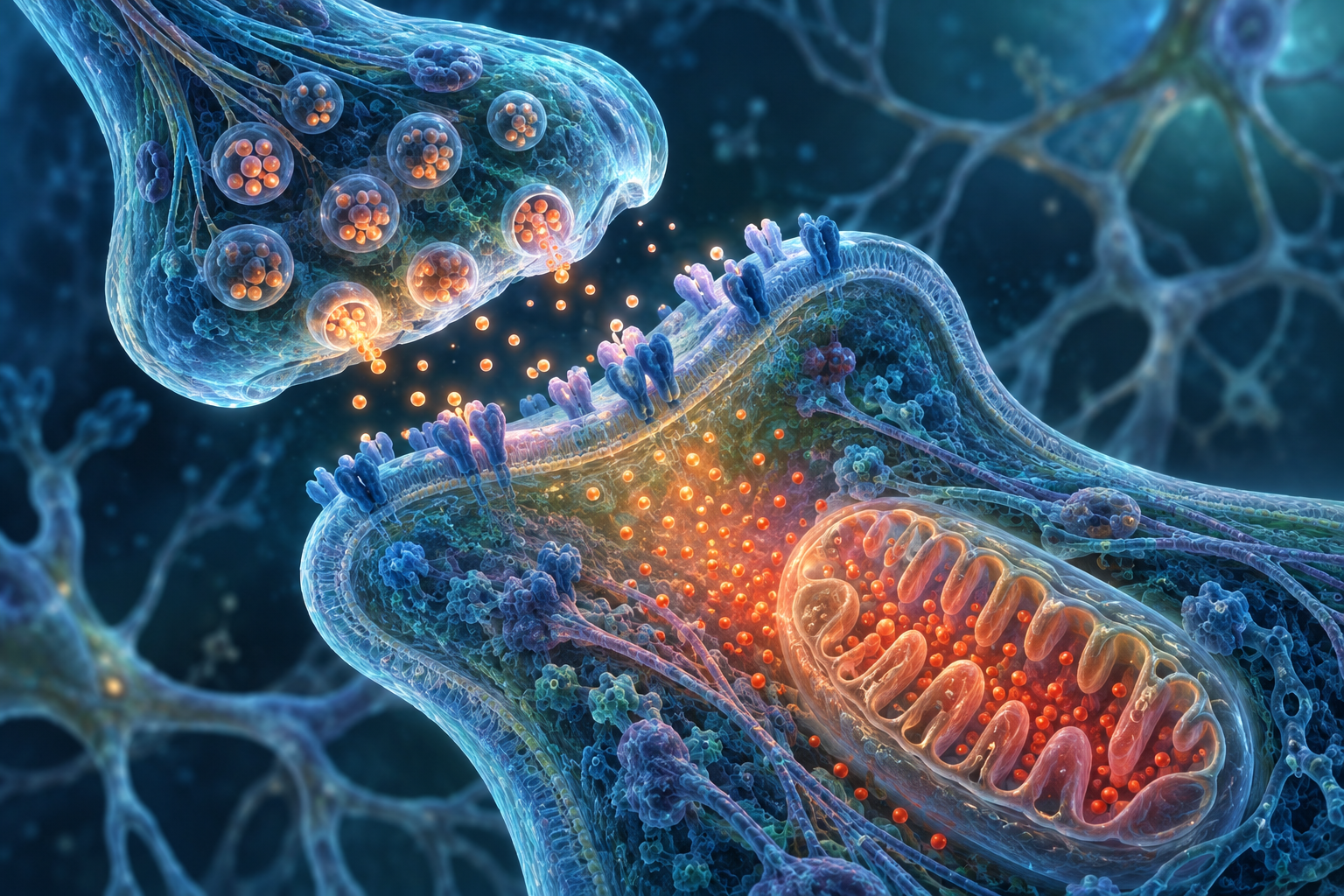
Glutamate is necessary for normal signaling, learning, and plasticity, but excessive prolonged activation of its receptors can damage a neuron. When energy supply is disrupted, transmitter reuptake deteriorates and extracellular glutamate accumulates. NMDA receptors can conduct Ca²⁺, while the involvement of AMPA receptors depends, among other things, on subunit composition: not every AMPA channel is equally permeable to calcium. Accordingly, the diagram does not draw all glutamate receptors as identical openings for an unlimited calcium flow.
Increasing cytosolic Ca²⁺ changes the activity of calmodulin, calpain proteases, and neuronal nitric oxide synthase, nNOS. The NO produced can react with superoxide: NO• + O₂•⁻ → ONOO⁻. Mitochondrial calcium overload and redox damage to membranes then aggravate the energy failure, creating a positive feedback loop. Damage to proteins and lipids in this setting accompanies disruption of ion gradients and activation of cell death signaling.
Melatonin and some of its metabolites have been studied as modifiers of particular steps in this process. For example, enzyme experiments and a model involving brain tissues investigated the effects of AMK and melatonin on nNOS activity, whereas AFMK did not show the same effect in that system. Other models evaluated calcium handling, oxidative damage, Bax/Bcl-2, and caspases. Together, these studies explain the interest in neuroprotection, but do not establish obligatory sequential blockade of NMDA, calmodulin, nNOS, the apoptosome, and caspase-3 by any oral dose.
Professional interpretation must account for intervention timing and tissue availability. During ischemia, some processes develop rapidly, and a high concentration applied directly to a cell medium is not equivalent to exposure after a tablet. In addition, reducing a laboratory marker does not necessarily mean reducing disability or improving survival. Accordingly, melatonin is not presented here as an emergency stroke treatment, and signs of stroke require immediate standard medical care.
The Warburg effect and the context of a metabolic response
The Warburg effect means increased lactate production from glucose when oxygen is available, not a complete absence of mitochondrial respiration. A tumor cell can use glycolysis, the tricarboxylic acid cycle, and oxidative phosphorylation simultaneously, while also directing carbon into cellular building blocks. Pyruvate sits at a branch point: lactate dehydrogenase converts it to lactate, whereas PDH supplies an acetyl group for mitochondrial metabolism. Changes in PDK can influence this partitioning, but do not by themselves explain all tumor bioenergetics.
Primary studies of tumor models have investigated the relationship between PDK, hypoxic responses, and lactate production independently of melatonin. Studies of melatonin in cell lines also show that its effects can differ between healthy and tumor systems. In head and neck squamous cell carcinoma cells, researchers investigated enhancement of cisplatin and radiation effects accompanied by increased mitochondrial ROS, apoptosis, and autophagy. This result is especially useful in demonstrating why melatonin cannot be described as absolutely lacking prooxidant effects.
The hypothesis that local melatonin synthesis can be restored by changing PDK/PDH remains a separate research question. Confirming the entire chain requires measuring PDH phosphorylation, carbon flux, acetyl-CoA availability, and melatonin production in the same model. An observed reduction in lactate or change in PDK expression does not substitute for these measurements. Still less can such results establish that melatonin universally “reverses the Warburg effect” and treats malignant tumors.
Macrophage metabolism also changes during inflammation, but the M1/M2 classification is a simplified language for experimental states. Tissues contain diverse programs that change over time and depend on the environment and injury. Macrophage cultures have shown effects of melatonin and its metabolites on particular inflammatory responses, including COX-2, but this does not mean a guaranteed transformation of every proinflammatory macrophage into a reparative one. Clinical prescribing should be based on an outcome measured in the patient, not an idealized cellular phenotype switch.
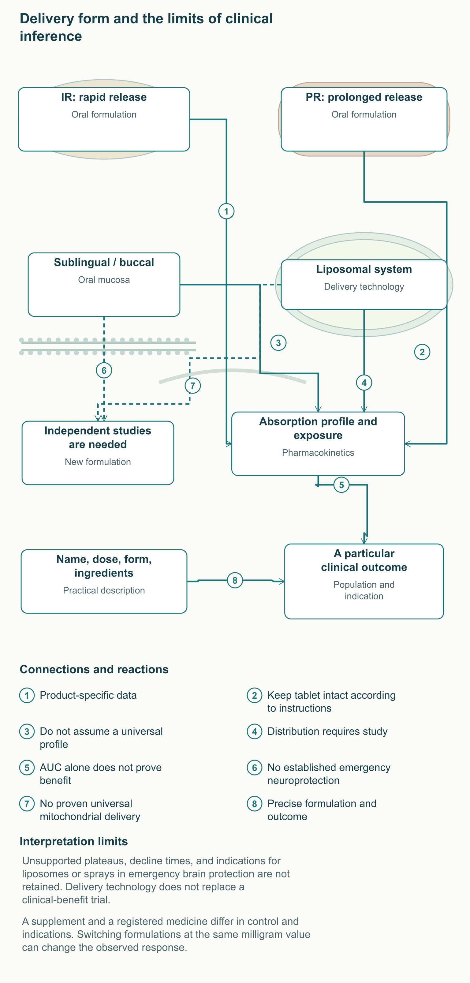
Pharmacokinetics: why IR and PR should not be combined
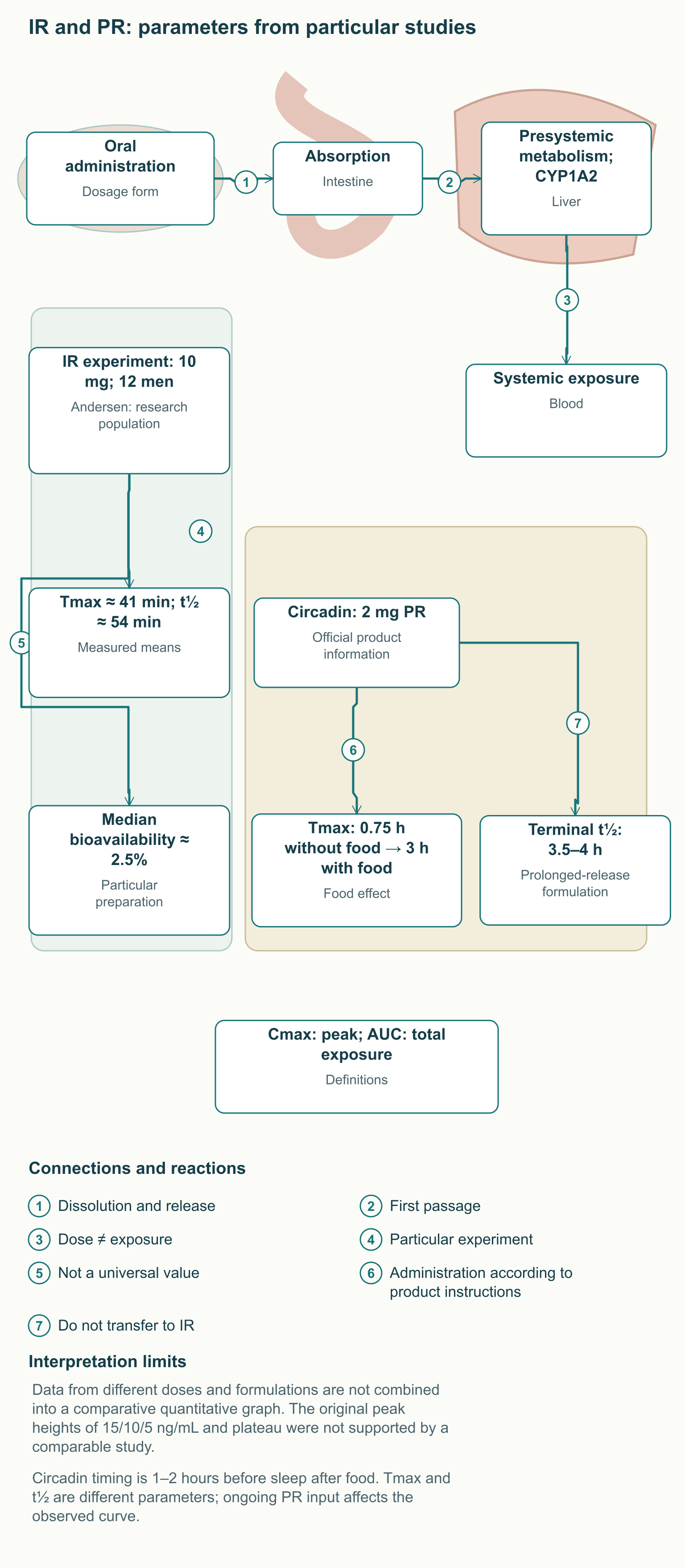
The amount of melatonin in a tablet is not the amount reaching systemic circulation. After oral administration, the substance is absorbed and undergoes substantial presystemic metabolism, while exposure depends on formulation, food, individual metabolism, and concomitant medicines. CYP1A2 is important in metabolism; the resulting 6-hydroxymelatonin is subsequently conjugated, including formation of 6-sulfatoxymelatonin, aMT6s. This urinary metabolite reflects a particular portion of systemic metabolism, rather than directly reflecting melatonin content in the mitochondrial matrix.
An immediate-release formulation, IR, is designed for faster delivery, whereas PR slows release from the dosage form. Tmax denotes the time to maximum concentration, Cmax the maximum itself, and AUC total exposure over the observed period. Half-life characterizes concentration decline under specified conditions and is not identical to the duration of the desired clinical effect. In PR, continued absorption also affects the observed curve, so a “long tail” cannot be explained solely by slow liver metabolism.
In a small study by Andersen and colleagues, 12 healthy men received 10 mg melatonin orally and intravenously on different days. For the oral preparation used, mean Tmax was about 41 minutes and mean half-life about 54 minutes; median absolute bioavailability was approximately 2.5%. This is a particular pharmacokinetic experiment with a dose high by chronobiological standards, not standard exposure for any supplement. The study also demonstrates why the same plasma peak cannot confidently be drawn for every person receiving the same tablet.
For Circadin 2 mg PR, the product information gives a different profile: food changes Tmax from approximately 0.75 to 3 hours, while terminal half-life is approximately 3.5–4 hours. These data concern a specific prolonged-release formulation and its administration conditions. They cannot be combined with the 10 mg IR experiment into one quantitative curve of “15 ng/mL versus 5 ng/mL” without a comparable study. The infographic therefore presents measured parameters and influencing factors rather than an invented concentration graph with precise but unsupported heights.
Melatonin, MTNR1B, and an evening glucose challenge
The timing signal of night intersects with metabolic regulation. MTNR1B encodes MT2, and melatonin signaling in pancreatic cells can affect insulin secretion through intracellular intermediates. Variants of this gene are associated with differences in glucose metabolism measures, but a genetic association does not prove that melatonin always causes diabetes. A short-term response to a challenge, long-term disease risk, and the effect of medicinal administration are separate research questions.
Garaulet and colleagues conducted a randomized crossover study involving 845 participants from a Spanish population with habitually late eating. Each underwent two evening tests with 75 g glucose after an eight-hour fast: four hours and one hour before habitual bedtime. This is a standardized oral glucose challenge, not a comparison of two complete dinners containing protein, fat, and fiber. The late test coincided with endogenous melatonin levels approximately 3.5 times higher; insulin AUC was 6.7% lower and glucose AUC 8.3% higher.
The adverse effect of the late challenge on glucose tolerance was greater in carriers of the G allele of the MTNR1B variant. This supports a causally coherent hypothesis about the coincidence of a high nocturnal signal and carbohydrate exposure, but does not establish a universally safe interval of 2.5–3 hours for all foods and formulations. The experiment itself did not administer a melatonin tablet or measure the emergence of diabetes after long-term treatment. Consequently, the result should not become a ban on all late meals or a requirement for genetic testing of every user.
In practice, professionals should discuss habitual sleep and meal times, diabetes or prediabetes, and the actual purpose of melatonin use. If a medicine has already been prescribed, eating schedules and glucose monitoring are selected in the context of the disease and the specific formulation. Circadin's instruction to take it after food cannot mechanically be overturned by an experiment involving endogenous melatonin and an OGTT. Where needed, changes in treatment or timing should be coordinated as a single strategy rather than assembled from contradictory general advice.
Drug interactions and safety
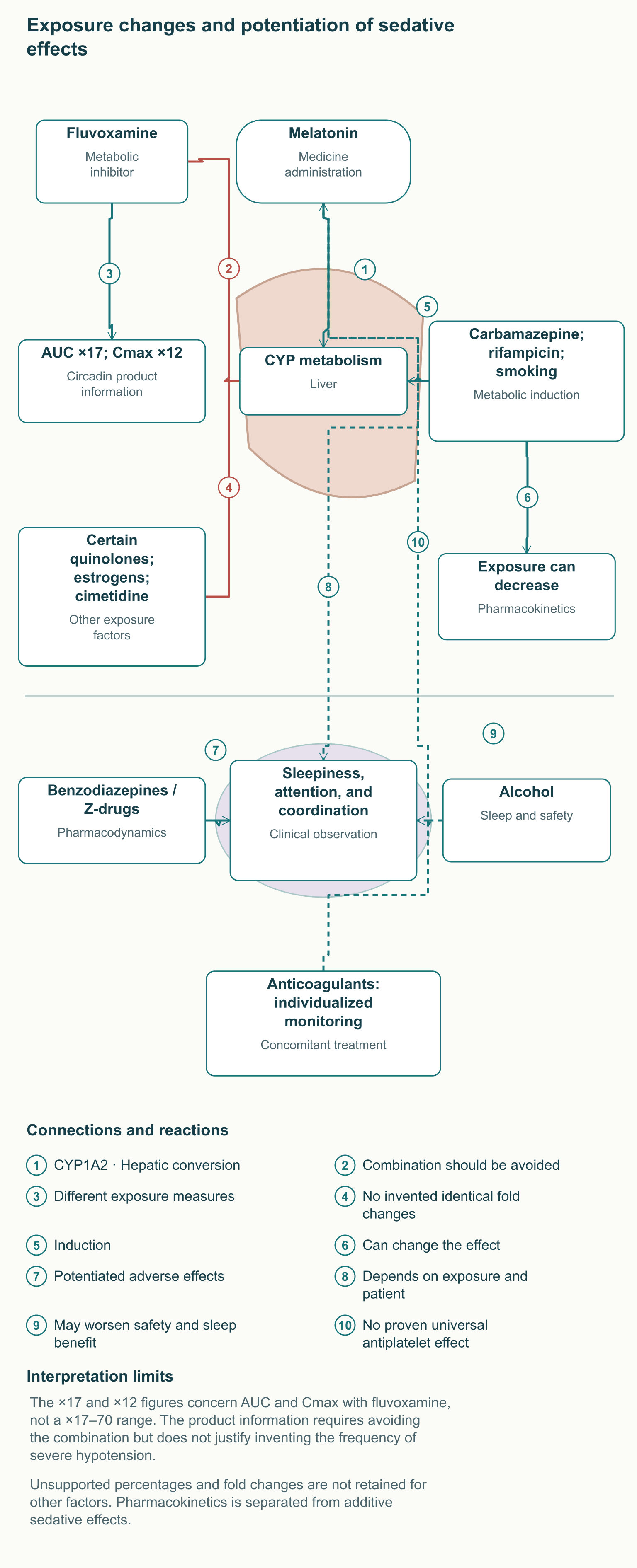
Fluvoxamine is an important example of a pharmacokinetic interaction. According to the Circadin product information, it increases melatonin AUC approximately 17-fold and Cmax 12-fold; the combination should be avoided. These are two distinct, precise measures, so they should not be merged into a “17–70-fold” range or accompanied by an invented frequency of severe hypotension. A substantial exposure change deserves attention even when the original tablet dose seems small.
Other metabolic inhibitors, including certain quinolones, can increase exposure, while estrogens and cimetidine are also mentioned in the product information as interaction factors. Conversely, carbamazepine, rifampicin, and smoking can reduce levels through enzyme induction. An identical fold change cannot be invented for each situation without a separate study. Starting or stopping smoking can also change the metabolic context, so a stable familiar dose does not always mean stable exposure.
A pharmacodynamic interaction is different from a concentration change. Benzodiazepines and certain Z-drug hypnotics may increase impairment of attention, coordination, and sleepiness when combined with melatonin; alcohol can reduce the expected sleep benefit and increase unsafe behavior. For warfarin and other medicines requiring precise monitoring, the combination should be discussed with the treating professional, but melatonin should not be declared a proven universal antiplatelet agent. The reason for caution and the observation plan should be specific to the medicine and patient.
Possible adverse events include sleepiness, dizziness, and headache, and the ability to drive safely must be considered after administration. Circadin use is limited in liver disease, caution is needed in kidney disease, and insufficient evidence affects recommendations in autoimmune disorders. Its product information also does not recommend use during pregnancy and breastfeeding because relevant data are inadequate. Information about one formulation does not become proof of equal safety for all high doses, all supplements, and all age groups.
Product quality matters in practice, especially for a supplement with a small stated amount. In Erland and Saxena's analysis of commercial products, melatonin content deviated from the label by −83% to +478%, and serotonin was found in some samples. This was a study of a particular market sample, not a claim that every modern product necessarily has such a deviation. It nevertheless shows why a registered medicinal formulation and manufacturing reliability matter just as much as calculating the desired tenths of a milligram.
Administration time: circadian disorder, insomnia, and jet lag
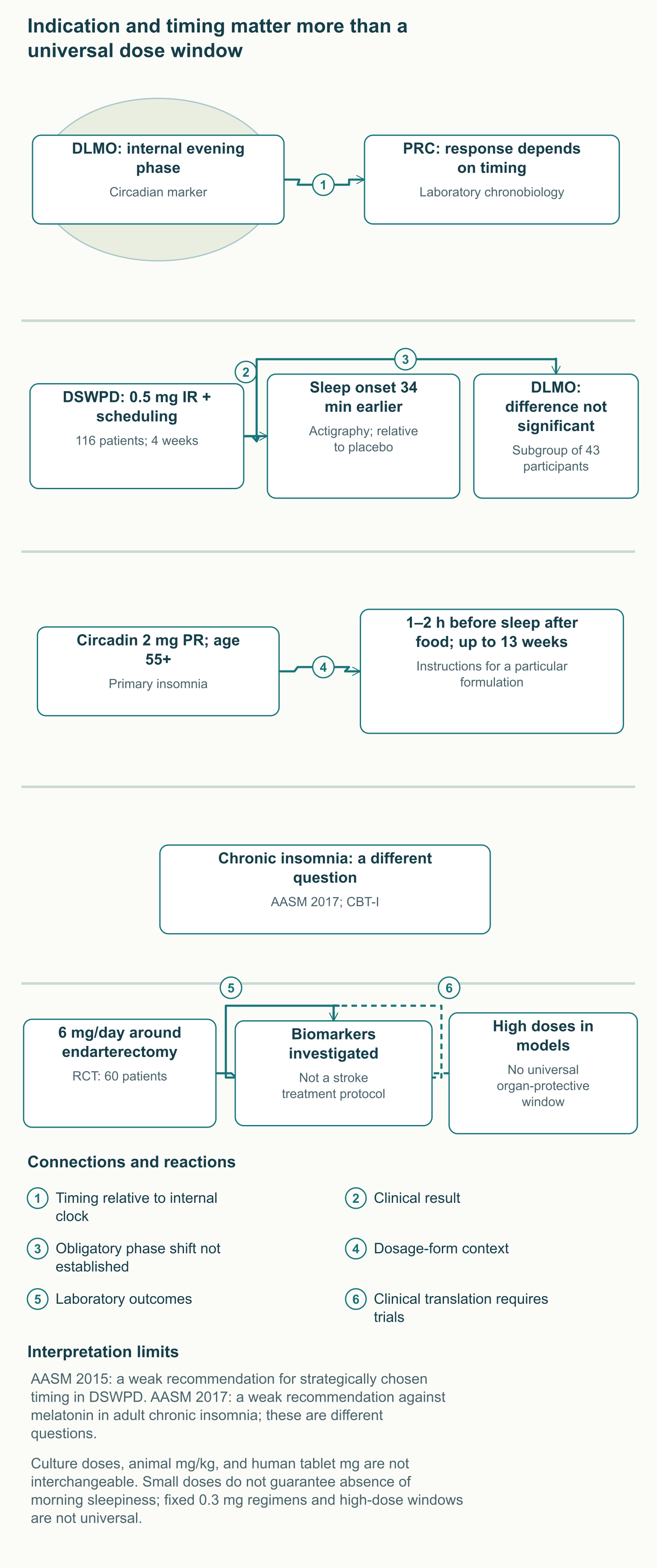
A chronobiotic changes biological clock phase, whereas an immediate effect on falling asleep is a different result. DLMO, dim-light melatonin onset, is one marker of internal phase. A phase response curve, PRC, shows how the direction and magnitude of a shift depend on administration time relative to that phase. Thus, the same dose can act differently at different biological times, and a fixed prescription of “10 p.m. for everyone” is not a complete chronobiological protocol.
In a human laboratory study, Burgess and colleagues compared phase responses to three successive days of 0.5 mg administration with previously obtained 3 mg data using the same protocol. Both doses could shift phase, but optimal timing relative to the endogenous rhythm differed. This explains why increasing the dose does not replace choosing the time. Nor does it mean that every patient should receive 0.3 mg exactly four to five hours before sleep: time relative to habitual bedtime and time relative to DLMO are different reference points.
In delayed sleep-wake phase disorder, DSWPD, a person is usually able to sleep normally on their own late schedule but has difficulty falling asleep and awakening earlier. In its 2015 guideline, the AASM made a weak recommendation for strategically timed melatonin in the relevant patients. A weak recommendation indicates limited certainty and the need to consider circumstances, not an absence of physiological rationale. It does not establish one universal dose or automatically apply to every complaint of “I cannot fall asleep.”
Sletten's trial enrolled 116 patients with DSWPD and confirmed delayed melatonin timing relative to desired sleep. They received 0.5 mg fast-release melatonin or placebo one hour before desired bedtime for four weeks, alongside behavioral scheduling. Actigraphic sleep onset was 34 minutes earlier relative to placebo; the 95% confidence interval corresponded to approximately 8–60 minutes earlier onset. Actigraphy estimates sleep from movement and is not polysomnography, so describing this result as PSG is incorrect.
In a subgroup of 43 participants, the difference in DLMO change between groups was not statistically significant. The trial therefore supports the clinical effect of this combined regimen, but does not demonstrate a pronounced phase shift in every patient. Some of the benefit could have resulted from facilitating sleep onset at the selected time, while behavioral scheduling was included in both groups. Long-term efficacy and safety beyond the four weeks studied were not established by this trial.
The clinical question in adult chronic insomnia is different: sleep remains difficult despite sufficient opportunity and an appropriate schedule. The 2017 AASM guideline contains a weak recommendation against melatonin for sleep-onset or sleep-maintenance difficulties in this context. This does not cancel the recommendation for DSWPD or ban all registered formulations in every country. For chronic insomnia, the AASM separately recommends multicomponent cognitive behavioral therapy for insomnia, CBT-I, which addresses behavior and persistent mechanisms maintaining the disorder.
For jet lag, travel direction, the number of time zones, local scheduling, and light exposure matter. Melatonin has been studied in this context, but administration time cannot be reduced to one clock time regardless of the route. Inappropriate timing may cause unwanted sleepiness or interfere with the required direction of adaptation. A practical plan should therefore take internal time and the intended destination schedule into account, not merely a package label saying “for travel.”
Clinical doses and investigational organ protection
Small doses of melatonin can produce a meaningful signaling effect because the receptor system is sensitive to low concentrations. However, the term “physiological dose” depends on age, formulation, and individual pharmacokinetics. In a small study by Zhdanova in adults over 50, 0.1, 0.3, and 3 mg were compared with placebo in week-long periods, administered 30 minutes before sleep; 15 participants had normal sleep and 15 reduced sleep efficiency. Polysomnography showed improved sleep efficiency in the relevant group, while 3 mg could maintain elevated melatonin into daytime hours.
This experiment explains the interest in small doses, but does not make 0.3 mg mandatory treatment for every sleep problem. Its population and duration differ from the DSWPD study and from prolonged-release studies. Circadin has a registered regimen of 2 mg PR for primary insomnia with poor sleep quality in patients aged 55 or older: one to two hours before bedtime after food, for up to 13 weeks. This regimen concerns a particular medicine and indication; an arbitrary 2 mg immediate-release tablet is not its equivalent.
High doses in cellular or organ protection research address a different objective, but there is no proven universal boundary between “chronobiotic” and “organ-protective” dose ranges. Blood melatonin content does not automatically establish the concentration at a mitochondrial target. Milligrams per kilogram in a mouse, a micromolar concentration in culture, and milligrams in a human tablet are not interchangeable exposure units. The assumption of receptor desensitization with every 3–10 mg dose should likewise not be presented as a clinical rule without appropriate evidence.
Zhao's study investigated the neuroprotection hypothesis both in an animal ischemia-reperfusion model and in a small randomized trial of 60 patients undergoing carotid endarterectomy. The human component used 6 mg daily from three days before surgery to three days afterward and assessed inflammatory and oxidative biomarkers. This is an important example of a transition from a model to humans, but biomarkers surrounding elective vascular surgery are not equivalent to proof of acute stroke treatment. The study cannot be used to prescribe 10–20 mg to all patients to prevent neurological disability.
Mouse models of sepsis have also investigated the NLRP3 inflammasome, a protein system involved in maturation of inflammatory signals. Comparing ordinary and genetically modified animals helps clarify the contributions of NLRP3 and NF-κB to tissue responses to injury and melatonin administration. In these experiments, involvement of functional RORα in a particular transcriptional response does not prove direct melatonin binding to that nuclear protein. Changes in these pathways provide a research rationale, but do not establish a standard dose for sepsis, ARDS, or a so-called cytokine storm in humans.
The same limitation applies to oncology, sepsis, and other areas in which melatonin is being investigated as an adjunctive intervention. Doses of 3–20, 10–40, or 50 mg from heterogeneous publications do not create one evidence-based “metabolic protection” protocol. Such a protocol requires evidence on a particular disease, formulation, combination with standard treatment, clinical outcomes, and adverse events. Convincing cellular mechanisms support research, but do not replace its findings or justify abandoning effective treatment.
Delivery forms and product selection
A dosage form has a function: controlling delivery rate and reproducibility of exposure. For IR and PR, it is important to know not only the dose, but also administration instructions, food effects, and whether the tablet can be modified. A prolonged-release medicine generally should not be arbitrarily crushed if this disrupts its intended release. When melatonin with a particular profile is needed, the formulation should be chosen according to the clinical objective rather than the assumption that a faster peak is necessarily more useful.
Form
What it changes
What a clinical conclusion requires
Oral IR
Faster release; the profile depends on the product
A study of the relevant population, timing, and outcome
Oral PR
More prolonged delivery from the dosage form
Evidence for the particular medicine and compliance with its instructions
Sublingual or buccal
May change the absorption route and presystemic metabolism
Its own pharmacokinetic data; emergency neuroprotection cannot be promised
Liposomal and other systems
Potentially changes distribution and stability
Studies of safety, delivery, and clinical benefit
A sublingual spray or liposomal package does not prove delivery to the brain at a protective concentration, and does not turn a product into an emergency treatment. This requires direct distribution data, followed by studies of the relevant clinical outcome. Technological novelty may be interesting, but development stages cannot be skipped because of the marketing term “bioavailable.” Even a substantial increase in AUC does not by itself establish a better balance between benefit and adverse effects.
The choice between a supplement and a medicine also depends on local regulation and available indications. Professionals should record the exact name, melatonin amount, release characteristics, and accompanying ingredients. In a combination with botanical extracts or other sedating substances, the effects can no longer be attributed to melatonin alone. Switching products while retaining the same “2 mg” label can change the observed response, so evaluation must consider both formulation and product origin.
Laboratory measures and practical observation
DLMO is useful when circadian phase needs clarification, but measuring it requires an appropriate protocol. Saliva or blood samples are collected serially under controlled dim light, starting sufficiently early relative to the expected rise. Sampling frequency, acceptable illumination, and the onset threshold are chosen according to the laboratory method and objective. A universal “7–11 p.m., every hour” window can miss the rhythm of someone with pronounced delay, and recent exogenous melatonin can interfere with interpretation.
Urinary aMT6s is used as a measure of melatonin metabolism over a defined interval. The result depends on collection time, sample completeness, and normalization, including creatinine adjustment in appropriate protocols. It does not directly reveal how much substance was synthesized within a neuronal mitochondrial matrix. Neither a low nor a high measure diagnoses universal “mitochondrial melatonin deficiency” or determines an organ-protective dose.
Malondialdehyde, 8-OHdG, and the GSH/GSSG ratio are used in research questions concerning oxidative damage and redox state. Different methods have different specificity, and sample handling itself can change the measured value. These measures do not constitute a mandatory observation panel for ordinary melatonin use in a sleep disorder. Glucose and HbA1c are assessed for metabolic indications, not because everyone needs expanded laboratory monitoring after their first tablet.
Practical care begins by describing the complaint: desired and actual sleep times, nighttime awakenings, daytime sleepiness, light exposure, shift work, and current medicines. A sleep diary helps distinguish a persistently late rhythm from an irregular schedule and assess change without substituting one measure for another. Snoring with breathing pauses, pronounced sleepiness, depressive symptoms, or other signs of disease require a separate diagnostic assessment. Melatonin should not conceal a cause that can and should be treated differently.
Before starting, it is helpful to define one or more specific outcomes: sleep-onset time, ability to wake on the required schedule, sleep quality, and daytime functioning. Timing adherence, formulation, tolerability, and the relationship of changes to the behavioral schedule are then assessed. If there is no result, the mechanistic promise of “mitochondrial protection” does not justify indefinitely increasing the dose. A coordinated decision to continue, modify, or discontinue treatment rests on the indication and observed response, preserving the distinction between interesting biology and demonstrated clinical benefit.
FAQ
Is melatonin really produced only in the pineal gland?
No. Melatonin and enzyme systems have been detected outside the pineal gland, including in tissue and mitochondrial models. However, the evidence does not allow universal human proportions of 5% and 95% to be calculated. The circulating nocturnal signal and local measurements require different interpretative methods.
Can a blood test determine the mitochondrial melatonin reserve?
A blood or saliva assay describes the measurable systemic signal at a particular time. Urinary aMT6s reflects part of metabolism over the collection period. None of these measures directly quantifies melatonin in a neuronal mitochondrial matrix.
Why does timing matter more than increasing the dose in delayed sleep phase?
The phase response depends on exposure time relative to the internal clock, so the same dose at different times can produce different shifts. DSWPD treatment uses strategically chosen timing together with scheduling and light management. It cannot be reduced to one administration time for everyone.
How do 0.5 mg IR and 2 mg PR differ?
They differ not only in amount but also in release, exposure, and the indications studied. The 0.5 mg regimen in a DSWPD trial cannot be substituted for Circadin 2 mg PR instructions for patients aged 55 or older. Formulation and timing are selected according to the clinical objective and evidence for the particular product.
Must everyone leave three hours between food and melatonin?
A universal interval has not been established. The study of 845 participants compared two times for a 75-g glucose challenge at different endogenous melatonin levels, not every dinner and every medicine. With diabetes and prescribed treatment, timing is coordinated individually, also taking the formulation's instructions into account.
Can melatonin be used for emergency brain protection?
Experimental effects on redox pathways, receptors, and cell death do not establish tablet or spray efficacy in acute stroke. Small biomarker studies around surgery do not replace trials of functional outcomes. Signs of stroke require immediate standard medical care.
Which combinations particularly matter?
Fluvoxamine substantially increases melatonin exposure, and the Circadin product information recommends avoiding the combination. Sedative medicines can increase sleepiness and impaired coordination. Enzyme inducers and inhibitors, anticoagulants, and comorbid conditions require discussion of a specific regimen.
Is an antioxidant laboratory panel mandatory during use?
A mandatory MDA, 8-OHdG, and GSH/GSSG panel has not been established for ordinary use in sleep disorders. These measures more often address research questions and depend on the method. Observation primarily considers the indication, sleep timing, daytime functioning, tolerability, and clinically justified measures.