Huperzine A: acetylcholinesterase and limits of neuroprotection — NutriFit
Huperzine A: acetylcholinesterase and limits of neuroprotection
A detailed account of reversible AChE inhibition, pharmacokinetics, NMDA, mitochondria, Nrf2 and APP. Dementia trials are distinguished from cell and animal findings, with safety, interactions and practical assessment of memory complaints.
Huperzine A is a pharmacologically active alkaloid that reversibly inhibits acetylcholinesterase. This mechanism is considerably better established than its ability to protect the human brain against neurodegeneration. Cell cultures and animal studies have reported changes in calcium signaling, mitochondrial metabolism, antioxidant responses and amyloid precursor protein processing. These findings cannot be combined into a promise of a universal “neuroprotective shield”: the model, concentration, administration route and clinical outcome matter for each effect.
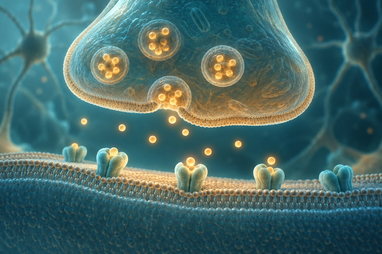
Interest in the compound is understandable in Alzheimer disease. Cholinergic projections from the basal forebrain, including the nucleus basalis of Meynert, contribute to cortical function and memory; damage to these networks reduces acetylcholine availability. By slowing the breakdown of the remaining transmitter, an inhibitor may enhance signaling in functioning synapses. It does not automatically replace lost neurons or address every cause of cognitive decline. Symptomatic effects, cellular markers and slowing disease progression therefore need separate discussion.
The molecule and its botanical source
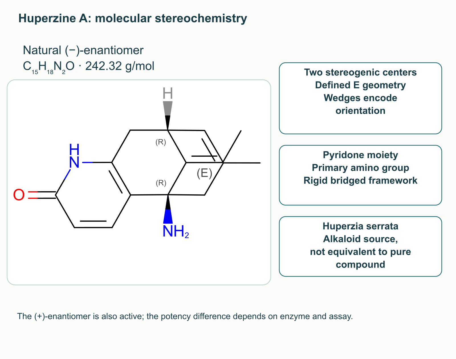
Huperzine A was isolated from Huperzia serrata, a lycophyte known in Chinese tradition as Qian Ceng Ta. It is a sesquiterpene alkaloid with the formula C₁₅H₁₈N₂O and a molar mass of approximately 242.32 g/mol. Its rigid bridged framework contains a pyridone moiety, a primary amino group, two stereogenic centers and an exocyclic double bond with defined geometry. An illustration needs an actual structural formula: a chain of arbitrary boxes conveys neither its molecular structure nor its stereochemistry.
The natural (−)-enantiomer is more potent in several cholinesterase assays, but the (+)-enantiomer is not entirely inactive. The difference depends on the enzyme and experimental conditions, so a study using another species’ enzyme cannot establish universal “human” constants for both forms. Similarly, a botanical extract, a purified alkaloid and a multi-ingredient supplement are not interchangeable. Extracts may contain other cholinesterase inhibitors, and the actual alkaloid content requires separate measurement.
Acetylcholinesterase architecture
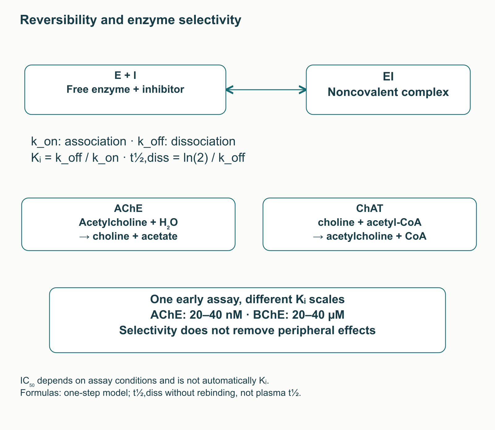
Acetylcholinesterase, or AChE, hydrolyzes acetylcholine to choline and acetate. This reaction terminates transmitter action, while choline returned to the nerve terminal can be reused for synthesis. Choline acetyltransferase transfers an acetyl group from acetyl-CoA to choline. Maintaining this substrate cycle does not mean that adding choline necessarily improves the clinical effects of huperzine.
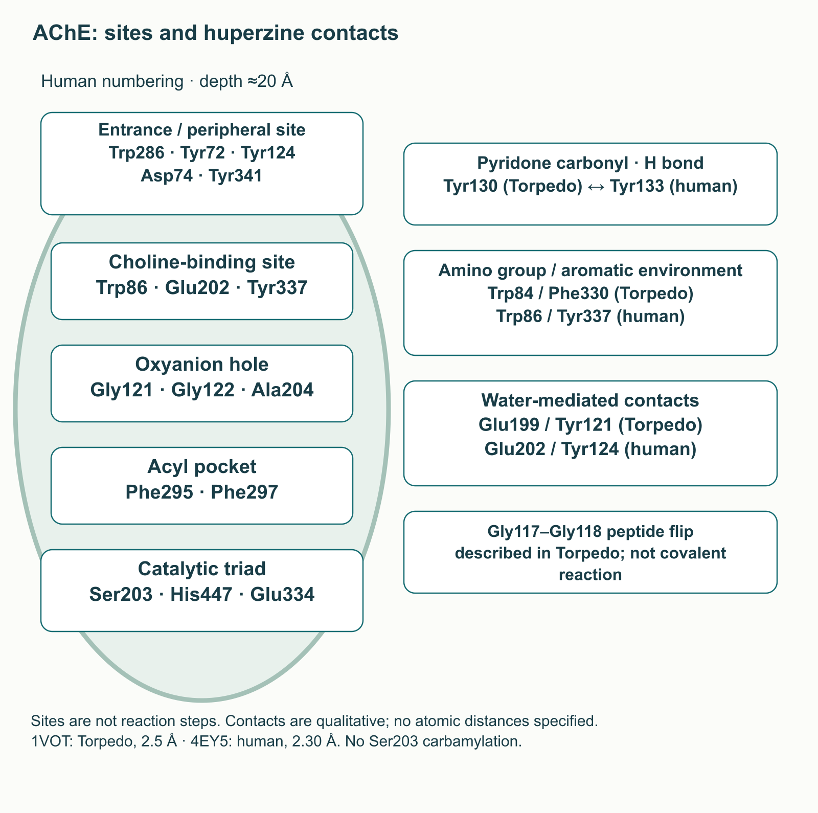
The AChE active center lies at the bottom of a narrow gorge roughly 20 Å deep. In the human enzyme, Ser203, His447 and Glu334 form the catalytic triad. Recognition of the substrate’s choline moiety involves Trp86, Glu202 and Tyr337. The oxyanion hole containing Gly121, Gly122 and Ala204 stabilizes a reaction intermediate, while Phe295 and Phe297 constrain the acyl pocket. A peripheral site near the entrance includes Trp286, Tyr72, Tyr124, Asp74 and Tyr341. The historical term “anionic” does not mean recognition is entirely mediated by negatively charged residues: aromatic interactions are important.
These sites perform different functions; they are not consecutive steps in a metabolic pathway. Butyrylcholinesterase, BChE, is a distinct enzyme rather than simply a peripheral form of AChE. Its more spacious acyl pocket is associated, among other features, with Leu286 and Val288 at positions corresponding to the aromatic AChE residues. This affects its substrates and inhibitors without making BChE a universal detoxification system.
Binding, reversibility and the meaning of constants
Crystal structures confirm noncovalent huperzine binding deep within the AChE gorge. An early complex with Torpedo californica enzyme was studied at 2.5 Å resolution; a human enzyme complex was later obtained at 2.30 Å. Residue numbering differs between species, and identical numbers do not guarantee equivalent positions. Descriptions of contacts therefore need to identify the enzyme concerned.
Structural studies highlight the pyridone carbonyl contact with Torpedo Tyr130, corresponding to human Tyr133, interactions of the protonated amino group with the aromatic environment of Torpedo Trp84/Phe330, and the participation of water. The corresponding human aromatic positions are Trp86/Tyr337. Water-mediated contacts involve the surroundings of Torpedo Glu199 and Tyr121, corresponding to human Glu202 and Tyr124. This network must not be replaced with an arbitrary direct amino-group–Glu202 salt bridge, nor should the inhibitor carbonyl be drawn as an ordinary oxyanion-hole substrate. A Gly117–Gly118 peptide-bond flip has also been described in Torpedo complexes. This is a protein rearrangement, not covalent attachment of the inhibitor to Ser203.
The minimal binding model is E + I ⇌ EI, where E is free enzyme, I is inhibitor and EI is their complex. The association constant k_on has units of concentration⁻¹·time⁻¹; the dissociation constant k_off has units of time⁻¹. For a simple one-step model, Kᵢ = k_off/k_on. Once rebinding is prevented, the time for the amount of complex to fall by half is t½,diss = ln(2)/k_off. These formulas describe molecular interactions rather than the duration of a tablet’s effects.
IC₅₀ is the concentration that reduces activity by half in a particular assay. It depends on substrate and enzyme concentrations, incubation time and measurement method, and cannot be equated with Kᵢ without specifying conditions. With tight binding, a substantial fraction of inhibitor can be enzyme-bound, so its free concentration differs from the amount added. An early mammalian cholinesterase study reported Kᵢ values around 20–40 nM for AChE and 20–40 µM for BChE. This illustrates selectivity in that experiment, rather than providing universal potency values for the human brain.
Compound
Chemical and enzymatic profile
Considerations for comparison
Huperzine A
Sesquiterpene alkaloid; reversible noncovalent inhibitor, predominantly of AChE in several assays
Clinical evidence and supplement quality differ from licensed medicines; certain mechanistic effects require high concentrations
Historically important, with a substantial hepatotoxicity problem; comparison does not demonstrate the absence of this risk for every other compound
IC₅₀ values from incomparable experiments cannot rank these drugs’ clinical efficacy. AChE selectivity also does not eliminate peripheral effects: AChE contributes to autonomic regulation and neuromuscular transmission. Enhancing acetylcholine signaling may affect cognition, digestion and heart rhythm at the same time.
Absorption, the barrier and elimination
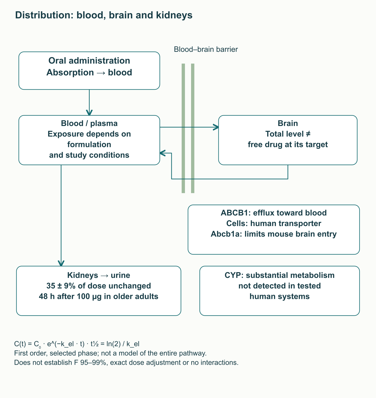
Huperzine A can be measured in blood after oral administration, but comparing different oral formulations establishes relative bioavailability rather than proving that 95–99% of a dose is absorbed. An early study of six volunteers given a single 0.99 mg dose found a peak at approximately 80 minutes and an elimination half-life around 4.8 hours. A later formulation study in healthy Chinese men reported a half-life of approximately 12.1 hours for the reference formulation. Different populations, doses, methods and models should not be collapsed into one mandatory regimen.
The blood–brain barrier is not merely a filter based on molecular size and lipophilicity. In cells expressing human ABCB1, huperzine was a substrate; in mice, deleting Abcb1a increased the brain-to-plasma concentration ratio. ABCG2 did not play a similarly predominant role in that study. The claim that huperzine does not interact with P-glycoprotein is therefore incorrect. Nevertheless, cells and knockout mice do not establish the magnitude of an interaction with a particular medicine in humans.
In a metabolism study involving older participants given a single 100 µg dose, approximately 35 ± 9% of the dose was recovered unchanged in urine over 48 hours. Under the tested human microsome and hepatocyte conditions, substantial metabolism, inhibition or induction of several CYP enzymes was not detected. This conflicts with claims of established predominant human metabolism through CYP1A2, CYP2D6 and CYP3A4. Unchanged urinary recovery is not a complete dose mass balance, and negative laboratory CYP findings do not exclude all interactions. Reduced renal function raises an exposure question worthy of clinical consideration, but these data do not yield a precise dose adjustment.
Elimination is often illustrated using C(t) = C₀·e^(−k_el·t) and t½ = ln(2)/k_el. C₀ is the concentration at the start of the selected phase and k_el its decline constant. The simplified model assumes first-order kinetics and does not automatically describe absorption, distribution or changing renal function. Plasma t½ differs from dissociation of the EI complex, and total brain levels differ from free concentrations at a receptor. None of these measures alone establishes dosing frequency or a “mandatory break.”
NMDA: an actual experiment and a concentration gap
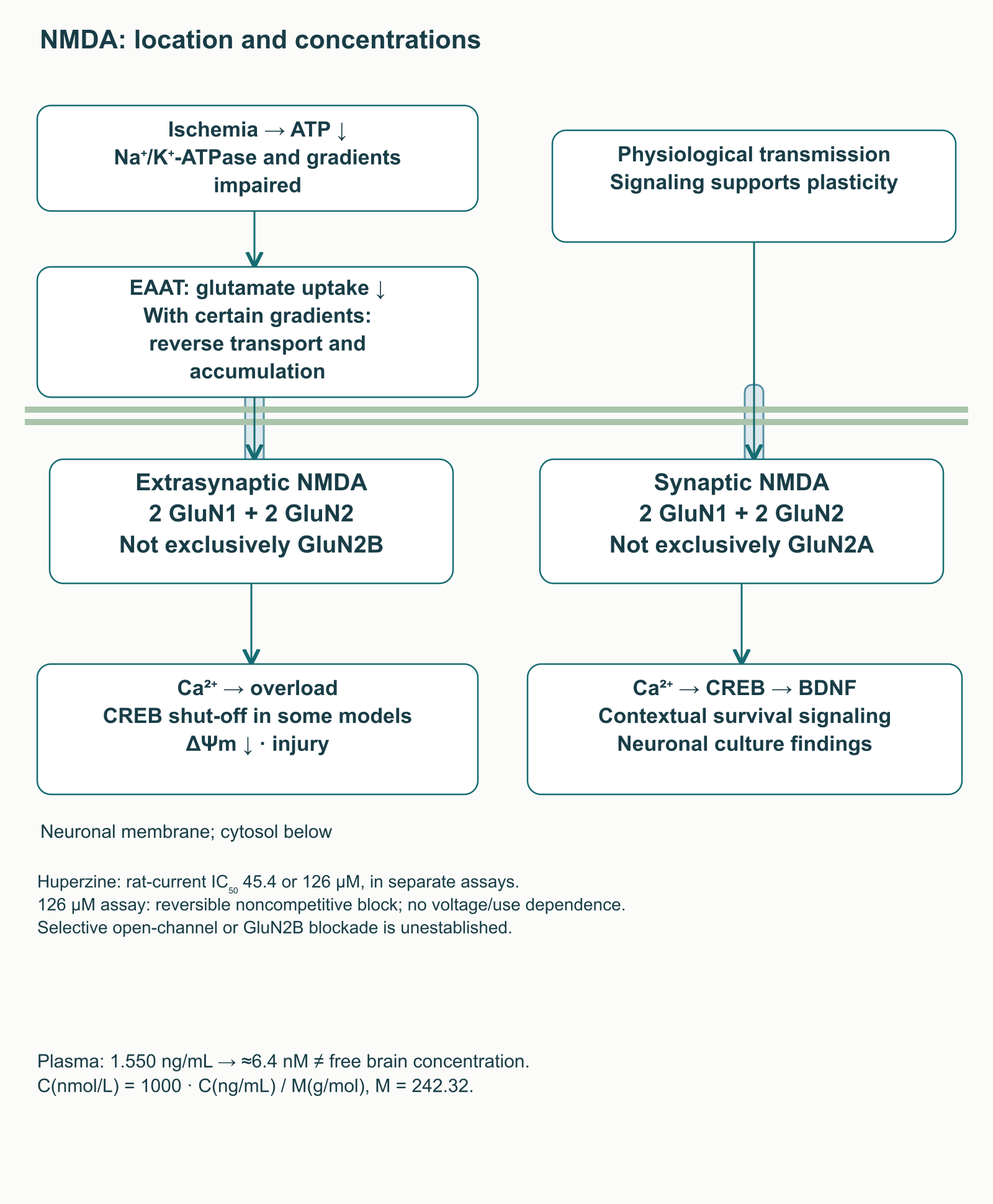
During ischemia, energy depletion impairs Na⁺/K⁺-ATPase function and ionic gradients. EAAT-mediated glutamate uptake becomes insufficient, with reverse transport possible under certain gradients. Prolonged NMDA receptor activation and Ca²⁺ influx can initiate damaging processes. Physiological NMDA signaling, however, is necessary for plasticity; suppressing all such transmission cannot itself be considered beneficial.
A conventional NMDA receptor contains two GluN1 and two GluN2 subunits, with some complexes containing different GluN2 subunits. The distinction between synaptic and extrasynaptic signaling cannot be reduced to “GluN2A is always beneficial, GluN2B is always harmful.” In neuronal cultures, synaptic stimulation supported CREB and BDNF expression, whereas certain extrasynaptic activation patterns switched off CREB and were associated with loss of mitochondrial potential. This is a context-dependent relationship, not a diagnostic map of every receptor in the human brain.
Huperzine reduced NMDA-induced currents in rat neurons, but at concentrations much higher than the nanomolar range typical for AChE inhibition. Current IC₅₀ was 45.4 µM in one experiment and 126 µM in another. The latter study found reversible, noncompetitive inhibition without detectable voltage or use dependence. Consequently, selective blockade of only open channels or only GluN2B cannot be drawn as an established mechanism. Changes in MK-801 radioligand binding in another system are also not equivalent to current inhibition.
In the oral formulation study, the mean peak plasma concentration of the reference product was 1.550 ng/mL. Conversion uses C(nmol/L) = 1000·C(ng/mL)/M(g/mol): with M = 242.32, this is approximately 6.4 nM. Comparing this with 45.4–126 µM reveals a gap of several orders of magnitude, but does not measure free drug in the brain. The practical conclusion is limited: direct NMDA effects in these experiments cannot be declared an established mechanism of benefit from ordinary oral use.
Mitochondria and apoptosis: two distinct membrane events
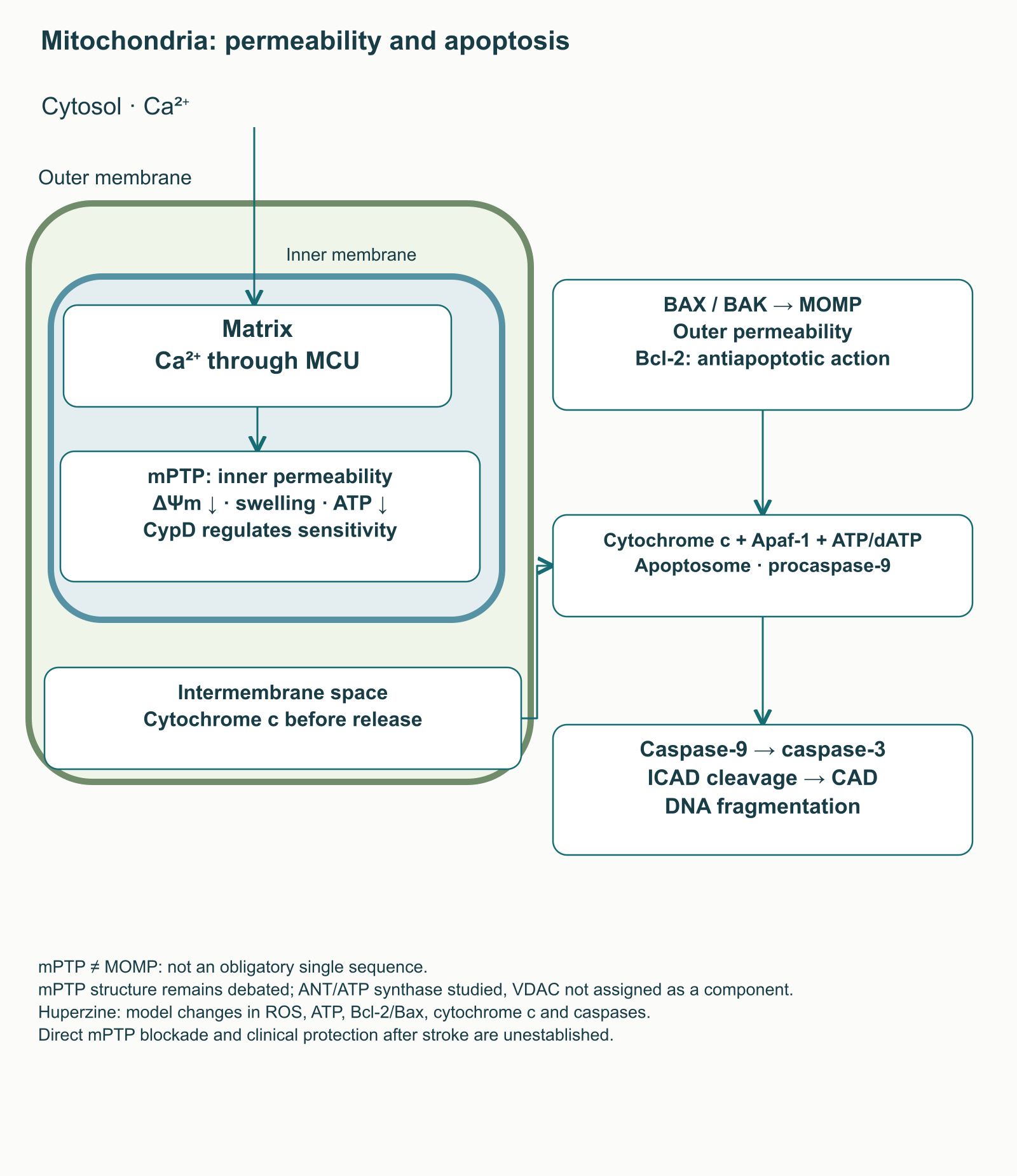
Ca²⁺ can move from cytosol into the mitochondrial matrix through the MCU complex in the inner membrane. Moderate uptake contributes to metabolic regulation, whereas calcium overload and oxidative stress can disrupt energy coupling. Opening the mitochondrial permeability transition pore, mPTP, affects the inner membrane: selectivity is lost, ΔΨm falls, and swelling and ATP depletion may follow. Current models do not justify portraying mPTP as a definitively established three-component structure of VDAC, ANT and cyclophilin D. CypD regulates sensitivity; the roles of ANT and ATP synthase components remain under investigation, and pore organization is still debated.
This should be distinguished from MOMP: increased outer mitochondrial membrane permeability involving BAX and BAK. Cytochrome c then leaves the intermembrane space for the cytosol. Together with Apaf-1 and ATP/dATP, it contributes to apoptosome assembly and procaspase-9 activation, followed by caspase-3 activation. Cleavage of the inhibitor ICAD releases CAD nuclease, which can fragment DNA. mPTP and MOMP can interact in injured cells, but they are not an obligatory single sequence in every form of apoptosis.
In serum-deprived rat cortical neurons, huperzine pretreatment was associated with changes in Bcl-2/Bax, less cytochrome c release and less caspase-3 activation. In PC12 cells exposed to the Aβ₂₅–₃₅ fragment, researchers reported reduced reactive oxygen species accumulation and improved energy measures. Mitochondrial studies after middle cerebral artery occlusion in rats also exist. They support an experimental hypothesis without proving direct huperzine binding to mPTP or reduced disability after human stroke.
Antioxidant responses and neurotrophic signaling
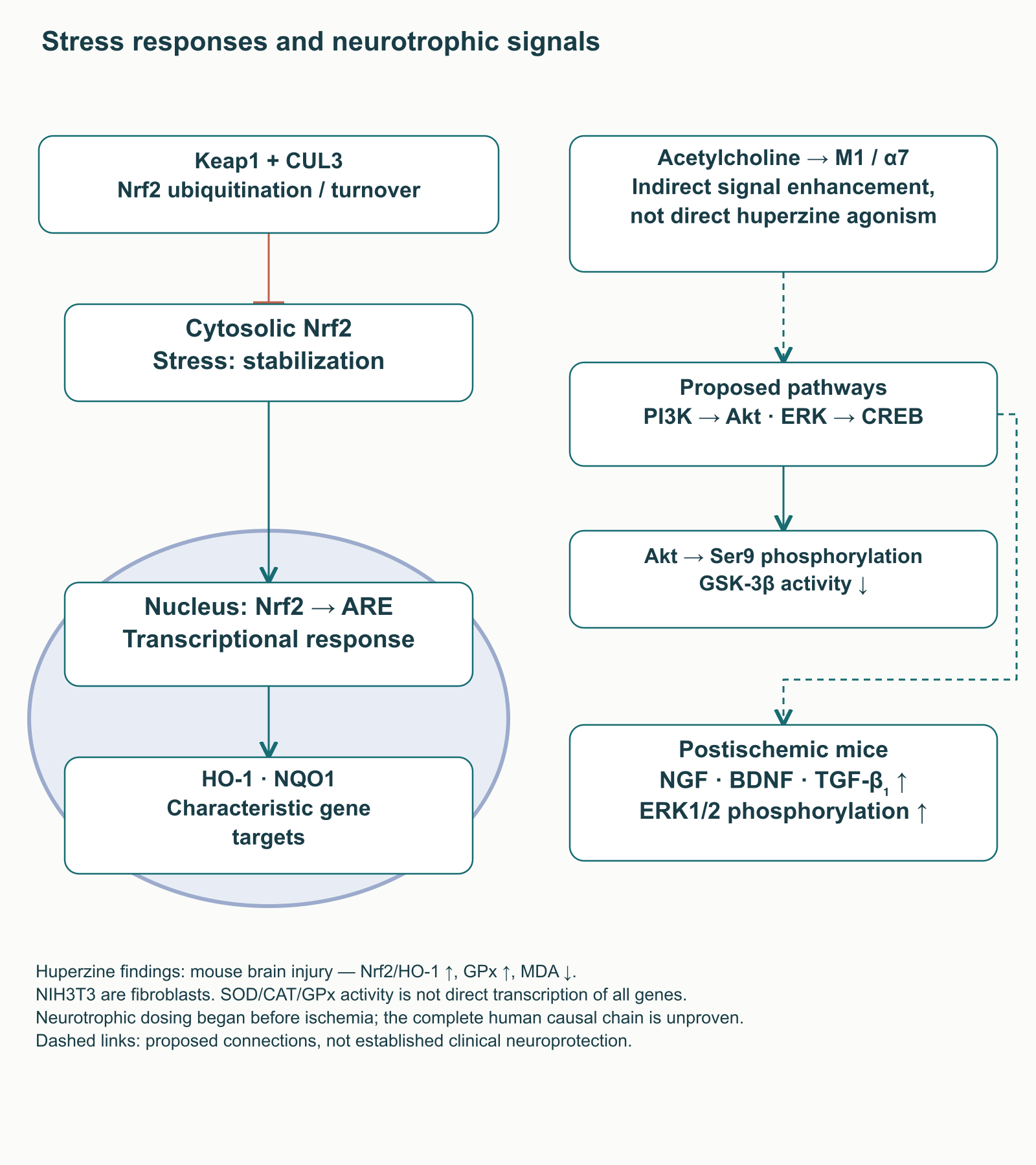
Nrf2 is a transcriptional regulator of cellular stress responses. Normally, Keap1 and the CUL3 system promote its ubiquitination and turnover; when the response is activated, Nrf2 accumulates in the nucleus and regulates genes through ARE elements. HO-1 and NQO1 are characteristic targets. Changes in SOD, catalase or glutathione peroxidase activity do not mean that every experiment has established direct Nrf2-mediated transcriptional regulation of all these enzymes.
In a mouse brain injury model, intraperitoneal huperzine administration was associated with increased nuclear Nrf2 and HO-1, increased glutathione peroxidase activity and reduced MDA, a lipid oxidation marker. Other data come from irradiated NIH3T3 fibroblasts, which are not human neurons. Such findings belong to a specific model, administration route and exposure duration. They do not establish supplement treatment for traumatic brain injury or huperzine as a direct universal Nrf2 activator.
The neurotrophic findings require similar care. After transient ischemia in mice, huperzine was associated with increased NGF, BDNF and TGF-β₁ and ERK1/2 phosphorylation; dosing began before surgery. Proposed M1/α7, PI3K–Akt, ERK and CREB pathways help formulate hypotheses about signaling and protein expression. Akt can also inhibit GSK-3β through Ser9 phosphorylation. However, a complete sequence from huperzine through every receptor and kinase to neuronal restoration has not been demonstrated in humans. Enhancing acetylcholine signaling differs from direct huperzine agonism at M1 or α7, and changes in BDNF or tau phosphorylation cannot replace a functional clinical outcome.
APP processing and the limits of the antiamyloid hypothesis
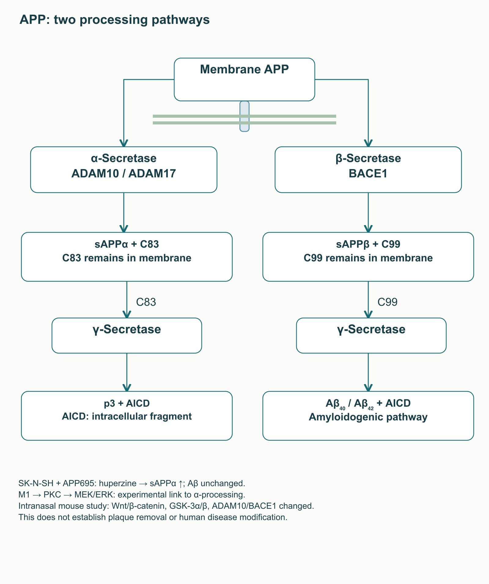
APP is a membrane-associated amyloid precursor protein processed through several routes. α-Secretase cleavage within the Aβ sequence produces soluble sAPPα and the membrane fragment C83; subsequent γ-secretase action produces p3 and the intracellular fragment AICD. BACE1 cleavage produces sAPPβ and C99, after which γ-secretase releases Aβ, including Aβ₄₀ and Aβ₄₂, and AICD. These routes require separate enzymes and products in a diagram rather than a single “less amyloid” arrow.
In human SK-N-SH neuroblastoma cells overexpressing APP695, huperzine increased sAPPα release. The effect depended on M1, PKC and MEK/ERK signaling and was associated with α-secretases, including ADAM10; ADAM17 also participated in relevant experiments. Aβ generation in that system did not change. Increased sAPPα therefore does not automatically prove reduced Aβ, much less disappearance of plaques in a patient.
A separate study in transgenic mice using prolonged intranasal administration reported changes in Wnt/β-catenin signaling, GSK-3α/β, ADAM10, BACE1 and amyloid measures. A different model and administration route may produce a different result without resolving the clinical question. Neither that experiment nor cellular APP processing establishes disease-modifying effects of oral huperzine in humans.
Clinical research findings
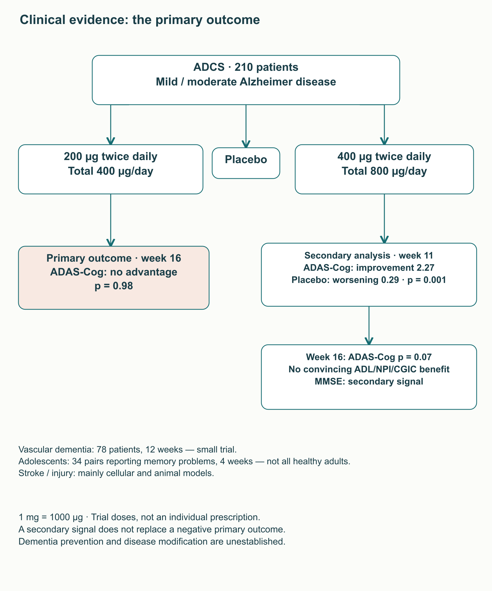
The most informative American placebo-controlled trial enrolled 210 patients with mild to moderate Alzheimer disease. Its primary question concerned 200 µg twice daily, or 400 µg/day, and change in ADAS-Cog at week 16. That prespecified endpoint showed no significant advantage over placebo. A secondary group received up to 400 µg twice daily, or 800 µg/day, following gradual escalation.
At week 11, this higher-dose group improved by 2.27 ADAS-Cog points compared with a 0.29-point deterioration with placebo; at week 16, the ADAS-Cog difference no longer reached the usual significance threshold. There was a secondary MMSE signal, but no convincing advantage in activities of daily living, neuropsychiatric symptoms or global clinical assessment. Selecting a dose, outcome and time after viewing results increases the risk of overinterpretation. These findings justify further research rather than demonstrating that doubling the dose ensures meaningful clinical benefit.
Systematic reviews present a more favorable overall picture but combine heterogeneous studies, often with methodological weaknesses. An early review included six trials and 454 participants; a broader 2013 review included 20 and 1,823. The 2014 analysis included eight Alzheimer trials with 733 participants: these numbers do not describe the 2013 review. Limitations in randomization, blinding, duration and publication selection reduce confidence. A pooled memory-test result is not evidence of preserved independence, quality of life or slower neurodegeneration.
Population
What was actually studied
Limits on practical interpretation
Alzheimer disease
Short trials and a larger study with a negative primary result; some secondary cognitive signals
Dementia prevention and disease modification remain unestablished; huperzine does not automatically replace standard treatment
Vascular dementia
A small 78-patient trial: 0.1 mg twice daily for 12 weeks, with positive cognitive and functional scale changes
One small sample does not establish a universal effect in all vascular disorders; long-term outcomes remain insufficiently studied
Stroke and brain injury
Mainly cellular and animal injury models
Animal lesion reductions cannot be transferred to human recovery or justify delaying emergency treatment
People without established dementia
An old, small four-week trial in 34 matched pairs of adolescents reporting memory problems
This does not establish safe intelligence enhancement in all healthy adults or recommend use in children
Trial doses and receptor adaptation
A published dose describes a research intervention rather than an individual prescription. Confusing units is particularly dangerous here: 1 mg = 1000 µg, so 0.1 mg is 100 µg. The American trial escalated doses according to protocol, with monitoring of participants and tolerability. It does not establish a universal ladder of “50 µg for attention, 200 for memory, 800 for neuroprotection,” or a safe regimen for healthy people. Nor does it establish a mandatory combination with a choline donor.
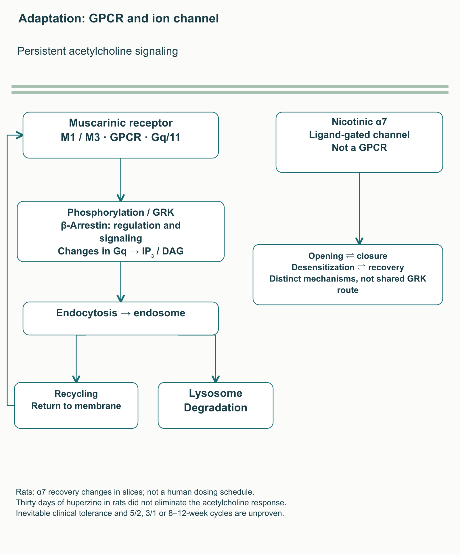
Persistent changes in cholinergic signaling can alter receptor sensitivity, but different receptor families adapt differently. Muscarinic receptors are GPCRs: under particular conditions, phosphorylation and interactions with GRKs and β-arrestins contribute to signaling changes and endocytosis. Receptors may return to the surface or undergo degradation. Arrestins can also participate in signaling themselves, so treating them simply as off switches is inadequate.
The nicotinic α7 receptor is a ligand-gated ion channel, not a muscarinic GPCR. Its desensitization should not be drawn as the same universal GRK–arrestin sequence. In rat hippocampal slices, AChE inhibitors changed acetylcholine exposure duration and α7 recovery from desensitization; this does not establish a fixed human schedule of breaks. Moreover, in rats, 30 days of huperzine administration did not eliminate the observed extracellular acetylcholine increase. Inevitable clinical tolerance therefore cannot be presented as established.
Neither “five days on, two days off,” alternating weeks, nor a mandatory break after 8–12 weeks has adequate direct support in these data. If tolerability changes or apparent benefit disappears, a clinician revisits the diagnosis, concomitant medicines, product and original goal. Self-directed escalation or cycling does not resolve uncertainty about efficacy.
Safety and interactions
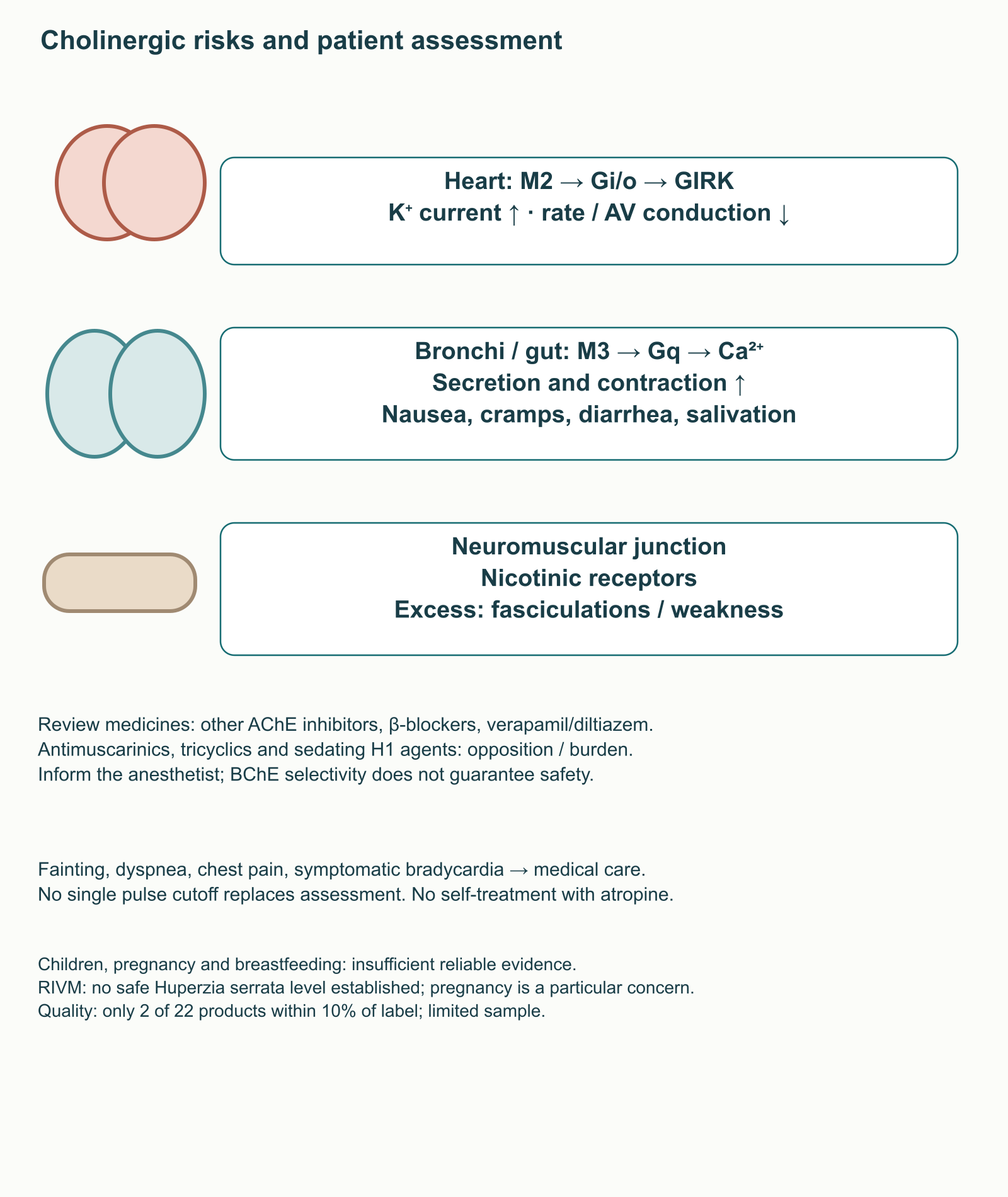
Adverse effects are consistent with increased cholinergic transmission: nausea, vomiting, diarrhea, abdominal cramps, salivation, sweating and sleep disturbance may limit tolerability. Via M2 receptors and Gi/o signaling, acetylcholine increases GIRK potassium current and can slow heart rate and atrioventricular conduction. M3/Gq/Ca²⁺ signaling increases secretion and smooth-muscle contraction, including in the bronchi. Marked excess can also produce nicotinic neuromuscular symptoms. AChE selectivity does not remove these risks.
Combination
Reason for caution
What a clinician should establish
Other cholinesterase inhibitors
Potential additive cholinergic effects
Duplication with donepezil, rivastigmine or galantamine; adding treatment without the treating clinician’s assessment is unjustified
β-Blockers
Potential additional slowing of heart rate
Metoprolol, bisoprolol, atenolol or other drugs; symptoms, baseline pulse and conduction abnormalities
Verapamil or diltiazem
Additive effects on rhythm and conduction
Indication, other rate-slowing medicines, and the need for clinical assessment and ECG
Atropine, scopolamine and other antimuscarinic agents
Pharmacodynamic opposition
Why they are prescribed; atropine for poisoning is a medical intervention, not a means of compensating for a supplement at home
Tricyclic antidepressants and sedating H1 antihistamines
Anticholinergic burden may impair cognition
The entire medication list and potential treatment review; huperzine is not a universal “antidote”
Succinylcholine and anesthesia
Anesthetic interactions are described for cholinesterase inhibitors; direct huperzine data are limited
Inform the anesthetist about the product, dose and last administration; preserving BChE does not guarantee no risk
These mechanistic and class-based warnings do not establish identical risks for every combination. Particular attention is warranted with bradycardia, sick sinus syndrome, AV conduction abnormalities, asthma or COPD, peptic ulcer disease, impaired urinary outflow and seizure disorders. Without a specific licensed product’s instructions, these should not be declared one international list of absolute contraindications. Reliable data for pregnancy, breastfeeding and unsupervised use in children are insufficient. RIVM’s assessment separately warns about Huperzia serrata preparations, particularly in pregnancy, and establishes no safe consumption level.
Fainting, profound weakness, breathing difficulty, chest pain or symptomatic slowing of heart rate require timely medical care. A single pulse threshold, such as 55/min, cannot replace assessment of symptoms, baseline status and ECG. The absence of serious events in a small short trial also does not exclude rare or long-term complications. Product quality matters: only two of 22 analyzed supplements contained huperzine within 10% of the labeled amount. The sample is limited, but illustrates why a label is not a dose measurement.
Applying this knowledge professionally
For memory complaints, a clinical question is a more useful starting point than a list of “nootropic” mechanisms. A clinician establishes onset and progression, effects on everyday functioning, sleep, mood, alcohol and other substances, sensory impairments and anticholinergic medicines. That picture guides investigation and treatment. Persistent or progressive decline, acute confusion and focal neurological symptoms need the appropriate medical pathway; supplements should not delay it.
If a patient already uses huperzine, the record should distinguish purified compound, Huperzia serrata extract and a combination product, and document active-ingredient mass, units, manufacturer, composition and possible medication duplication. Pulse, symptoms, renal function and ECG indications are assessed according to the clinical situation rather than an invented universal protocol. Benefit discussions should center on a specific goal, such as preserving a particular daily activity, with a preselected monitoring approach. A single test-score change may reflect practice, fluctuations or measurement characteristics.
When reading a paper, a clinician asks whether participants were human, which product and route were used, whether exposure is comparable, which endpoint was primary and whether functioning improved. These questions are especially important for huperzine: a well-established AChE mechanism does not erase a negative primary clinical result, and NMDA, Nrf2, APP and mitochondrial findings do not become proven brain restoration. Current dementia recommendations include other standard medicines; decisions about huperzine require assessment of evidence, the specific product’s status and individual risk. A defensible interpretation investigates promising mechanisms without promising universal neuroprotection or converting laboratory concentrations into human prescriptions.
FAQ
Is human neuroprotection established?
Reversible AChE inhibition is established. NMDA, Nrf2, APP and mitochondrial findings are largely experimental; a major clinical trial had a negative primary outcome.
Why is IC₅₀ not a tablet dose?
IC₅₀ belongs to specified laboratory conditions. Dose, plasma level, free brain concentration and clinical benefit are distinct measures.
Are fixed cycles and choline required?
Adequate direct evidence for fixed breaks or mandatory choline donors is lacking. Receptor adaptation does not establish a universal schedule.
Can huperzine be added to donepezil?
Unsupervised combination is unjustified because cholinergic effects may add up. The treating clinician should review medicines, symptoms and bradycardia risk.
Which symptoms require medical care?
Fainting, profound weakness, breathing difficulty, chest pain and symptomatic slowing of heart rate need timely medical assessment. A single pulse cutoff cannot replace it.