ADMA: metabolism, DDAH1, and cardiovascular risk — NutriFit
Asymmetric dimethylarginine: ADMA metabolism and vascular risk
ADMA release from methylated proteins, DDAH1 and DDAH2 catalytic evidence, AGXT2, arginine transport, and NOS inhibition. Clinical associations, measurement methods, and why a reference interval is not a treatment threshold.
Asymmetric dimethylarginine, ADMA, is an endogenous methylated derivative of L-arginine. It can competitively inhibit nitric oxide synthases, NOS, connecting protein turnover, amino-acid transport, and endothelial function. Higher concentrations occur in several cardiovascular, renal, and metabolic disorders. A biochemical mechanism, an association with clinical outcomes, and a clinically useful diagnostic test nevertheless represent different levels of evidence.
ADMA has a basis for investigation as both a marker and a possible participant in disease. Infusion experiments show that changing its concentration can affect hemodynamics, while observational studies associate it with certain outcomes. Neither establishes that one blood result explains a disease, calculates an individual's risk, or defines a treatment program. Distinguishing the plasma measurement from free ADMA in a particular cell close to NOS is especially important.
Chemical differences between ADMA, SDMA, and monomethylarginine
Arginine has a guanidino group at the end of its side chain; this is not a ring. Methylation of its terminal nitrogen atoms produces several derivatives. In ADMA, both methyl groups occupy the same terminal nitrogen. In symmetric dimethylarginine, SDMA, one occupies each terminal nitrogen. These are positional isomers with identical molecular formulas and different methyl-group arrangements. L-NMMA, monomethyl-L-arginine, contains only one methyl group.
Compound
Formula of the free base
Molar mass, g/mol
Important functional distinction
L-arginine
C₆H₁₄N₄O₂
174.20
Substrate for NOS and other metabolic pathways
L-NMMA
C₇H₁₆N₄O₂
188.23
Endogenous NOS inhibitor and DDAH1 substrate
ADMA
C₈H₁₈N₄O₂
202.25
Endogenous NOS inhibitor; metabolized by DDAH1 and AGXT2
SDMA
C₈H₁₈N₄O₂
202.25
Not a conventional direct NOS inhibitor; linked to amino-acid transport and kidney function
At physiological pH, the guanidino moiety is predominantly positively charged. Methylation changes hydrogen-bond donor availability, hydrophobicity, and protein recognition rather than simply eliminating charge. Thus equal masses do not imply equal biological effects. An analytical method must also distinguish ADMA from SDMA.
ADMA and L-NMMA can inhibit endothelial eNOS, neuronal nNOS, and inducible iNOS. Inhibition constants depend on the isoform, substrate concentration, and experimental conditions; one Ki is not a universal constant for every tissue. For SDMA, effects on arginine availability or inflammation observed in particular models must be distinguished from direct competitive inhibition of NOS.
Protein residues are methylated before free ADMA is released
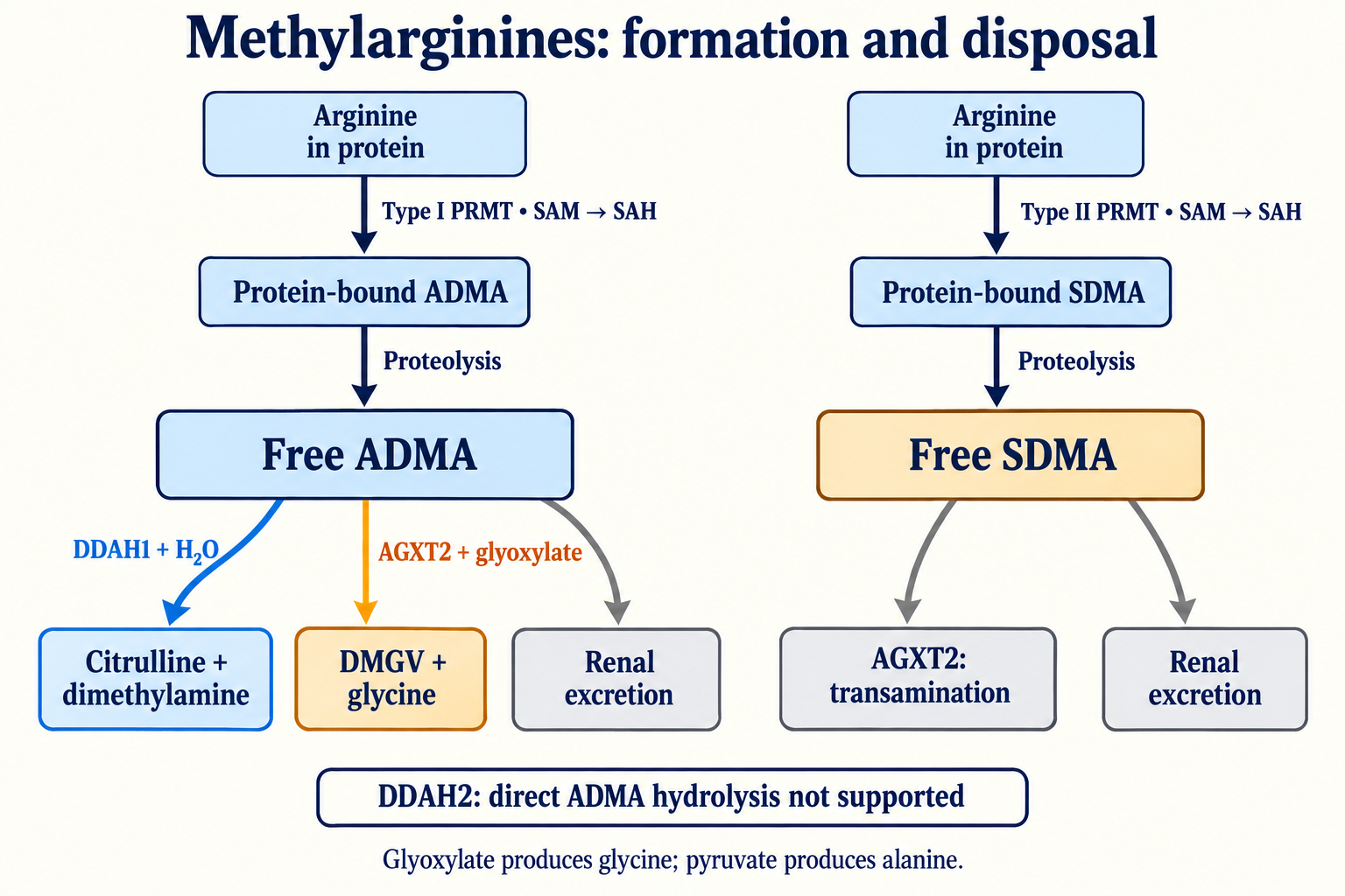
The principal source of ADMA is degradation of proteins containing methylated arginine residues. Protein arginine methyltransferases, PRMTs, transfer a methyl group from S-adenosylmethionine, SAM, to arginine within a protein. The donor becomes S-adenosylhomocysteine, SAH. This pathway does not directly convert free dietary or circulating arginine into free ADMA.
Type I PRMTs produce a monomethylated residue and subsequently asymmetric dimethylation. PRMT1 belongs to this group, but type I is not synonymous with PRMT1 alone: other members have distinct substrates and cellular roles. Type II enzymes, including PRMT5 and PRMT9, produce symmetric dimethylation through a monomethylated intermediate. PRMT7 is classified as type III, characterized by monomethylation (Zurita-Lopez et al., 2012); Yang and colleagues (2015) established PRMT9 as type II. Differences in protein-sequence recognition mean that a single recognition motif cannot be assigned to every PRMT.
Arginine methylation regulates protein interactions, RNA processing, transcription, chromatin, and signaling complexes. It is part of normal physiology. The amount of free ADMA ultimately released depends on more than PRMT expression: the methylated protein repertoire, protein synthesis and degradation, membrane transport, and subsequent methylarginine disposal all matter.
The discussion of SAM and methyl-group metabolism provides useful context for the origin of these groups. Increasing SAM availability does not linearly determine plasma ADMA. SAM/SAH balance, enzyme regulation, tissue, and protein turnover intervene. One-carbon metabolism and homocysteine metabolism intersect with this system without replacing it.
Proteolysis and the appearance of free ADMA
The ubiquitin–proteasome system degrades many intracellular proteins. Proteasomes generate peptide fragments; additional peptidases release amino acids and methylarginines. Lysosomal degradation and autophagy also contribute to protein turnover. Proteins and organelles delivered to lysosomes are broken down by hydrolases, including cathepsins.
Faster proteolysis may increase the precursor flux, but free ADMA concentration reflects the balance between formation and removal. Inflammation, catabolism, sepsis, and other severe illnesses can change several processes simultaneously: enzyme expression, organ function, transport, and distribution volume. An identical ADMA increase cannot therefore be predicted whenever protein degradation accelerates.
In a human metabolism and infusion study, Achan and colleagues (2003) estimated production at approximately 300 µmol of ADMA daily, with about 250 µmol undergoing DDAH-dependent metabolism. This is a historical estimate under particular conditions, not a fixed daily quantity for every person. Converting 300 µmol using ADMA's molar mass gives approximately 60.7 mg. A daily flux estimate is not the amount present in plasma at one instant.
A cellular pool can be represented by a balance equation:
dtdA=P−vDDAH1−vAGXT2−Jout+Jin.
Here A is the amount of free ADMA in the selected cellular compartment, P its production through proteolysis, v the metabolic reaction rates, and J the membrane fluxes. All terms require consistent units of amount per time. A concentration balance additionally requires the compartment volume. A plasma balance must incorporate tissue release, distribution, and excretion; cellular export alone does not destroy ADMA within the body.
In an extremely simplified model with constant production P and linear removal kA, the steady-state amount is P/k. Reducing k by 30% would increase the amount by 1/0.7, approximately 1.43-fold, rather than automatically two- to fourfold. Nonlinearity, alternative disposal pathways, and feedback may modify the actual response. A universal half-life or accumulation factor cannot be assigned without defining the model and conditions.
Transport and why plasma does not describe the NOS microenvironment
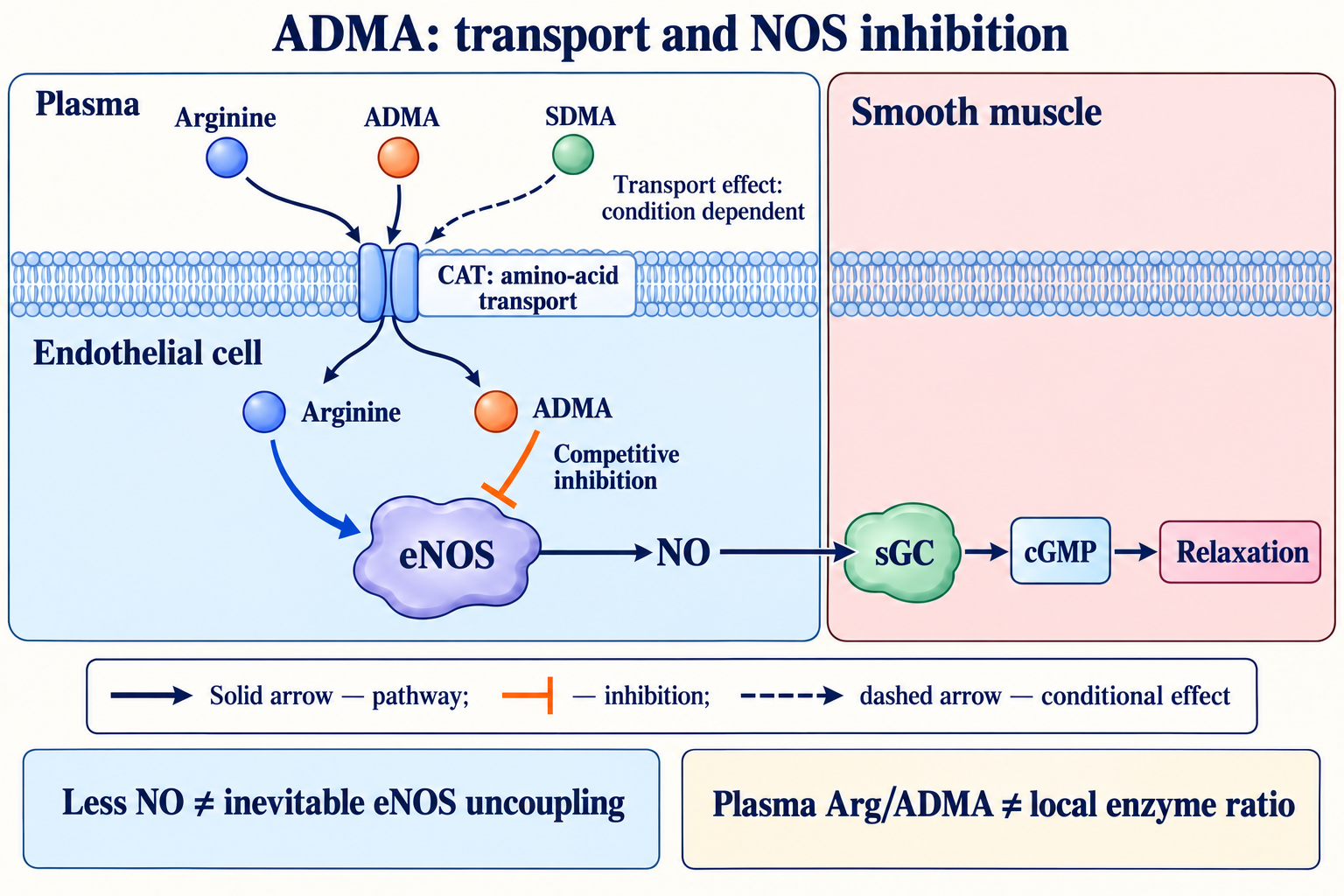
Cationic amino acids are transported by system y⁺ proteins, including CAT1, encoded by SLC7A1. CAT2A and CAT2B are variants of SLC7A2, rather than products of two separate genes; CAT3 is encoded by SLC7A3. Their tissue expression and kinetics differ. CAT1 is important in many cell types, including endothelium, while CAT2 variants participate in hepatic metabolism and responses of activated cells.
Transport is predominantly sodium independent and influenced by electrochemical conditions and concentrations on both sides of the membrane. Trans-stimulation can occur when an intracellular substrate accelerates uptake of an external substrate. However, treating the system as obligatory exchange of one arginine molecule for one ADMA molecule under every condition is an oversimplification.
Arginine and methylarginines interact with transport systems, but transport competition is not equivalent to NOS binding. SDMA can interfere with arginine availability in certain settings without acting as an equivalent direct NOS inhibitor. Submicromolar plasma concentrations cannot simply be substituted into experiments using much higher concentrations of transport competitors.
The arginine paradox describes the observation that additional arginine sometimes changes NO production despite total intracellular arginine exceeding the enzyme's Km. Possible explanations include compartmentalization, local transport complexes near eNOS, competition from methylarginines, and citrulline recycling. Total arginine in a cellular extract does not necessarily reveal the accessible substrate concentration at the active site.
DDAH1 and DDAH2 have different catalytic evidence
Dimethylarginine dimethylaminohydrolase 1, DDAH1, hydrolyzes ADMA and L-NMMA. ADMA yields L-citrulline and dimethylamine; L-NMMA yields L-citrulline and methylamine. SDMA is not a conventional substrate for this hydrolase. DDAH is an enzyme, not a transcription factor.
ADMA+H2ODDAH1L-citrulline+dimethylamine
Older schemes commonly linked DDAH1 mainly with neuronal NOS and DDAH2 with endothelial NOS, treating both proteins as equivalent ADMA hydrolases. Such a rigid division does not adequately reflect tissue distribution or current catalytic evidence.
Ragavan and colleagues (2023) compared recombinant proteins, cellular manipulations, and mouse models in a multicenter study. The convergent results did not support DDAH2's ability to metabolize ADMA, whereas DDAH1 was active. A reduction in ADMA following altered DDAH2 expression in older cellular studies therefore cannot establish direct hydrolysis by that protein.
DDAH2 remains biologically important. It has been linked to vascular remodeling, angiogenesis, signaling, and metabolic processes. These effects can be independent of direct ADMA metabolism or mediated through other systems. A protein's name does not establish its reaction, and a metabolite change after a genetic manipulation does not by itself identify the chemical mechanism. Evaluating the pathway requires catalytic measurements, control of other enzymes, and an appropriate model.
DDAH1 structure and reaction chemistry
DDAH has a repeated α/β architecture around a central catalytic region. A catalytic cysteine and other residues support nucleophilic chemistry, acid–base transfers, and active-site geometry. Cysteine attack on the guanidino carbon forms a covalent intermediate; release of a methylated amine and subsequent hydrolysis yield citrulline and regenerate the enzyme. This intermediate should not be casually described as an ordinary carbonyl thioester.
Human DDAH1 catalytic residues are commonly designated Cys274, His173, and Asp127 when numbering from the initial methionine; structural publications also use numbering offset by one residue. This must not be mixed with bacterial numbering or used to replace the human aspartate with the glutamate described in a bacterial enzyme.
Structural record 1H70 represents the C249S mutant of Pseudomonas aeruginosa DDAH with citrulline, associated with Murray-Rust and colleagues (2001). It is not a human DDAH1 structure. A human citrulline complex is represented by 2JAI, associated with Leiper and colleagues (2007). Species, mutation, and numbering matter when transferring structural conclusions about catalytic residues.
Michaelis–Menten kinetics provide a useful simplified description:
v=Km+[A]Vmax[A].
[A] is ADMA concentration in the reaction system, Km is a parameter of the enzyme preparation and conditions, and Vmax is the limiting rate for the specified amount of active enzyme. At concentrations well below Km, the dependence becomes approximately linear. Km, kcat, and pH effects measured with one recombinant preparation do not determine ADMA clearance in every tissue. Active protein abundance, oxidative modifications, substrate access, and alternative metabolism also matter.
Redox regulation and the limits of a feedback model
DDAH1's catalytic cysteine makes it sensitive to certain thiol modifications. Leiper and colleagues (2002) demonstrated S-nitrosylation and altered DDAH activity experimentally. Formation of an S-nitrosothiol requires appropriate nitrosating chemistry; it is not an obligatory direct reaction of every NO molecule with every thiol.
Thiol oxidation may involve a sulfenic form, mixed disulfides with glutathione, and other states. Some modifications can be reversed by cellular reducing systems; deeper oxidation may be difficult to reverse. The actual pathway, rate, and effect on protein degradation nevertheless require measurements. A simple stoichiometric illustration of an initial oxidation step is:
RSH+H2O2→RSOH+H2O.
This does not mean that all cellular DDAH1 reacts through that pathway or that every RSOH modification is irreversible. An unbalanced reaction in which a single peroxynitrite molecule immediately converts a thiol to a sulfonate while also producing nitrite is unsuitable for describing the chemistry.
Oxidative stress can reduce DDAH1 activity, while increased ADMA can restrict NO formation. Under particular conditions, this can sustain adverse feedback. The system is not required to increase geometrically: AGXT2, transport, organ metabolism, and protein repair remain operative. Effects of oxidized LDL, homocysteine, cytokines, or high glucose depend on the model and do not demonstrate identical suppression of every DDAH protein in every tissue.
GPX4 and membrane lipid peroxidation describe an adjacent redox mechanism. GPX4 reduces lipid hydroperoxides; the present pathway concerns arginine availability and enzymatic ADMA metabolism. Their intersection through oxidative stress does not make the pathways interchangeable or establish that increasing GPX4 activity necessarily normalizes ADMA.
AGXT2: mitochondrial metabolism and alternative amino acceptors
Alanine-glyoxylate aminotransferase 2, AGXT2, is a mitochondrial enzyme that requires pyridoxal phosphate, PLP, the active coenzyme form of vitamin B6. Expressed in kidney and liver, it has broad substrate specificity; β-aminoisobutyrate and other compounds are important substrates alongside methylarginines.
Unlike DDAH1, AGXT2 performs transamination rather than hydrolysis of the guanidino group. Transfer of ADMA's α-amino group to glyoxylate produces the corresponding α-keto acid, commonly called DMGV, and glycine. If pyruvate is the acceptor, it becomes alanine rather than glycine:
These are alternative reactions using different acceptors, not a sequence producing both amino acids simultaneously. SDMA can also undergo AGXT2-dependent metabolism to its corresponding symmetric α-keto derivative. Glycine is not a product of DDAH1. Its appearance in the glyoxylate reaction does not establish that oral glycine accelerates ADMA removal.
Rodionov and colleagues (2010) studied human AGXT2 experimentally and demonstrated lower ADMA and protection of NO formation from its inhibitory effect. This supports the pathway's existence without establishing a universal proportion of ADMA disposal through AGXT2 in all humans. AGXT2 variants may change several substrates; an association with SDMA or β-aminoisobutyrate cannot automatically be converted into a proven effect on ADMA or cardiovascular outcomes.
DMGV is measurable in biological fluids and is also investigated as a metabolic marker. It is not merely final harmless waste with a known elimination rate for every individual. Similarly, sufficient vitamin B6 supports PLP-dependent reactions, but raising its intake does not guarantee faster AGXT2 activity or clinical risk reduction.
Kittel and colleagues (2014) studied 400 healthy volunteers: AGXT2 variants rs37369 (Val140Ile) and rs16899974 (Val498Leu) affected SDMA and/or β-aminoisobutyrate, but neither variant affected plasma ADMA. This illustrates why a genetic enzyme change cannot automatically be translated into greater ADMA or clinical risk.
Kidney and liver combine metabolism with excretion
Some ADMA is excreted by the kidney, while a substantial amount undergoes enzymatic metabolism. The kidney contributes more than filtration: it participates in methylarginine metabolism. The liver can also extract and metabolize ADMA. Thus an increase in chronic kidney disease cannot be explained exclusively by reduced glomerular filtration.
SDMA is more closely related to kidney function and often rises as filtration falls. Nevertheless, AGXT2 metabolism prevents it from being treated as an absolutely nonmetabolizable compound always cleared by one route at a fixed 95% fraction. Production, distribution, and patient-specific circumstances affect concentration. In human medicine, SDMA does not automatically replace standard kidney assessment; veterinary criteria should not be transferred to humans.
Renal impairment also changes the uremic environment, protein turnover, inflammation, and enzyme activity. Together, these may promote ADMA accumulation and connect renal dysfunction with endothelial abnormalities. Effects of vascular function on renal perfusion make the system bidirectional. No fixed ADMA fold increase universally defines a kidney-disease stage, and the ADMA/SDMA ratio cannot independently locate a metabolic block in the liver or kidney.
Competitive NOS inhibition and the arginine paradox
In a simplified competitive model, arginine and ADMA compete for enzyme binding. The initial rate is:
[S] denotes available L-arginine, [I] ADMA, and Ki the inhibition constant in that system. Apparent Km increases, whereas Vmax remains unchanged in this simple model when substrate saturation is sufficient. This is a description of isolated kinetics, not a guarantee of unchanged maximal NO production in a living cell. Transport, NOS abundance and condition, cofactors, oxygen, reducing equivalents, and NO consumption intervene.
NOS converts L-arginine into NO and citrulline through N-hydroxy-L-arginine. Electron transfer proceeds from NADPH through flavin centers to heme and requires suitable cofactor conditions, including tetrahydrobiopterin, BH4. For eNOS, calcium, calmodulin, phosphorylation, and localization are important. Describing the entire cycle as ADMA simply blocking oxygen access to iron or necessarily displacing BH4 is not justified.
Increasing arginine can weaken competitive inhibition under certain conditions. That does not establish supplementation benefit: transport is limited, arginases and other pathways use arginine, and the local substrate/inhibitor ratio can differ from the plasma ratio. A change in NO metabolites is likewise not equivalent to fewer infarctions or deaths.
Lower NO production is different from NOS uncoupling
During productive eNOS activity, electron flow supports NO synthesis. BH4 deficiency or oxidation, thiol modifications, and certain other conditions can divert part of the flow toward superoxide; this is called NOS uncoupling. ADMA can participate in an environment associated with endothelial dysfunction, but inhibiting NOS does not automatically make every enzyme molecule produce superoxide.
Depending on conditions, an inhibitor may reduce some NOS-dependent radical fluxes as well as NO. The scheme “ADMA occupies the site, therefore all eNOS immediately becomes a superoxide source” is not universal. Demonstrating uncoupling requires appropriate measurements and control of alternative reactive-oxygen sources, including NADPH oxidases and mitochondria.
NO reacts with superoxide to form peroxynitrite:
NO∙+O2∙−→ONOO−.
This both consumes bioavailable NO and produces a reactive species. Oxidation of BH4 and proteins may intensify impaired vascular signaling, but the effect depends on the site of formation, antioxidant systems, and competing reactions. Plasma ADMA does not directly measure endothelial superoxide, BH4 content, or peroxynitrite production.
Vascular signaling and hemodynamics
NO activates soluble guanylate cyclase in smooth-muscle cells, increasing cGMP and influencing vascular relaxation. It also contributes to platelet regulation, leukocyte–endothelial interactions, and vascular remodeling. Reduced NO availability can increase flow resistance and alter endothelial protective functions. Sympathetic activity, endothelin, prostanoids, local metabolites, and other mechanisms nevertheless regulate vascular tone simultaneously.
Achan and colleagues (2003) performed a randomized, double-blind infusion experiment in 12 healthy men. Compared with placebo, ADMA altered heart rate, cardiac output, mean blood pressure, and systemic vascular resistance. This is direct human evidence that changing ADMA can affect hemodynamics under defined conditions. It does not describe lifelong effects of naturally elevated ADMA or determine an individual risk boundary from one blood test.
Renal and cerebral microcirculation are sensitive to impaired NO signaling without depending on that pathway alone. An identical fall in arteriolar diameter or cerebral perfusion cannot be derived for every patient from one ADMA concentration. NO availability, blood pressure, and local autoregulation can affect organs differently.
Endothelial senescence and inflammatory signals
Scalera and colleagues (2004) studied cultured endothelial cells repeatedly exposed to ADMA. Under these conditions, senescence-associated β-galactosidase and telomere shortening increased; telomerase activity declined, redox measures changed, and MCP-1 and interleukin-8 secretion were affected. This links ADMA with an experimental endothelial phenotype rather than proving that plasma ADMA accurately measures the biological age of human vessels.
DNA-damage responses, cell-cycle arrest, and the senescence-associated secretory phenotype provide a framework for discussing chronic stress. Every p53/p21 step, all SASP components, and a precise loss of telomerase activity in humans cannot be attributed to one experiment unless directly measured. Cell culture differs from an intact vessel in surrounding cells, flow, repair processes, and exposure duration.
Likewise, an inflammatory association does not make ADMA the sole cause of NF-κB activation or monocyte recruitment. Inflammation may precede the ADMA change or accompany a common underlying disorder. Intervention direction and control of alternative mechanisms matter.
Findings from cardiovascular studies
Results depend on population, outcome, and assay. High ADMA in patients with established disease may have a different prognostic value from the same measurement in the community. A hazard ratio comparing extreme thirds of a distribution cannot be converted directly into an individual's absolute event probability.
Primary study
Design and population
Meaning of the finding
Limit of inference
Schulze et al., 2006, CARDIAC
Multicenter case–control study: 131 coronary-disease cases and 131 matched controls
Association with existing coronary disease
Not a prospective dialysis cohort or intervention trial
Schnabel et al., 2005, AtheroGene
Prospective coronary-disease cohort; ADMA measured in 1874 participants
Higher baseline concentrations associated with cardiovascular death or nonfatal infarction
Association in established disease does not define a universal threshold
Böger et al., 2009, Framingham
3320 community participants followed for about 10.9 years
ADMA and Arg/ADMA associated with all-cause mortality
ADMA, arginine, and their ratio were not associated with incident cardiovascular disease
Tang et al., 2008
132 patients with chronic systolic heart failure
Methylarginines related to diastolic measures; ADMA associated with adverse outcomes
Small specialized cohort; association does not prove benefit from ADMA-guided treatment
Dückelmann et al., 2008
118 patients with acute decompensated heart failure
ADMA did not identify subsequent clinical events; NT-proBNP was informative in this sample
A negative finding demonstrates dependence on the clinical setting
In AtheroGene, the highest versus lowest ADMA third had HR 2.48, 95% CI 1.52–4.06; the association persisted in models including additional biomarkers. This compares risk within a study, rather than establishing that ADMA above 0.7 doubles everyone's risk. In Framingham, the adjusted association with all-cause mortality was HR 1.21 per standard deviation, 95% CI 1.07–1.37, but incident cardiovascular disease was not associated. Different outcomes should remain distinct instead of being merged into a universal prediction claim.
Positive findings in coronary disease, after vascular interventions, or in heart failure support continued biomarker research. They do not mean that ADMA replaces troponin in acute myocardial injury, NT-proBNP in heart-failure assessment, or established chest-pain evaluation. Clinical implementation requires reproducibility, external validation, added value beyond an accepted model, and evidence that the result changes decisions to patients' benefit.
Heart, kidney, and brain: related but distinct consequences
In heart failure, reduced NO signaling may affect vascular loading, myocardial relaxation, and pulmonary resistance. Cellular cGMP, calcium-handling, and contractile-protein pathways make the relationship biologically plausible. They do not establish that every case of diastolic dysfunction results from direct ADMA action on SERCA2a or that ADMA is uniformly two- to threefold elevated in all heart-failure forms. Kidney function, treatment, severity, and comorbidities influence it.
In renal disease, elevated ADMA can reflect impaired metabolism and contribute to endothelial dysfunction. Studies in renal failure associate it with outcomes, but blood pressure, fluid balance, lipids, anemia, mineral metabolism, and the underlying disorder also account for substantial risk. A laboratory decrease does not demonstrate improvement in those outcomes.
Neuronal and endothelial NOS participate in nervous-system signaling and neurovascular coupling. ADMA can interact with these mechanisms, but plasma concentration is not its concentration in a neuron or cerebral endothelial cell. Pikula and colleagues (2009) associated ADMA with certain MRI signs of vascular brain injury in Framingham. These observational findings do not establish that ADMA directly causes white-matter disease, impaired glymphatic transport, or dementia in a particular patient.
Insulin resistance, hyperglycemia, and homocysteine
Stühlinger and colleagues (2002) found an association between plasma ADMA and impaired insulin-mediated glucose disposal. In part of the study, pharmacological improvement in insulin sensitivity accompanied a reduction in ADMA. This is consistent with overlapping metabolic and vascular pathways without establishing causal direction in every patient or making ADMA an independent diagnostic criterion for diabetes.
High glucose can change reactive-oxygen production, PKC signaling, DDAH1 expression or activity, and other processes in experimental models. DDAH1 remains an enzyme, rather than a transcription factor initiating synthesis of free ADMA. Greater free ADMA can involve protein methylation, degradation, and reduced disposal. Kidney function and treatment add further variation; diabetes does not produce an identical ADMA elevation in everyone.
Homocysteine is related to the methionine cycle, the redox environment, and vascular measures. Methionine-loading experiments have demonstrated ADMA and endothelial-function changes, but these are specific experimental conditions rather than ordinary food intake. Folate and B12 support homocysteine remethylation, while B6 supports transsulfuration and other PLP-dependent reactions. Correcting confirmed deficiency has its own value. Lowering homocysteine does not automatically demonstrate normalized ADMA or prevention of cardiovascular events through this pathway.
Measurement: chromatography, mass spectrometry, and ELISA
ADMA is measured using liquid chromatography with fluorescence or mass-spectrometric detection, and immunoassays. Validated LC-MS/MS can quantify arginine and methylarginines together if selectivity, calibration, matrix effects, and accuracy are demonstrated. ADMA and SDMA have the same mass, so a shared precursor signal alone does not distinguish them.
Chromatographic resolution and product-ion selection must suit the actual procedure. An ADMA transition near m/z 203→46 may detect dimethylammonium, whereas an SDMA signal near 203→172 reflects loss of a neutral fragment, not methylammonium with a mass of 172. These examples are not universal instrument settings. Isotope-labeled internal standards help account for losses and matrix effects without eliminating the need for validation.
Derivatization is an option rather than an obligatory part of every LC-MS/MS method. The HILIC approach described by Martens-Lobenhoffer and Bode-Böger demonstrates a chromatographic alternative to claims that every assay requires a butyl ester. Quantification limit, coefficient of variation, and run time belong to a particular procedure; they are not fixed properties of an ADMA test.
Competitive ELISA is convenient for large studies, but antibodies, sample preparation, cross-reactivity, and matrix influence results. A possible bias relative to chromatography cannot be expressed as one compulsory percentage correction for all kits. Even weak cross-reactivity with a much more abundant compound can matter; the actual effect requires evaluation of the specific kit. Different platforms should not be compared against one threshold table without checking comparability.
Interpretation requires units, specimen type, storage conditions, method, and laboratory reference information. Longitudinal comparisons ideally use the same validated procedure. Time of day, food, acute illness, and medication can affect context, but this does not establish a validated circadian treatment protocol or one universal sampling hour.
A reference interval is different from a disease boundary
Schwedhelm and colleagues (2009) established LC-MS/MS ADMA reference values in 1126 nonsmoking Framingham participants without clinical cardiovascular disease, hypertension, diabetes, or obesity. Mean concentration was 0.52 µmol/L, with SD 0.11; the 2.5th and 97.5th percentile limits were 0.311 and 0.732 µmol/L.
These describe a distribution in a selected sample measured by a particular method. They do not define optimal, predisease, severe-risk, or compulsory-supplement categories. Someone outside an interval does not necessarily have disease, and someone inside is not necessarily protected against an event. Age, illness, assay, and the clinical question matter more than applying one universal scale.
Plasma Arg/ADMA is calculated using consistent units:
R=[ADMA][Arg],0.5μmol/L60μmol/L=120.
The ratio is dimensionless. It represents the balance of the measured components, not a direct measurement at the eNOS active site. A value below 100 does not imply inevitable NOS uncoupling. Another research measure, the global arginine bioavailability ratio, Arg/(ornithine+citrulline), includes metabolic products. The names, formulas, and interpretations must remain distinct.
Measure
Question it can help investigate
What it does not independently establish
ADMA
Balance of formation and disposal of an endogenous NOS inhibitor
Disease cause, degree of eNOS uncoupling, or a personal treatment regimen
SDMA
Methylarginine metabolism and relation to kidney function
Direct NOS inhibition, exact GFR, or a universal diagnosis
L-NMMA
Another endogenous inhibitor and protein turnover
An ADMA equivalent with the same mass and identical thresholds
L-arginine
Substrate availability in the measured specimen
Local NOS availability or expected supplementation benefit
Arg/ADMA
Balance of two plasma components
Compulsory vascular damage below one boundary
ADMA/SDMA
Different profiles of two isomers
Location of a DDAH1, AGXT2, liver, or kidney defect
Citrulline
Intestinal–renal metabolism and several production pathways
The specific rate of DDAH1 or NOS alone
Homocysteine
One-carbon metabolism in context
ADMA production or the need for an identical vitamin regimen
Arginine and citrulline: a substrate hypothesis with clinical limits
Arginine comes from food and endogenous synthesis. Oral availability depends on intestinal and hepatic metabolism, and arginase competes for its use. Citrulline participates in the intestinal–renal axis and can supply arginine; this is a separate pathway rather than direct destruction of ADMA.
Argininosuccinate synthetase, ASS1, combines citrulline with aspartate, consuming ATP to AMP and pyrophosphate. Argininosuccinate lyase, ASL, subsequently produces arginine and fumarate:
Conversion depends on tissue and enzyme availability. Claims that citrulline always avoids every loss and guarantees greater local eNOS substrate are too strong. In a double-blind crossover trial, Schwedhelm and colleagues (2008) compared week-long arginine and citrulline regimens in 20 healthy volunteers. Citrulline increased plasma arginine and certain NO-related signaling measures, but no regimen improved FMD over baseline. Pharmacokinetic changes do not establish improved cardiovascular outcomes in patients.
VINTAGE MI, Schulman and colleagues (2006), is particularly instructive. It randomized 153 patients after a first ST-elevation myocardial infarction to L-arginine or placebo for six months. Expected improvements in vascular stiffness and ejection fraction were not demonstrated; six deaths occurred in the arginine group versus none with placebo, prompting enrollment closure. This limits the translation of substrate logic into post-infarction treatment. It does not demonstrate identical harm from dietary arginine or citrulline in every population, but it rules out automatic supplementation solely to improve Arg/ADMA.
Thiols, antioxidants, and vitamin cofactors
N-acetylcysteine can provide cysteine for glutathione synthesis. It is not itself an enzyme catalyzing that synthesis and is not established as a universal means of restoring DDAH1 in every patient. A change in cellular redox conditions and clinical risk reduction are separate outcomes.
Alpha-lipoic acid and its reduced form are discussed in relation to thiol chemistry and redox regulation. A free supplement differs from a lipoyl residue covalently attached to energy-metabolism enzymes. Thermodynamic feasibility does not guarantee delivery of the necessary amount to DDAH1's catalytic cysteine in a tissue. A diagram of enzyme oxidation cannot set a clinical dose.
Ascorbate can affect redox processes, including preservation of NOS cofactor conditions in some models; tocopherols help interrupt lipid-oxidation chains. Selenium is incorporated through selenocysteine into selenoproteins, including certain glutathione peroxidases, rather than acting as a free metal cofactor for every antioxidant enzyme. GPX enzymes differ in substrates and localization. These mechanisms do not establish that a mixture of vitamins and selenium selectively removes ADMA or is safe at any dose.
Folate, B12, and B6 status should be considered in relation to diet, illness, and clinical indications. Deficiency and excess are different circumstances; prolonged high-dose B6 can cause neuropathy. Neither PLP-dependent AGXT2 nor a homocysteine–ADMA association establishes the same high-dose vitamin combination for everyone.
Pharmaceutical and plant compounds: mechanisms do not select treatment
PPARγ signaling can change metabolic conditions and protein expression, and some studies have explored its relationship to ADMA. Telmisartan blocks the AT1 receptor and has additional experimental properties; thiazolidinediones affect insulin sensitivity. Hypothesized DDAH regulation does not make either a universal first choice for elevated ADMA. Indications, contraindications, and demonstrated clinical outcomes belong to the disease and specific drug.
Statins lower LDL and cardiovascular risk in appropriate populations. Their effects on ADMA can differ across studies; a fixed 20% reduction is not an obligatory class effect. Changes in DDAH1 or PRMT in a cell model do not replace established lipid mechanisms and clinical trial evidence.
FXR-dependent regulation of hepatic metabolism and other transcriptional pathways remain research topics. Altering DDAH1 expression in a model does not create an established indication to use an FXR agonist for ADMA reduction. Tissue, exposure, safety, and clinically meaningful outcomes require evidence.
Resveratrol, curcumin, and other polyphenols have proposed antioxidant, inflammatory, and signaling effects. Cell and animal results depend on concentration and bioavailability. They do not establish a universal percentage reduction in ADMA or prevention of human events. Combination products make it harder to identify the responsible component; composition, interactions, and tolerability matter independently.
Activity, diet, and NOS-independent NO production
Blood flow and shear stress affect endothelial signaling, including eNOS, Akt, and transcriptional regulation. Physical activity improves several cardiovascular-health measures, but ADMA responses depend on population and intervention. A 15–20% reduction after a fixed number of weeks is not obligatory. Increased DDAH2 expression after exercise also does not demonstrate more direct ADMA hydrolysis by that protein.
Diet quality, adequate protein and micronutrient intake, avoiding smoking, and managing metabolic factors matter in broader prevention. Their benefit need not depend entirely on ADMA. A Mediterranean dietary pattern or reducing certain processed foods is not a standardized methylarginine detoxification program with a guaranteed laboratory result.
Dietary nitrate can contribute to nitrate–nitrite NO formation, partly bypassing NOS. This depends on nitrate processing, microbial activity, and tissue conditions. It provides an alternative signal source rather than directly removing ADMA. It cannot be assumed to compensate fully for every eNOS defect or remove the need to treat hypertension and other illnesses.
Daily regulation of metabolism, vascular function, and activity makes chronobiology an interesting research subject. A morning substrate/evening antioxidant scheme providing round-the-clock ADMA protection is not supported as a universal protocol. Medication timing should follow the agent's own evidence and clinical purpose rather than an imagined sequence of molecular reactions.
Using ADMA knowledge in clinical assessment
An elevated measurement is considered alongside kidney and liver function, blood pressure, lipids, glycemia, inflammation, and clinical findings. Method, laboratory reference interval, sampling circumstances, and the reason for testing should first be clarified. An unexpected result does not independently justify a supplement bundle or replacing necessary treatment.
Clinical question
Information that supports decisions
ADMA's possible role and limit
Overall cardiovascular risk
Age, pressure, smoking, lipids, diabetes, family and clinical history; an appropriate validated model
Potential additional research marker without a universal treatment threshold
Renal dysfunction
Creatinine, estimated GFR, albuminuria, and cause of injury
May reflect altered metabolism; does not independently stage disease
Acute chest pain or decompensation
Clinical evaluation, ECG, accepted biomarkers, and indicated imaging
Does not replace troponin, NT-proBNP, or urgent assessment
Possible nutrient deficiency
Diet, symptoms, risk factors, and relevant laboratory findings
Does not automatically establish B6, folate, B12, or antioxidant deficiency
Response to intervention
Prespecified clinical outcome, tolerability, and a validated measure
Lower ADMA alone does not establish improved prognosis
Flow-mediated dilation and pulse-wave velocity help investigate vascular function but depend on protocol, equipment, blood pressure, age, and other factors. A single FMD percentage is not a universal boundary for ADMA action. Greater vascular stiffness also does not identify which metabolite caused it. The connection among a blood test, functional assessment, and treatment decision needs separate validation.
Preparation method and evidence boundaries
This is a narrative scientific examination of ADMA, with targeted verification of disputed mechanisms, structural records, and clinical findings on 8 October 2026. Primary bibliographic records, accessible abstracts and indexed passages of primary publications, and open RCSB structure pages were used. Some full texts were unavailable; the corresponding conclusions rely on accessible primary records and abstracts.
An exhaustive systematic search, independent GRADE assessment, formal external peer review, and clinical protocol validation were not conducted. Numbers, designs, and publication names are included where the corresponding records supported verification; unsupported universal cutoffs and treatment promises were removed. The strongest conclusions concern ADMA release from methylated proteins, direct DDAH1 hydrolysis, alternative AGXT2 metabolism, and ADMA's ability to inhibit NOS. Prognostic utility, local compartment effects, and benefits of specifically changing ADMA in humans remain questions for defined populations and clinical studies.
FAQ
Are ADMA and SDMA the same compound?
They are positional isomers of equal mass with different methyl-group arrangements. ADMA directly inhibits NOS; SDMA is not a conventional direct inhibitor and is more closely related to kidney function.
Does DDAH2 break down ADMA?
A modern multicenter study did not demonstrate direct ADMA hydrolysis by DDAH2. Its biological effects should be distinguished from DDAH1 catalysis.
Is there a universal dangerous ADMA concentration?
This evidence does not establish one universal diagnostic or treatment boundary. A reference interval, study threshold, and individual risk have different meanings.
Should elevated ADMA be treated with arginine?
The measurement alone does not justify supplementation. Competitive kinetics do not guarantee clinical benefit; negative findings and the safety signal in VINTAGE MI after infarction are particularly relevant.
Can LC-MS/MS and ELISA use one threshold table?
Not without verifying comparability. Selectivity, matrix, calibration, sample preparation, and the particular kit or analytical method affect results.
Does lowering ADMA prove lower risk?
No. A biomarker change differs from improved clinical outcomes; the specific intervention and population require study.