
GPX4 and ferroptosis: glutathione, selenium and membrane lipid protection
How GPX4 reduces lipid hydroperoxides, why GSH and selenium matter, how parallel defenses work and where the clinical limits lie.
How GPX4 reduces lipid hydroperoxides, why GSH and selenium matter, how parallel defenses work and where the clinical limits lie.
Glutathione peroxidase 4 (GPX4) reduces phospholipid hydroperoxides, keeping membrane lipid peroxidation below a level at which cells lose viability. The enzyme links selenium, cysteine and glutathione metabolism to lipid composition, iron availability and mitochondrial function. Ferroptosis arises from a particular combination of oxidizable substrates, catalytically accessible iron and inadequate protection; it is not synonymous with every form of oxidative stress.
GPX4 mechanisms provide a rationale for investigating tissue protection and tumor vulnerabilities. However, an inhibitor's cellular effect, a change in an oxidation marker and an improvement in a patient's condition represent different levels of evidence. One cannot substitute for another.
Dixon and colleagues introduced the term “ferroptosis” in 2012 for an iron-dependent form of regulated death induced by erastin in the cells they studied. Yang and colleagues linked susceptibility to this process to GPX4 in 2014. This is not a new subtype of apoptosis: the central injury involves accumulation of oxidized membrane lipids. Nevertheless, death pathways can coexist in one tissue, and the outcome depends on the cell model, culture medium and measurement time.
| Process | Main system under investigation | What helps distinguish mechanisms |
|---|---|---|
| Apoptosis | Caspases, BCL-2 and mitochondrial cytochrome c release | Caspase activation, nuclear morphology and appropriate pathway suppression |
| Necroptosis | RIPK3 and MLKL in an appropriate context | MLKL phosphorylation/activation and genetic confirmation |
| Pyroptosis | Gasdermins and inflammatory proteases | Gasdermin cleavage, pore formation and inflammatory signals |
| Ferroptosis | Iron, phospholipid oxidation, GPX4 and parallel defenses | Combined lipid, functional and genetic findings, including prevention of death by appropriate inhibitors |
| Cuproptosis | Copper and lipoylated mitochondrial proteins | Evidence of copper dependence and protein injury; distinct from iron-dependent lipid oxidation |
Cuproptosis is a distinct copper-dependent mechanism. In Tsvetkov and colleagues’ 2022 study, copper bound lipoylated TCA-cycle components, including DLAT; aggregation of these proteins and loss of iron–sulfur proteins were associated with proteotoxic stress. Mitochondrial respiration, FDX1 and lipoylation mattered in the studied cells. This does not justify labeling every form of copper toxicity cuproptosis or extending experimental inhibitor selectivity to all tissues.
Failure of one caspase inhibitor to protect cells does not prove ferroptosis. An isolated increase in MDA, a decrease in GSH or staining with a single probe is also insufficient. More persuasive evidence combines lipid oxidation, iron dependence, rescue by lipid radical traps and altered susceptibility after interventions in GPX4 or lipid metabolism. Inhibitors have selectivity limitations too, making independent approaches important.
A hydroperoxide group makes part of a fatty-acid chain more polar. An oxidized chain may move toward the bilayer surface, alter neighboring lipid packing and create permeability defects. Subsequent reactions produce truncated lipids and reactive aldehydes. Accumulation of these changes disrupts membrane barrier function and can lead to rupture.
In some ferroptosis models, mitochondria become smaller, lose cristae and develop dense membranes. These features are neither universal nor a diagnosis on their own. Pore size, the fraction of oxidized lipids and bilayer thinning depend on the model and cannot define a single death threshold for every cell. Mitochondrial metabolism is also not equally necessary for every experimental form of ferroptosis.
GPX1–GPX8 have been described in humans. They differ in location, catalytic residue, accessible substrates and reducing partners. GPX4 is particularly important for its ability to reduce complex lipid hydroperoxides, including phospholipids, without first removing the fatty acid from the membrane. This reaction makes it a central constraint on ferroptosis.
| Member | Characteristic | Limitation of the comparison |
|---|---|---|
| GPX1 | Selenoprotein acting on soluble cellular peroxides | Activity against H₂O₂ does not equal protection of membrane phospholipids |
| GPX2 | Selenoprotein prominent in epithelial tissues | Expression and protection depend on the tissue |
| GPX3 | Secreted extracellular glutathione peroxidase | Does not replace intracellular GPX4 |
| GPX4 | Monomeric selenoprotein with access to lipid hydroperoxides | Its importance also depends on substrates and parallel systems |
| GPX5 and GPX6 | Associated with reproductive/sensory tissues, among others; GPX5 contains Cys, human GPX6 contains Sec | GPX6 properties differ between species |
| GPX7 and GPX8 | Cys-containing endoplasmic-reticulum proteins associated with oxidative folding | Membership in the family does not imply identical use of GSH |
Alternative transcription and translation initiation provide cytosolic, mitochondrial and nuclear GPX4 forms. Targeting sequences alter protein distribution; exact length and residue numbering depend on the form examined. Cytosolic GPX4 supports widespread lipid protection, while mitochondrial GPX4 contributes to protection in that compartment and sperm maturation.
During spermatogenesis, GPX4 does more than act as a conventional peroxidase. The mitochondrial form contributes to structural formation in the sperm midpiece, whereas the nuclear form is associated with chromatin packaging and protein-thiol oxidation. These roles must be distinguished: the nuclear form cannot automatically be treated as the sole builder of the mitochondrial capsule.
Selenocysteine (Sec) enters the protein during translation. The UGA codon, normally a stop signal, is read as Sec when the appropriate machinery and a SECIS element in the mRNA's 3′ untranslated region are present. Specialized tRNA^[Ser]Sec, SBP2 and the eEFSec factor participate.
First, SerRS attaches serine to this tRNA. PSTK phosphorylates the attached serine using ATP, and SEPSECS then converts it to Sec using selenophosphate formed with the involvement of SEPHS2. Only after this preparation can Sec-tRNA deliver the amino acid to the ribosome. Selenium intake therefore does not act as instant attachment of a free “cofactor” to an already completed GPX4 protein.
Selenium deficiency changes synthesis of different selenoproteins unequally. This hierarchy depends on mRNA regulation, translation and tissue needs; it cannot be represented by one constant fraction of preserved GPX4. Dietary context and excess-intake risks are discussed in the article on selenium function and safety.
Selenol and thiol groups differ in acidity and reactivity; free-amino-acid pKa values do not automatically determine a protein active site's properties. In GPX4, the environment around Sec, including Gln, Trp and Asn, helps organize catalytic reactions. Lipid-substrate access matters alongside the chemistry of a single atom.
In experiments by Ingold and colleagues, replacing Sec with Cys retained enough activity for normal mouse embryogenesis but made the enzyme and cells more vulnerable to peroxide stress. Survival of particular interneurons depended on Sec-containing GPX4, and severe seizures developed. Sec-to-Cys substitution therefore cannot be equated with complete Gpx4 knockout or described as inevitably killing every embryo. Sec's resistance to overoxidation also does not mean GPX4 cannot be inactivated.
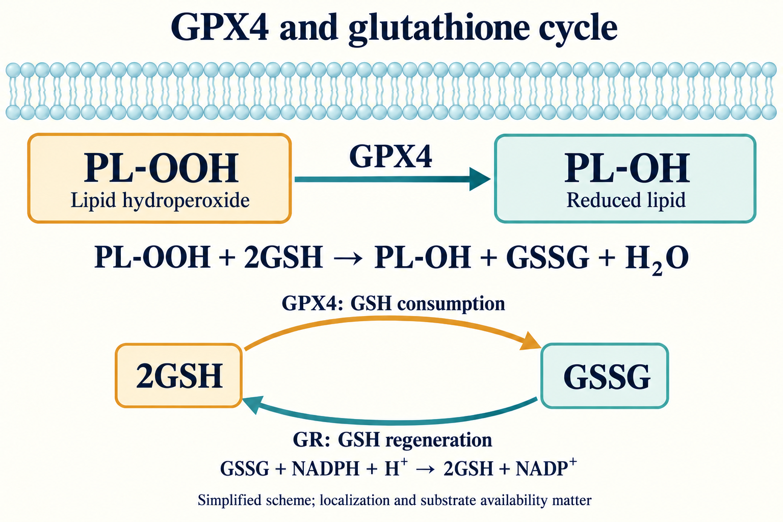
Let PL-OOH denote a phospholipid hydroperoxide, PL-OH the reduced product and Enz-Se⁻ the active enzyme. Simplified cycle steps are:
Overall:
Glutathione reductase (GR) returns GSSG to its reduced form:
GPX4 thus consumes GSH, while sustaining the cycle requires NADPH. Flux depends on multiple substrate concentrations, location and enzyme availability. It cannot be reduced to a universal “zero Km” or one GSH concentration. GPX4 removes hydroperoxide substrate, whereas lipophilic radical traps primarily interrupt radical-chain propagation: their actions complement one another.
System x_c⁻ consists of the light transport subunit SLC7A11 and heavy subunit SLC3A2, linked by a disulfide bond. It exchanges extracellular cystine for intracellular glutamate at a 1:1 ratio. Imported cystine is reduced to two cysteine molecules; this step also requires reducing resources. Cysteine then supplies GSH synthesis.
High extracellular glutamate can hinder cystine uptake, and cystine deprivation in the medium changes experimental outcomes. During amino-acid deprivation, GCN2 phosphorylates eIF2α and promotes the ATF4 response. Nrf2, regulated through the KEAP1 system, supports expression of protective genes, including elements of cystine and GSH metabolism. p53 can suppress SLC7A11, but its influence on ferroptosis depends on cell context and other targets; it is not a universal “p53 on—ferroptosis on” switch.
GSH is the tripeptide γ-glutamyl-cysteinyl-glycine. Glutamate-cysteine ligase (GCL), composed of catalytic GCLC and modifier GCLM subunits, produces γ-glutamylcysteine. Glutathione synthetase (GSS) adds glycine:
The unusual γ-linkage makes GSH less accessible to many conventional peptidases but does not make it indestructible: turnover includes γ-glutamyltransferase and other pathways. GSH provides feedback regulation of GCL, while GCLM changes the complex's kinetic properties. Synthesis depends on amino-acid supply, ATP and enzyme regulation, so increasing one precursor does not guarantee increased protective flux.
Most synthesis occurs in the cytosol, from which GSH enters other compartments. This is crucial for mitochondria: their membrane restricts exchange, and reducing resources and lipid injury are local. Wang and colleagues' 2021 study demonstrated that SLC25A39 is necessary for mitochondrial GSH import in mammalian cells. The older attribution of direct transport exclusively to the dicarboxylate/2-oxoglutarate carriers SLC25A10 and SLC25A11 cannot be treated as an established alternative to these findings.
In the endoplasmic reticulum, an oxidizing environment supports protein folding and disulfide formation. A single GSH/GSSG measurement, however, does not describe the entire redox state. The pair's potential depends on absolute concentrations, [GSH]²/[GSSG] and pH; whole blood cannot substitute for measurements in a particular membrane or mitochondrion. No universal redox-potential threshold diagnoses ferroptosis in a patient.
Erastin and its derivative IKE are used to disrupt cystine supply; sulfasalazine can also affect system x_c⁻. Sorafenib's effect on this system depends on the model: it is a multitarget anticancer drug rather than a universally selective inhibitor of the ferroptosis axis. With sufficient alternative cysteine sources, cells can partly bypass exchanger blockade, for example through transsulfuration.
RSL3 and ML162 are used as direct GPX4 inhibitors; ML210 undergoes intracellular conversion to a reactive inhibitor. Efficacy and selectivity depend on chemical structure, concentration and exposure time; none of these names denotes a ready, safe human treatment. FIN56 combines changes in GPX4 and CoQ metabolism, whereas FINO2 affects iron and lipid oxidation with indirect disruption of GPX4 protection. Different intervention points help design an experiment but require confirmation of the actual target and death mechanism.
Polyunsaturated fatty acids (PUFA) contain bis-allylic positions from which hydrogen is relatively easy to abstract. Arachidonic acid (AA, 20:4) and adrenic acid (AdA, 22:4) have several such vulnerable sites. AA- and AdA-containing phosphatidylethanolamines have been extensively studied as substrates of ferroptotic oxidation. Nevertheless, the list is not restricted to these two fatty acids or one phospholipid class: lipidomes and oxidation pathways vary between cells.
Monounsaturated fatty acids (MUFA), including oleate, alter membrane composition and can reduce the proportion of readily oxidized PUFA. Synthesis through SCD1 and lipid incorporation involving ACSL3 matter here. This is a change in the substrate pool, not proof that a MUFA molecule directly traps every radical. Saturated chains are less susceptible to this oxidation route, but a cell containing them does not become immune to every injury.
Replacing hydrogen with deuterium at bis-allylic positions can slow bond cleavage through the kinetic isotope effect. Deuterated PUFA are therefore studied as a way to reduce lipid-oxidation propagation. The reaction is slowed rather than chemically impossible; effect size, tissue incorporation and clinical benefit require separate measurements.
ACSL4 activates several long-chain fatty acids, with preference for particular PUFA. After acyl-CoA formation, LPCAT3 participates in lysophospholipid reacylation in the Lands cycle:
In studied models, ACSL4 loss changes the lipidome and reduces susceptibility to particular ferroptosis inducers. This is an important causal observation, not a rule that every ACSL4-deficient cell is fully protected. Other enzymes, available fatty acids and independent routes to oxidizable lipids can alter the dependency.
Lipoxygenases introduce oxygen into suitable lipid substrates. In Wenzel and colleagues' work, the ALOX15/PEBP1 complex changed substrate-directed oxidation and supported oxidized PE formation. PEBP1 is also known as a signaling-protein regulator; its state and interactions can connect signaling to lipid chemistry.
This mechanism is not the obligatory sole beginning of ferroptosis. Lipoxygenases matter in some models, whereas nonenzymatic reactions and other oxidation sources matter in others. Suppression by a compound called a “LOX inhibitor” does not on its own prove a causal role for LOX: some such compounds also trap radicals.
Let LH denote the parent lipid, L• its radical, LOO• a peroxyl radical and LOOH a hydroperoxide. A simplified chain includes:
The first equation shows one route to reactive-radical formation involving iron; actual reactions depend on ligands, environment and metal accessibility. The last step transfers injury to a neighboring lipid. GPX4 reduces the hydroperoxide reservoir that can feed this chain, while radical traps hinder its continuation.
Oxidized-lipid breakdown produces malondialdehyde (MDA), 4-hydroxynonenal (4-HNE) and other electrophilic products. They modify proteins and can alter enzyme and signaling functions. These are signs of lipid injury, but they are not specific to ferroptosis. Their presence requires explanation of origin, location and relation to actual cell death.
Transferrin mainly carries Fe³⁺ and binds the TfR1 receptor. Following endocytosis, endosomal acidification facilitates iron release; STEAP3 contributes to reduction to Fe²⁺, and DMT1 to transport across the endosomal membrane. Iron then supplies enzymes, mitochondria and stores or replenishes the labile pool.
The labile iron pool (LIP) comprises exchangeable iron bound to relatively accessible ligands, rather than a solution of completely uncoordinated free ions. Its participation in oxidative reactions depends on coordination and compartment. Total iron, serum ferritin and local catalytic accessibility are not equivalent. High ferritin alone therefore neither proves ferroptosis nor establishes an indication for a chelator.
When iron availability is low, IRP proteins bind IRE elements in mRNA and change synthesis of storage and transport proteins. IRP1 can switch between an IRE-binding state and an aconitase function containing an iron–sulfur cluster. IRP2 stability is linked to the FBXL5 system and cell state. Different IRE positions in mRNA allow iron uptake and storage to be coordinated.
Ferritin is a shell containing 24 FTH1 and FTL subunits. FTH1 provides ferroxidase activity, and the complex retains mineralized iron, reducing availability for unwanted reactions. Maximum shell capacity does not mean that every molecule is filled equally. Storage is useful only in dynamic balance: iron must be mobilized for normal metabolism, while excess accessible metal can amplify injury.
NCOA4 recognizes ferritin and helps deliver it for lysosomal degradation, a process called ferritinophagy. Breakdown of the protein shell releases iron for reuse. This pathway supports physiological turnover and, under particular conditions, increases susceptibility to ferroptosis.
Mobilizing a store does not inevitably produce an “explosive release” and death: the result depends on autophagic flux, iron export and utilization, lipid composition and antioxidant protection. Experiments must distinguish NCOA4 presence, actual ferritin degradation and changes in accessible iron. Lysosomal inhibition affects many processes and is not a selective clinical switch for ferritinophagy.
Bersuker and Doll's 2019 studies showed that FSP1, previously known as AIFM2, can suppress ferroptosis independently of direct GPX4 catalysis. N-myristoylation helps localize FSP1 to membranes. Using NAD(P)H, the enzyme reduces ubiquinone to ubiquinol, which traps lipid peroxyl radicals:
FSP1 maintains reduced CoQ, whereas GPX4 reduces hydroperoxides: they control different chain steps. One pathway may reduce dependence on the other in a particular cell, but it does not guarantee complete compensation in every tissue.
The mevalonate pathway contributes to CoQ isoprenoid-unit synthesis and Sec-tRNA modification. Interventions can therefore affect several protective mechanisms simultaneously. Experimental statin sensitivity is not a reason to stop prescribed treatment or to regard a statin as a proven cause of clinical ferroptosis. The distinction between endogenous CoQ function, supplementation and therapeutic benefit is discussed in coenzyme Q10, ubiquinol and clinical limits.
Dihydroorotate dehydrogenase (DHODH) in the inner mitochondrial membrane oxidizes dihydroorotate to orotate during pyrimidine synthesis and transfers electrons to the CoQ pool. In Mao and colleagues' 2021 model, this flux maintained reduced CoQ and limited mitochondrial lipid oxidation in parallel with GPX4.
Its protective importance depends on mitochondrial state and other pathways. A DHODH inhibitor also disrupts pyrimidine synthesis, so an observed effect cannot automatically be attributed solely to ferroptosis. Restoring pyrimidine supply and measuring lipid injury separately help distinguish mechanisms. Cardiolipin matters for the mitochondrial membrane, but DHODH should not be described as directly reducing only cardiolipin hydroperoxides.
GCH1 initiates tetrahydrobiopterin (BH₄) synthesis from GTP. Kraft and Soula's 2020 studies linked this system to ferroptosis resistance. BH₄ can contribute to radical trapping, while lipid composition and interactions with other antioxidant systems also change.
BH₄ oxidation requires consideration of biopterin-pool recovery, including DHFR and NADPH involvement in the relevant pathway. Synthesis, regeneration and location jointly determine protection. These findings do not make BH₄, CoQ or GSH universally interchangeable medicines.
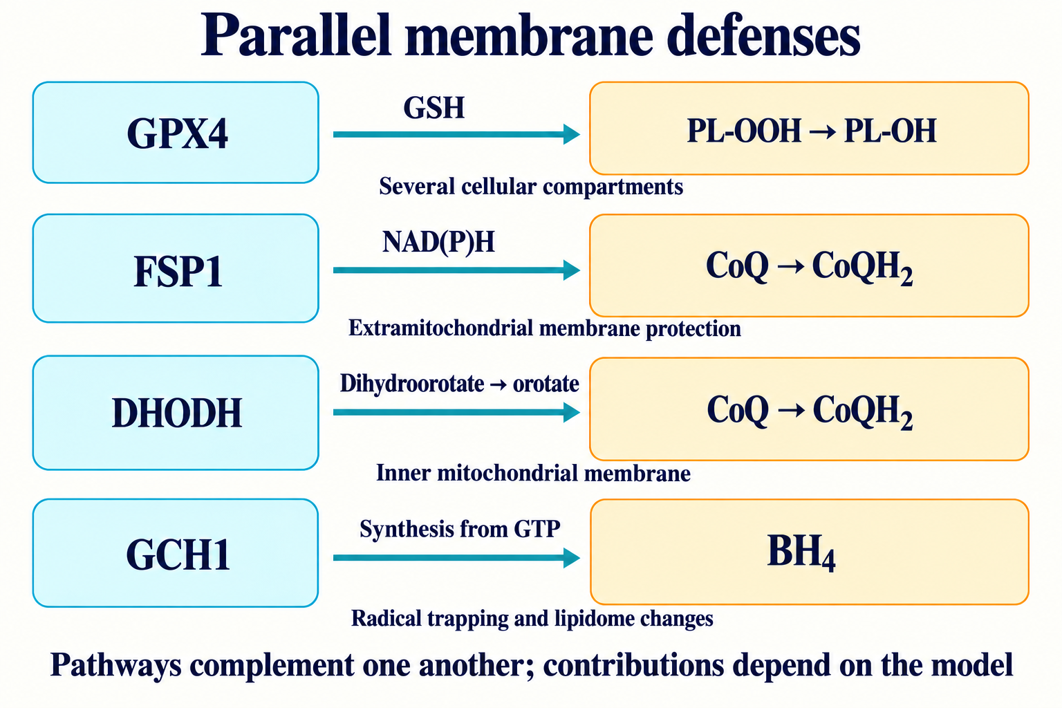
| Defense axis | Main reaction/flux | Relevant location | Supported conclusion |
|---|---|---|---|
| x_c⁻ → GSH → GPX4 | Cysteine supply and PL-OOH reduction | Plasma-membrane exchanger, cytosolic GSH synthesis; GPX4 in several compartments | Central dependency in many models; susceptibility depends on alternative supply and defenses |
| FSP1 → CoQH₂ | CoQ reduction and radical trapping | Membrane localization important for extramitochondrial protection | Parallel defense established in studied models |
| DHODH → CoQH₂ | Dihydroorotate oxidation coupled to CoQ reduction | Inner mitochondrial membrane | Connection to local protection and pyrimidine synthesis |
| GCH1 → BH₄ | Biopterin synthesis, radical trapping and lipidome changes | Depends on component and substrate distribution | Additional system rather than a clinical replacement for GPX4 |
The brain combines high oxygen demand, PUFA-rich membranes and uneven iron distribution. Parkinson's and Alzheimer's research examines iron accumulation, GSH changes, lipid oxidation and neuronal injury. ALS studies also investigate iron, SOD1/TDP-43 proteins and motor-neuron susceptibility. Conditional Gpx4 deletion in mice or protection by an experimental inhibitor helps establish a mechanism's role in a model, but does not prove that it explains an entire human disease.
The negative clinical result of FAIRPARK-II (Devos and colleagues, 2022) is especially important. The trial involved 372 people with early Parkinson's disease who had not previously received levodopa. Over 36 weeks, total MDS-UPDRS scores worsened by 15.6 points with deferiprone and 6.3 with placebo, a difference of 9.3 points (95% CI 6.3–12.2). MRI-assessed iron declined more with deferiprone, but this did not deliver clinical benefit. Serious adverse events included agranulocytosis and neutropenia.
This result illustrates the limitations of a surrogate marker: reducing iron in a brain region does not equal improving function. It does not refute every possible role of ferroptosis, but contradicts the notion that removing iron automatically treats Parkinson's disease. Interventions must be judged by clinical outcomes and safety, rather than biochemical target engagement alone.
Ischemia alters energy supply and redox balance; oxygen restoration during reperfusion can intensify lipid oxidation in the presence of accessible iron. Ferroptosis inhibition reduced injury to myocardium, kidneys and other tissues in experimental models. Effect size depends on the protocol: a percentage reduction from one experiment cannot be transferred to all heart attacks, strokes or transplants.
Studies of doxorubicin cardiotoxicity, including Fang and colleagues' 2019 work, implicate iron and lipid oxidation. HMOX1 regulation, heme breakdown and mitochondrial changes can participate. Doxorubicin also acts through other mechanisms, and cell rescue in a particular model does not establish complete prevention of patient cardiomyopathy.
Cold ischemia and subsequent donor-organ reperfusion can disrupt ATP-dependent metabolism and allow injuries to accumulate. Investigating lipid radical traps in perfusion media has translational interest. Experimental tissue preservation, clinical transplant suitability and long-term organ function require separate confirmation. Injured lipids can affect innate immunity, but post-transplant inflammation should not be explained solely by ferroptosis.
Linkermann and Friedmann Angeli's 2014 studies investigated ferroptosis in renal tubular injury. Gpx4 loss, ischemia and iron-supply conditions create different susceptibilities; liproxstatin-1 was used as an experimental protective tool. In rhabdomyolysis, myoglobin and heme iron form part of a more complex renal injury process alongside vascular and tubular factors.
In liver disease, iron overload, metabolic abnormalities and oxidative injury may coexist, but hemochromatosis, MASH and toxic injury are not one ferroptotic diagnosis. In acetaminophen overdose, NAPQI consumes GSH; mitochondrial stress and necrotic injury also matter. GSH depletion alone does not justify labeling all injury GPX4-dependent ferroptosis or replacing established emergency treatment with experimental radical traps.
Viswanathan and Hangauer's 2017 studies identified GPX4 dependence in some cells with a mesenchymal state and drug-tolerant persisters. Changes in ZEB1, lipid composition and PUFA-preparing enzymes can create vulnerability to lipid oxidation. This supports investigating ways to target cells that survive conventional therapy.
Vulnerability is not identical across tumors and does not guarantee selectivity over normal tissue. Inhibitor delivery, chemical stability, tolerability and parallel defenses determine applicability. VHL loss and lipid-metabolic features in clear-cell renal carcinoma are also studied in this context; a mutation alone cannot replace susceptibility measurements.
Ubellacker and colleagues showed in melanoma models in 2020 that the lymphatic environment can protect disseminating cells from ferroptosis. Lipid composition, including oleate, GSH and iron availability differ between lymph and blood; MUFA incorporation can increase subsequent cell resistance. This illustrates an environmental influence on metastasis rather than a universal rule for every cancer or a rationale for dietary changes intended to control metastases.
Ferrostatin-1 and liproxstatin-1 are widely used experimental ferroptosis inhibitors. As lipophilic radical-trapping antioxidants (RTA), they slow chain oxidation propagation. Capturing LOO• produces an antioxidant radical less able to continue the chain; this is not equivalent to GPX4's catalytic reduction of PL-OOH.
Kinetic constants, effective concentrations and half-lives depend on medium, species, administration route and compound structure. Pharmacological cell rescue helps test a mechanism, but membrane access and adverse effects must be considered. Fer-1 and Lip-1 are not presented here as available standard drugs for preventing heart attacks, neurodegeneration or aging.
Deferoxamine, deferasirox and deferiprone have medical uses in particular iron-overload conditions. They differ in administration, distribution and safety profile and are not interchangeable remedies for “oxidative stress.” Deferasirox requires attention to kidney and liver function; deferiprone carries neutropenia and agranulocytosis risks requiring treatment-specific hematological monitoring. Deferoxamine also has its own risks, including complications of inappropriate use and excessive iron removal.
Removing physiologically necessary iron can cause harm. Indication, dose and monitoring depend on the established disease and specific drug. In particular, negative FAIRPARK-II findings do not support deferiprone as preventive antiferroptotic treatment for Parkinson's disease. Adverse-event frequencies cannot be generalized from one population to every other population.
Selenium is required to synthesize Sec-containing proteins, and cysteine and glycine supply GSH. These facts do not prove efficacy of a combined “antiferroptosis stack” of selenium, NAC, ubiquinol and astaxanthin in healthy people or patients. The supplied sources contain no clinical test of that combination with neurodegeneration, cardiac injury or lifespan outcomes.
Correction of established selenium deficiency differs from supplementation when intake is already adequate. Excess causes toxicity, and serum concentrations depend on form, distribution and sampling circumstances. A level of 110–130 μg/L cannot be declared a universal GPX4-treatment target. Organic and inorganic forms undergo different metabolism; laboratory chemistry involving ascorbate does not establish a mandatory dosing interval for every clinical situation without appropriate data.
N-acetylcysteine (NAC) can supply cysteine and is used for specific medical indications. Its action depends on delivery, metabolism and baseline state; raising a plasma precursor does not guarantee GSH recovery in the required compartment. GSH esters are used in delivery models, and SAMe is linked to transsulfuration; these mechanisms do not validate arbitrary therapeutic doses or product equivalence.
CoQ participates in membrane redox metabolism, but oral ubiquinol should not be described as a guaranteed electron donor for FSP1 or DHODH: the enzymes reduce CoQ using their own electron sources, and delivery of a supplement to the relevant pool requires evidence. Astaxanthin, tocopherols and tocotrienols are studied as lipophilic antioxidants; bilayer positioning and radical-trapping activity do not establish clinical ferroptosis prevention. Every intervention requires attention to indications, safety and interactions with current therapy, particularly in cancer.
C11-BODIPY 581/591 is used to assess oxidation in cell experiments through a change in the red-to-green fluorescence ratio. It reflects the oxidative environment and depends on staining, illumination and analysis conditions. Its signal cannot independently prove ferroptotic death.
F₂-isoprostanes, MDA and 4-HNE adducts help assess lipid oxidation but are not unique to this mechanism. Mass-spectrometric analysis of specific oxidized phospholipids provides more detailed substrate information. Even precise product measurement cannot replace functional and causal confirmation.
Serum selenium, erythrocyte glutathione-peroxidase activity and whole-blood GSH/GSSG assess different aspects of physiological state; erythrocyte GPX activity is not a direct assay of brain membrane GPX4. Ferritin, transferrin saturation and T₂*/R₂* MRI are used to assess iron metabolism/accumulation for appropriate indications. Neither their combination nor a fixed transferrin-saturation value creates a validated universal diagnostic profile of “ferroptotic disease.”
Comparing molecular axes is useful when experimental tools are kept distinct from authorized treatment:
| Axis/process | What an intervention changes | Typical experimental tool | What else must be checked |
|---|---|---|---|
| SLC7A11/GSH | Cysteine and reducing-resource supply | Erastin, IKE; genetic SLC7A11 changes | Medium composition, transsulfuration, cysteine availability and actual GSH |
| GPX4 | Lipid hydroperoxide reduction | RSL3, ML162, ML210; Gpx4 manipulation | Target engagement, lipid injury, cell rescue and normal-tissue toxicity |
| ACSL4/LPCAT3 | Preparation of PUFA-containing phospholipids | Genetic interventions; pharmacological tools with limited selectivity | Actual lipidome and alternative fatty-acid incorporation routes |
| ALOX15/PEBP1 | Enzymatic formation of oxidized lipids in a suitable model | Complex-component manipulation, LOX inhibitors | Direct enzyme effects separately from radical trapping |
| NCOA4/ferritin | Mobilization of stored iron | NCOA4 manipulation and lysosomal-flux analysis | Ferritin degradation, local LIP and nonspecific autophagy effects |
| FSP1/CoQ | Regeneration of a membrane radical trap | FSP1 manipulation, iFSP1 and related experimental compounds | Localization, CoQ state and dependence on GPX4 |
| DHODH | Pyrimidine synthesis and local CoQ reduction | Brequinar and other DHODH inhibitors in research | Separate contributions of pyrimidine shortage and mitochondrial lipid oxidation |
| GCH1/BH₄ | Biopterin pool and lipid resistance | Genetic changes, experimental inhibitors | BH₄ regeneration, lipidome and biopterin functions outside ferroptosis |
A practical sequence for evaluating a study starts by asking where the result was obtained: in a cell-free system, cultured cells, an animal or a patient. Next, establish whether actual death was measured and linked to lipid oxidation, iron and the relevant protective pathway. Then assess intervention selectivity, achievable exposure and consequences for normal tissues. Clinical claims ultimately require patient-relevant outcomes and safety.
Distinguishing cytoprotection from anticancer ferroptosis induction remains scientifically valuable. However, the sequence “elevated biomarker → prescribe a chelator, selenium and a radical trap” is not a validated clinical algorithm. Diagnosis and treatment must match a specific disease; an experimental strategy becomes therapy only after appropriate evaluation.
GPX4 does not protect membranes alone. Its activity requires appropriate selenoprotein synthesis, GSH delivery, NADPH-dependent regeneration and access to lipid substrates. ACSL4/LPCAT3 and other processes determine which chains enter membranes; iron and oxidation pathways determine injury formation; FSP1, DHODH and GCH1/BH₄ influence the cell's ability to limit the radical chain.
Consequently, the same inhibitor has different effects in different cells and environments. Ferroptosis helps explain particular models of tissue injury and identify vulnerabilities in some tumor states. Translating these mechanisms into medicine requires a clear target, local action, a verified clinical outcome and acceptable risk rather than a general promise of “antioxidant protection.”
This is an editorial adaptation of the supplied text and bibliography, not a systematic review. The original local package contained no saved article body or accepted illustrations. Mechanistic evidence, animal models and clinical findings have been distinguished; unsupported universal numerical thresholds and a self-treatment scheme have not been carried forward as established facts.
On October 8, 2026, three specific conflicts were selectively checked: the Sec-to-Cys substitution result in Ingold's study, SLC25A39 involvement in mitochondrial GSH import and the clinical outcome of FAIRPARK-II. The latter's DOI was corrected, and the necessary primary SLC25A39 study was added. No comprehensive repeat literature search, audit of every link or formal GRADE assessment was performed. Shared references reflect the supplied bibliography and this focused check; inclusion does not imply that every statement was checked against the full text of every study.
During completion on October 8, 2026, the cuproptosis comparison was separately restored and Tsvetkov and colleagues’ 2022 primary study was selectively read in the Broad Institute author repository.
It reduces complex lipid hydroperoxides using GSH and limits accumulation of membrane injury.
No. These markers are nonspecific; research requires consistent functional, lipid and causal evidence.
Not automatically. Substrate requirements do not establish benefit from a universal supplement combination; deficiency correction and treatment have separate indications and risks.
In FAIRPARK-II, symptoms worsened more than with placebo despite reduced MRI-assessed iron.
Some tumor states are sensitive to loss of its protection. Applicability depends on selectivity, delivery, parallel pathways and normal-tissue safety.