Methylene blue and mitochondria: mechanisms and clinical limits — NutriFit
Methylene blue: redox cycling, the respiratory chain and the limits of clinical conclusions
The MB⁺/LMB mechanism, alternative electron transfer and limits of respiration measurements; tau, memory and vasoplegia studies distinguished from preclinical evidence, with MAO-A, G6PD and pharmacokinetic analysis.
Methylene blue is a medicinal dye with several distinct mechanisms that cannot be combined into a single “mitochondrial protocol”. In erythrocytes, its reduced form participates in converting methemoglobin to hemoglobin; in cellular and animal models, researchers have observed alternative electron transfer and changes in respiration; in the vascular wall, the compound can suppress NO–sGC–cGMP signalling. Dementia trials and small neuroimaging studies address different questions from experiments with isolated mitochondria. Dose, route of administration, oxidation state and drug interactions determine which mechanism can occur at all.
History: from dye to medicine
Heinrich Caro synthesized methylthioninium chloride in the late nineteenth century as an industrial phenothiazine dye. Paul Ehrlich used its ability to stain living structures, and his malaria experiments with Paul Guttmann became a historical example of early synthetic chemotherapy. However, selective staining of a parasite or neuron does not demonstrate infallible targeting of diseased tissue: dye accumulation depends on distribution, membranes, the redox environment and exposure time. The “magic bullet” concept matters to the history of pharmacology, but it does not establish modern clinical efficacy against malaria or neurodegeneration.
Treatment of acquired methemoglobinemia with a medicinal methylene blue preparation for established indications occupies a separate clinical role. Methemoglobin contains Fe³⁺ and does not carry oxygen like functional Fe²⁺ hemoglobin. The reduced dye, leucomethylene blue (LMB), can donate electrons to methemoglobin and return to its oxidized state. The physiological pathway for reducing methemoglobin in erythrocytes predominantly uses NADH and cytochrome-b₅ reductase CYB5R3, whereas an NADPH-dependent pathway is important for reducing administered methylene blue. This distinction explains why G6PD deficiency and NADPH availability are critical to the response. The reaction does not restore oxygenation instantaneously “within seconds” in every patient; its effect and monitoring depend on severity, cause and concomitant hypoxia.
Medical use in postoperative vasoplegia and shock is investigated separately from methemoglobinemia. Inhibition of soluble guanylate cyclase and changes in NO-dependent vasodilation offer a possible mechanism for increasing vascular tone; the role of iNOS and the overall effect depend on inflammation and hemodynamics. Availability of a medicine for one indication does not make every experimental use in vasoplegia or sepsis standard treatment. Likewise, neuronal culture and animal experiments do not establish therapies for Alzheimer’s or Parkinson’s disease.
The molecule and the MB⁺/LMB redox pair
Methylthioninium chloride is a cationic salt with a phenothiazine scaffold and two dimethylamino groups. The anhydrous salt has the formula C16H18ClN3S and a molar mass of approximately 319,85g/mol; the hydrate contains additional water, so substance content and the mass of a particular medicinal formulation must be distinguished. The conjugated π system explains visible absorption and the blue colour of the oxidized form. The absorption maximum and degree of dimerization depend on solvent, pH, ionic strength, concentration and optical methodology. Values of 664 and 605 nm may describe particular conditions, but “always dimers strictly above 10 μM” and “all nanomolar molecules are exclusively monomers” are not universal properties.
The two-electron transition can be represented schematically as MB++2e−+H+⇌LMB. MB⁺ is the coloured cation and LMB the reduced leuco form; in cells, the reverse reaction returns the carrier to its oxidized state. Formal charge, protonation and membrane–water partitioning affect reactant availability. The colour difference is analytically useful, but a colour change alone cannot measure electron flux through a mitochondrial complex. Claims about a strictly defined 110–115° dihedral angle, a pair of universal logP values and invariably rapid LMB diffusion across every biological barrier require measurements in a specified environment.
Reference conditions for this redox pair often give an E∘′ of about +0,011V at pH 7. This is not the potential of every cell: the actual potential depends on the MB⁺/LMB ratio, pH and binding; for a two-electron transition, its shift is described by the Nernst equation. The potentials of NAD⁺/NADH, flavins, Fe–S centres, ubiquinone and cytochrome c also depend on the protein environment. Individual Fe–S centres and membrane pools do not behave like free solutions. A positive difference in standard potentials makes the proposed transfer thermodynamically plausible, but does not “guarantee” a reaction without access to the centre, a suitable donor and an adequate exchange rate.
For any two-electron step, standard energy relates to potential difference through ΔG∘′=−nFΔE∘′, where n=2 and F is the Faraday constant. Substitution of the reference values −0,320, +0,011 and +0,254V does yield negative values for a hypothetical NADH→MB→cytochrome c chain. Yet summing these values describes the energetics of specified standard couples, not measured ATP yield or complete bypass of complex III within a neuron. In vivo, component activities and the proton gradient vary, and methylene blue may also be reduced by other enzymes and donate electrons to other acceptors, including oxygen. The image of an “irresistible electron sink” therefore misrepresents a reversible redox cycle.
Where alternative electron flow is possible
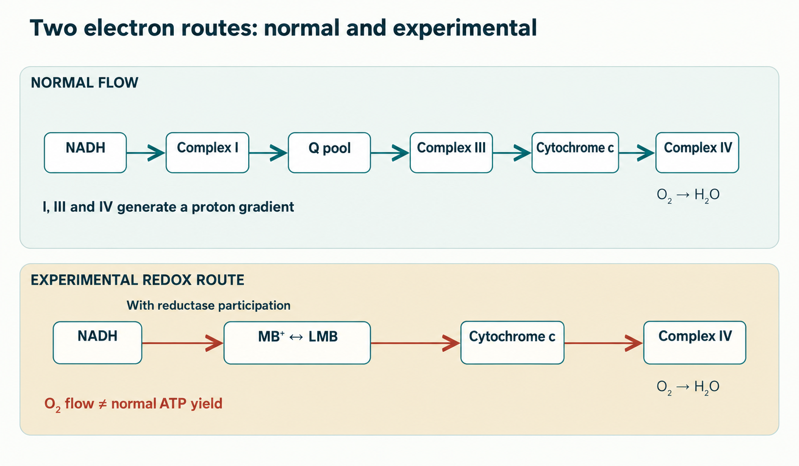
The inner mitochondrial membrane couples electron transfer to proton export from the matrix. Complex I receives NADH electrons at flavin mononucleotide (FMN), then transfers them through a chain of Fe–S centres to ubiquinone. Reduced ubiquinone feeds complex III, which transfers electrons to mobile cytochrome c; complex IV reduces oxygen to water. I, III and IV contribute to the proton motive force, and ATP synthase uses it to synthesize ATP. A simple balance for an electron pair from NADH in a fully coupled chain gives approximately 4, 4 and 2 translocated protons, respectively. This models idealized flow; actual yield depends on leaks, phosphate and adenine nucleotide transport, and cellular state.
The matrix arm of complex I, containing NDUFV1/FMN and Fe–S centres, is separate from its membrane ubiquinone-binding region; this complex architecture helps couple redox chemistry to proton pumping. Rotenone blocks the Q site and MPP⁺ disrupts complex I in toxicological models; succinate accumulation, Q-pool reduction and enzyme conformation also matter during ischemia and reperfusion. Structural models show several Fe–S centres, but their number and transfer sequence are meaningful only for the specified species and structure, and cannot be fixed for every enzyme variant. “Complex I blockade” also does not mean an immediate zero membrane potential: other substrates, complex II, ATP hydrolysis and membrane condition alter the response.
Wen and colleagues studied methylene blue as an alternative electron carrier in experimental systems. The proposed pathway involves reduction of MB⁺ by available donors near the matrix side of complex I or other flavoproteins, followed by donation to cytochrome c or another acceptor. NAD(P)H-dependent reductases, including NQO1, may participate in redox cycling in a suitable cell; a particular enzyme’s involvement must be examined by tissue, inhibitors and genetic experiments. The equation FMNH2+MB+→FMN+LMB+H+ schematically represents one possible route, but does not demonstrate direct MB⁺ contact with NDUFV1 in all mitochondria or establish a universal Km of 0.8–1.5 μM. The parallel reaction NADH+MB+→NAD++LMB requires an enzymatic route or confirmed chemical kinetics: comparing potentials alone does not establish its rate in the matrix.
If LMB does reduce cytochrome c downstream of the Q pool, electrons bypass part of the usual I→Q→III route. But “completely bypasses complexes I and III” is too strong: obtaining electrons from FMN entails participation of at least part of complex I, while conventional flow through III in the same cell need not stop. The cytochrome c route requires access to the oxidized protein on the appropriate side of the inner membrane and preserved complex IV activity. Cytochrome c transfers one electron at a time; one two-electron MB/LMB turnover could reduce two molecules. Demonstrating this route in a cell requires an inhibitor profile and oxygen/ATP balances, not merely improvement in one fluorescent readout.
In complex IV, cytochrome c passes an electron to CuA, then heme a and the catalytic heme a₃–CuB centre, where oxygen is reduced to water. Even if alternative transfer supports this segment, bypassing proton pumping by I and III leaves fewer gradient-generating protons per electron pair than usual. Possible maintenance of respiration under rotenone therefore cannot automatically be equated with restoration of normal ATP yield. Seahorse OCR measurements reflect oxygen consumption, but require separation of ATP-linked respiration, proton leak, nonmitochondrial O₂ consumption and direct oxidation of the reduced dye by oxygen. Claims that “10–100 nM always increases OCR”, “ATP becomes 1.0–1.5 per pair” and “ΔΨm remains at −130…−150 mV”, without a model and calibration methods, do not constitute a clinical property of the molecule.
In rotenone experiments, an alternative electron acceptor may shorten the time certain redox centres remain reduced and decrease superoxide formation. However, ROS arise both during forward flow with complex I blockade and during reverse electron transfer from a reduced Q pool at high membrane potential; location and rate depend on substrates and oxygen. Moreover, LMB can reoxidize with oxygen and, under some conditions, itself participate in a prooxidant cycle. Experiments showing lower ROS markers in a specific cell culture cannot establish a universal “70–90% reduction” across all tissues or complete prevention of reperfusion injury.
Inner-membrane cardiolipin helps organize respiratory complexes and cristae; when damaged, it can interact with cytochrome c and contribute to its peroxidase activity. I–III–IV supercomplexes represent one organizational level, but obligatory “tunnelling” of every electron between their components remains context dependent. If MB treatment reduces peroxidative injury in a model, it may indirectly preserve the membrane; direct retention of the Met80–heme bond, complete blockade of peroxidase activity and preservation of a respirasome of specified composition require separate structural and biochemical measurements. The same limitation applies to the mitochondrial permeability transition pore, mPTP: calcium loading, oxidative stress and potential affect opening, but its precise molecular organization remains debated. “The pore is necessarily formed by a dimer of ATP-synthase c-rings and is closed at a specified ΔΨm” is not an established universal threshold. A link between MB and NCLX-dependent Ca²⁺ clearance remains a model-specific hypothesis until calcium and ATP fluxes are measured independently.
Biphasic effects: why the curve does not define a dose
Redox hormesis proposes that a weak reversible stress can trigger adaptive responses, whereas a larger burden disrupts electron balance. Such behaviour has been observed for methylene blue in several cellular and animal experiments, including work from Gonzalez-Lima’s group. Here, however, the “Arndt–Schulz law” describes a possible response curve in a particular model, not a pharmacological law with a single milligram-per-kilogram scale. The concentration at an enzyme after oral dosing cannot be inferred directly from a culture-medium concentration: salt form, reduction in blood, distribution, exposure and elimination all matter.
At low experimental exposure, repeated MB⁺ reduction and LMB oxidation can alter the NAD⁺/NADH ratio, electron availability to complex IV and cellular signalling. Increased cytochrome-c-oxidase activity in cortex or hippocampus after treatment may reflect enzyme quantity, substrate supply or tissue cellular composition; establishing mitochondrial biogenesis requires independent measures of organelle mass, mtDNA, complex assembly and respiratory function. The proposed NAD⁺/SIRT1→PGC-1α deacetylation→NRF1/NRF2→TFAM sequence is biologically plausible as a research hypothesis. No change in NAD⁺/NADH alone proves the entire causal cascade or formation of new cristae specifically due to the dye. Figures such as “20,000 electrons per day” and a 30–40% activity increase, without a specified method, cannot become properties of the medicine.
At greater exposure, competing rates of dye reduction, electron donation to physiological acceptors and reaction with oxygen change. Autoxidation can be represented in simplified form as LMB+O2→MB∙+O2∙−+H+, but formation and fate of the intermediate radical require radical analysis of the particular system; successive arrows do not establish universal cellular stoichiometry. Potential consequences include consumption of NADPH reducing equivalents, impaired GSH regeneration by glutathione reductase, lipid peroxidation and respiratory uncoupling. Under other conditions, the oxidizing dye can increase the methemoglobin fraction—paradoxically for an agent used to treat methemoglobinemia. These effects depend on tissue, dose and time; “above 5–10 μM” and “above 5–10 mg/kg” do not define a general safety boundary. Contraindications and interactions cannot be offset by choosing a supposedly hormetic dose.
Tau protein, the synapse and neurodegeneration
Tau (MAPT) binds axonal microtubules and helps organize their dynamics; tauopathies alter its phosphorylation, localization, conformation and aggregation propensity. Important elements include PHF6* (275VQIINK280) and PHF6 (306VQIVYK311) in the microtubule-binding domain, together with the R2/R3 repeat context, tau isoform and structure of the particular filament. Hyperphosphorylation by GSK‑3β, CDK5 or MARK can reduce tau binding to microtubules, but “phosphorylation → toxic oligomer → paired helical filament” is not the only obligatory pathway. Soluble aggregates, tangles and tau loss of function address different experimental questions.
Wischik’s group showed that phenothiazine dyes can interfere with tau assembly and affect isolated filaments. Redox chemistry of the Cys291 and Cys322 thiols is one possible mechanism in a particular protein construct and environment. It does not justify depicting a single intramolecular Cys291–Cys322 bridge that invariably “shields” both PHF motifs and neutralizes every monomer: disulfide formation, intermolecular crosslinking and cysteine accessibility depend on isoform, protein concentration and oxidizing conditions. Cryo-EM structures of mature filaments reveal their folds, but do not automatically locate dye binding or describe disruption of existing filaments in a living neuron. In-vitro activity cannot be equated with an effect in a patient’s brain.
Hydromethylthionine (LMTM, LMTX) is a reduced medicinal form supplied as the bis(hydromethanesulfonate) salt and developed to have different pharmacokinetics from methylthioninium chloride. Changes in salt form and oxidation state cannot imply guaranteed “unrestricted” blood–brain barrier penetration or stable therapeutic concentration: clinical exposure must be measured and efficacy tested against prespecified endpoints. Phase III findings are discussed below; an attractive biochemical target does not repair a negative primary clinical result.
Cell cultures and animal models have also been used to study changes in Aβ aggregation, preservation of PSD‑95/synaptophysin and responses to calcium loading. Aβ oligomers can impair synaptic transmission and NMDA-dependent Ca²⁺ regulation, but direct MB binding to Phe19/Phe20, prevention of pore formation and subsequent complete preservation of ATP and exocytosis require separate structural, cellular and functional experiments; they cannot be presented as one confirmed causal chain. Plasticity does require ATP-dependent ion pumps and receptor-complex function, yet “80% of all brain ATP” as a fixed Na⁺/K⁺-ATPase share does not describe every neuron and activity state.
In parkinsonism models, rotenone and MPP⁺ damage complex I function; substantia nigra dopaminergic neurons are additionally vulnerable because of extensive axonal arborization and dopamine’s oxidative chemistry. Preservation of TH-positive cells, dopamine content or motor-task performance with dye treatment in a particular model indicates experimental protection within that protocol. These measurements do not confirm that direct complex I bypass necessarily caused the effect, or establish slower Parkinson’s disease progression in humans. MPTP models, rotenone toxicity and idiopathic disease have different timescales and pathogenesis.
Learning and fear-extinction experiments examine neuronal networks after MB administration. LTP in the Schaffer collateral–CA1 circuit, GluA1 phosphorylation, AMPA/NMDA signalling, CaMKII and ATP availability may change together, but improvement in one behavioural task does not prove the entire molecular route. NMDA receptor thiols are redox sensitive, and direct dye effects can be investigated with electrophysiology and mutagenesis; specified cysteine pairs and a 30–40% reduction in channel-opening probability in individual systems cannot be extrapolated to a patient’s brain. Likewise, AMPK–mTORC1–ULK1 signalling and PINK1/Parkin-mediated mitophagy describe mitochondrial quality-control mechanisms. Increased LC3B‑II or accumulated PINK1, without analysis of autophagic flux, lysosomal completion and functional organelle recovery, does not prove that the dye “cleanses the mitochondrial population”. Interpretation limits for synaptic and mitochondrial experiments are explored further in the article on acetyl-L-carnitine.
What was tested in humans
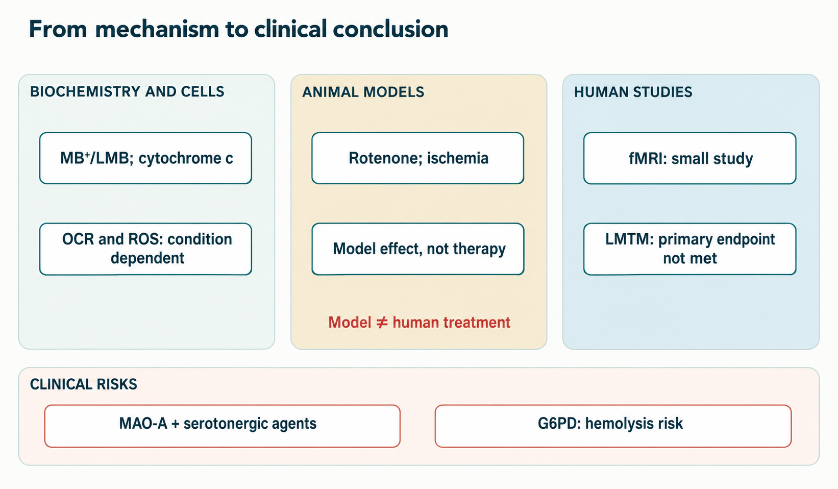
In a small randomized double-blind study, Rodriguez and colleagues examined a single methylene blue dose in healthy volunteers using functional MRI and attention/memory tasks. BOLD changes in several regions and a reported approximately 7% advantage in information recall apply to the particular tasks, post-dose timing and study sample. BOLD measures a hemodynamic correlate of neural activity, rather than direct electron flow through complex IV or ATP synthesis; MB’s own vascular effects complicate the energetic interpretation. Even a statistically significant difference in an experimental test in healthy people does not establish sustained cognitive improvement or dementia treatment. Claims of persistence for 48 hours and “accelerated metabolism” require separate methodological analysis: perfusion and BOLD measures are not direct measurements of metabolism.
A large LMTM trial in mild to moderate Alzheimer’s disease included hundreds of participants and assessed cognitive and functional measures against prespecified endpoints. In the overall randomized population, primary endpoints did not show the expected advantage of high doses over the low control dose. Subsequent analysis of participants receiving LMTM without other therapy generated a hypothesis that monotherapy effects might differ, but this changes comparison groups after randomization and is vulnerable to confounding. It cannot establish that cholinesterase inhibitors “cancelled” a true effect or that LMTM demonstrably slows cortical atrophy. Early phase II reports, LUCIDITY results and NfL analyses require separate assessment of design, published endpoints and multiple-comparison control. Without that assessment, “80% slower disease progression” or stabilization of MCI cannot be considered established.
In post-cardiac-surgery vasoplegia or critical illness, methylene blue has been used as an adjunct targeting NO–soluble guanylate cyclase–cGMP-dependent vasodilation. Proinflammatory iNOS induction increases NO production; NO binds the sGC heme in smooth-muscle cells and raises cGMP, after which PKG signals shift calcium handling and the contractile apparatus toward relaxation. MB can affect sGC and NOS and also has other redox effects. Higher blood pressure or lower vasopressor requirements in an individual study are hemodynamic endpoints, not evidence of reduced mortality or a universal sepsis treatment protocol. Bolus and infusion doses, SVRI increases and target MAP values from individual studies do not form a universal recommendation without the population, comparator, local protocol and interactions.
For ischemic stroke and traumatic brain injury, tMCAO and controlled cortical-impact models help assess lesion size, neurological behaviour, inflammation and barrier permeability. Timing relative to occlusion and reperfusion, model severity and infarct measurement methods strongly influence results. A smaller lesion in a rodent model cannot imply “50–65% efficacy” in humans. Microglia have many transitional states in these studies, so the binary phrase “M1 switched off” poorly describes their physiology.
Distribution, metabolism and the limits of pharmacokinetic generalization
After administration, MB⁺ is in dynamic equilibrium with LMB, binds blood components and distributes between aqueous and membrane phases. The degree of reduction varies in erythrocytes, endothelium, liver and other tissues; measuring only the blue cation in plasma may therefore fail to represent total exposure to the compound and its reduced form. Central nervous system penetration has been shown in experimental and pharmacokinetic studies, but the numerical brain/plasma ratio depends on species, sample preparation, time and whether MB, LMB and metabolites were measured together. Tabulated logP values for the two forms, obligatory OCT1/OCT2 transport and universal 10–20-fold accumulation in the human brain should not be treated as established for every regimen. Absolute bioavailability and distribution-volume estimates also cannot be transferred between formulations and administration routes without the study protocol.
NAD(P)H-dependent oxidoreductases can support phenothiazine-cation reduction in cells. NQO1 uses two-electron reactions for various quinone and other substrates; its specific participation in the MB cycle needs confirmation in the relevant tissue. CYB5R3 is particularly important for physiological NADH-dependent methemoglobin reduction through cytochrome b₅, but this does not mean it is invariably the principal reductase for administered MB in every cell. Thioredoxin reductase can also interact with redox-active substrates; its relative contribution depends on enzyme activity, available NADPH and competing substrates. A Km=1,2 μM or “75–85% of the cellular pool is always LMB”, without a defined enzyme assay and analytical method, is not a general biological constant.
Sequential N-demethylation of the phenothiazine scaffold produces related azure compounds and thionine. Azure B is trimethylated, Azure A dimethylated and Azure C monomethylated; the dyes and their leuco forms have different spectra and pharmacology. MB→Azure B→Azure A/C→thionine is useful as a map of possible transformations, but does not prove that every molecule strictly follows all steps in that order or that CYP1A2 and CYP2D6 exhaust the enzymes involved. Metabolite fractions depend on sampling time and method, so “Azure B = 15–20% of the circulating dose” cannot be retained without primary analysis. Conjugation and renal elimination likewise require distinction between free dye, reduced forms and analytically identified metabolites.
Excretion of coloured compounds can give urine a blue-green appearance; LMB can oxidize upon contact with air. Colour itself does not indicate how much of the dose reached the brain and is not a reliable quantitative marker of adherence, efficacy or safety. Generalized T1/2 values of 5–6.5 hours, “70–80% renally eliminated in a day” and 18–24 hours of CNS retention may refer to different analytical endpoints; without route, formulation and assay, they should not constitute one pharmacokinetic model.
Dangerous interactions and risk groups
Monoamine oxidase A inhibition is an independent methylene blue action, clinically much more important for safety than hypothetical “mitochondrial optimization”. MAO‑A metabolizes serotonin and other monoamines; combining its inhibition with drugs that enhance serotonergic transmission creates a risk of serotonin toxicity. Severe cases with SSRIs, SNRIs and other serotonergic agents are described in the literature. Clonus, hyperreflexia, agitation, hyperthermia and autonomic instability require urgent clinical assessment, but severity and onset vary; “always within 2–6 hours with temperature above 40 °C” would be false. Ki values and MAO‑A versus MAO‑B selectivity from one in vitro experiment cannot be treated as constants across all formulations and patients without examining conditions. Antidepressant withdrawal intervals depend on the drug, active metabolites, urgency and current prescribing information; independently stopping or replacing treatment to take MB is unacceptable.
G6PD deficiency limits NADPH production in erythrocytes, which lack mitochondria and rely heavily on the pentose phosphate pathway. With insufficient NADPH, reduction of administered dye and regeneration of reduced glutathione become difficult; methemoglobinemia treatment may fail and oxidative loading may worsen hemolysis. Hemoglobin damage, Heinz bodies, anemia and hemoglobinuria form a possible clinical sequence, but G6PD genotype and residual activity, methemoglobinemia cause and accompanying illness determine risk. Strict adherence to prescribing information and assessment of history and laboratory data belong to medical decision-making, not a universal online checklist. Genotype percentages and claims that testing is mandatory in every circumstance do not replace current protocols or the urgency of care.
Standardized medicinal products and laboratory dyes differ in identity, content, impurities, microbiological quality and labelling controls. Technical or aquarium dyes must not be used as medicines. Exact active-substance percentages and Pb/As/Cd/Hg/Zn limits differ between monographs and do not describe every technical sample. Specifications depend on jurisdiction, formulation and pharmacopoeia edition; without the relevant document, such figures offer false quality assurance. Verifying the registered formulation and its prescribing information matters more.
Pregnancy and the neonatal period require separate assessment of risk and potential benefit: severe fetal complications, including intestinal atresia, have been reported for intra-amniotic administration, but that route cannot be equated mechanically with every systemic exposure. Historical pregnancy categories “X/D” are not precise contemporary universal designations for methylene blue. In life-threatening conditions, the relevant specialist team decides according to current prescribing information. Discoloration of body fluids and skin, dye effects on optical measurements and possible pulse-oximetry interference also matter in practical monitoring.
Why these data do not define a “mitochondrial” regimen
A proposed daily 0.5–2 mg/kg protocol, weekly titration, compulsory fat-containing breakfast and “5 days on / 2 days off” cycles is not supported by the presented clinical trials as a means of cognitive enhancement or neurodegeneration prevention. Methemoglobinemia regimens and investigational vasoplegia protocols cannot be turned into outpatient instructions for healthy people. Even at low doses, interactions, G6PD deficiency and formulation issues remain. Comparing patients by eGFR, ALT/AST, blood counts and pregnancy may be clinically appropriate in a particular setting, but eGFR >60 mL/min/1.73 m² is not a universal diagnostic permission to use the drug.
Proposed “synergistic stacks” with coenzyme Q10, acetyl-L-carnitine, PQQ, NAD⁺ precursors and photobiomodulation have not been tested as one therapeutic combination for the outcomes discussed. Ubiquinone occupies the inner-membrane Q pool and transfers electrons from several dehydrogenases to III; it does not “support only complex II”. Acetyl-L-carnitine participates in acetyl-group metabolism, whereas long-chain acyl-group transport depends on CPT1–CACT–CPT2; the distinction is explained in the article on the carnitine shuttle and CPT1. Overlap between dye absorption and red-light wavelengths does not prove that transcranial illumination effectively excites the required concentration in neurons or doubles ATP. Combining MB and photobiomodulation requires consideration of light penetration, dosimetry, oxygen photochemistry and controlled clinical outcomes.
Agents can be compared by the investigated process step, rather than an unproven “optimizer ranking”:
Intervention or molecule
Investigated step
What the mechanism does not establish
Methylene blue
Reversible redox transfer in models; medicinal actions in methemoglobinemia and on NO–sGC
Complete safe bypass of I and III or dementia treatment
Coenzyme Q10
Endogenous membrane pool between several dehydrogenases and III
Restoration of any complex I blockade
Idebenone
Synthetic quinone with reductase-dependent pharmacology
Equivalence to ubiquinone in every tissue
PQQ
Redox and signalling responses in experimental models
Established vitamin status or clinical mitochondrial stimulation
NAD⁺ precursors
Effects on nicotinamide-cofactor availability
Automatic SIRT1/PGC‑1α activation or replacement of a respiratory complex
Methylene blue remains a valuable tool for studying the relationship between electron transport, vascular regulation and neuronal function. Clinical use must rely on the indication, registered formulation, individual contraindications and trial results for a specified outcome. Preclinical electron transfer may explain why research is rational, but cannot replace evidence of efficacy in Alzheimer’s disease, Parkinson’s disease, stroke or healthy “bioenergetic enhancement”. Further studies should connect measured MB/LMB tissue exposure with actual electron flux, ATP-linked respiration, interaction-related harm and clinically meaningful outcomes in defined populations.
How this review was prepared
This is a narrative review of methylene blue and mitochondrial redox metabolism based on a collection of 28 publications. In the October 4, 2026 edition, key studies on electron transfer, MAO-A inhibition and the clinical trial of a methylene blue derivative in Alzheimer’s disease were checked selectively. No exhaustive search, meta-analysis or formal assessment of each study’s risk of bias was performed; other bibliographic entries were not checked line by line against full texts.
Redox reactions, cellular or animal-model effects and clinical outcomes are distinguished. Concentration, chemical form and intervention regimen change interpretation; evidence for a medicinal derivative does not substitute for results for every methylene blue formulation. A mechanistic hypothesis does not establish universal efficacy or safety, and secondary and primary trial outcomes are assessed separately.
FAQ
Does methylene blue bypass complexes I and III in humans?
It supported alternative electron transfer during inhibition of these complexes in experiments. Complete direct bypass preserving normal ATP production in humans has not been established.
Is Alzheimer’s treatment with methylene blue or LMTM proven?
No. The large LMTM trial did not meet primary endpoints in the overall population; secondary monotherapy analyses do not replace a confirmatory trial.
Why is combining it with antidepressants dangerous?
Methylene blue inhibits MAO-A and many antidepressants enhance serotonergic transmission; the combination can cause severe serotonin toxicity.
Why does G6PD deficiency matter?
Erythrocytes need G6PD to generate NADPH. With deficiency, the drug may fail to reduce methemoglobin and promote hemolysis.
Can laboratory dye or a “mitochondrial” protocol be used?
No. Laboratory dye is not controlled as a medicine, and the presented data do not establish a universal outpatient protocol for enhancing mitochondrial function.