Leptin resistance: SOCS3, POMC, and the hypothalamus — NutriFit
Leptin resistance: hypothalamic inflammation, SOCS3, and POMC
Leptin and LEPR structure, JAK2/STAT3, PI3K/FoxO1, AMPK, POMC processing, and MC4R. How SOCS3, PTP1B, TCPTP, glia, ER stress, and brain delivery affect signaling; distinctions between animals, human obesity, and weight-loss adaptation.
Leptin connects energy stored in adipose tissue with neuronal systems controlling food intake, expenditure, and neuroendocrine adaptation. In common obesity, circulating leptin is often elevated while additional hormone produces less response than expected. Leptin resistance describes this reduced responsiveness. It encompasses several processes rather than a single diagnosis established by a blood test. Congenital leptin deficiency, receptor defects, diet-induced obesity in mice, and adaptation after human weight loss are distinct settings.
This article follows the hormone and receptor, intracellular pathways, arcuate neuronal populations, and mechanisms that weaken signaling. Most causal molecular evidence comes from cells and rodents. Human endocrine and imaging studies support relevant associations but do not establish the complete molecular chain in every patient. An experimental target does not by itself define a treatment protocol.
1. The molecule and endocrine signal
Cloning the mouse ob gene and its human homologue LEP established adipose tissue as part of endocrine weight regulation (Zhang et al., 1994). Human LEP lies at 7q32.1. Its 167-residue precursor loses a 21-residue signal peptide, leaving mature 146-residue leptin of approximately 16 kDa. White adipocytes provide most circulating hormone. Fat mass matters, but current energy balance, insulin, sex, and daily rhythms also affect secretion.
Leptin has a four-helix cytokine fold. Structure 1AX8 is an X-ray structure of leptin-E100, not of the entire receptor complex (Zhang et al., 1997). The disulfide connects Cys96 and Cys146 in mature leptin; mixing precursor and mature-protein numbering creates errors. The bond supports conformation, but individual mutations require experimental assessment rather than an assumption of complete inactivity.
Blood contains free leptin and leptin bound to soluble LEPR. Total concentration therefore does not equal brain availability. Considine et al. (1996) found a relationship with fat mass and higher concentrations in obesity. Their group averages are not universal reference limits, and elevated serum leptin does not directly measure hypothalamic STAT3 activity.
2. LEPR isoforms and architecture
The receptor identified by Tartaglia et al. (1995) belongs to class I cytokine receptors; human LEPR is at 1p31.3. Membrane isoforms share extracellular binding machinery but have different intracellular regions. Long LepRb supports canonical JAK2–STAT signaling. Short forms participate in distribution, internalization, and other functions rather than being merely inactive copies.
Soluble receptor can arise through alternative splicing and ectodomain shedding, with different relative contributions across species. Short isoforms are investigated in brain-barrier delivery, but assigning all human transport exclusively to LepRa/LepRc exceeds the evidence.
Extracellular architecture includes CRH1/CRH2 cytokine-receptor homology regions, an immunoglobulin-like domain, and fibronectin modules. CRH2 contributes to binding, and domain arrangement enables an active complex. Mancour et al. (2012) examined signaling-complex architecture. PDB 3V6O is an X-ray receptor-fragment/antibody structure, not full LepRb cryo-EM; the original citation conflated different studies. That fragment cannot establish one universal stoichiometry on living neuronal membranes.
3. JAK2, STAT3, and parallel branches
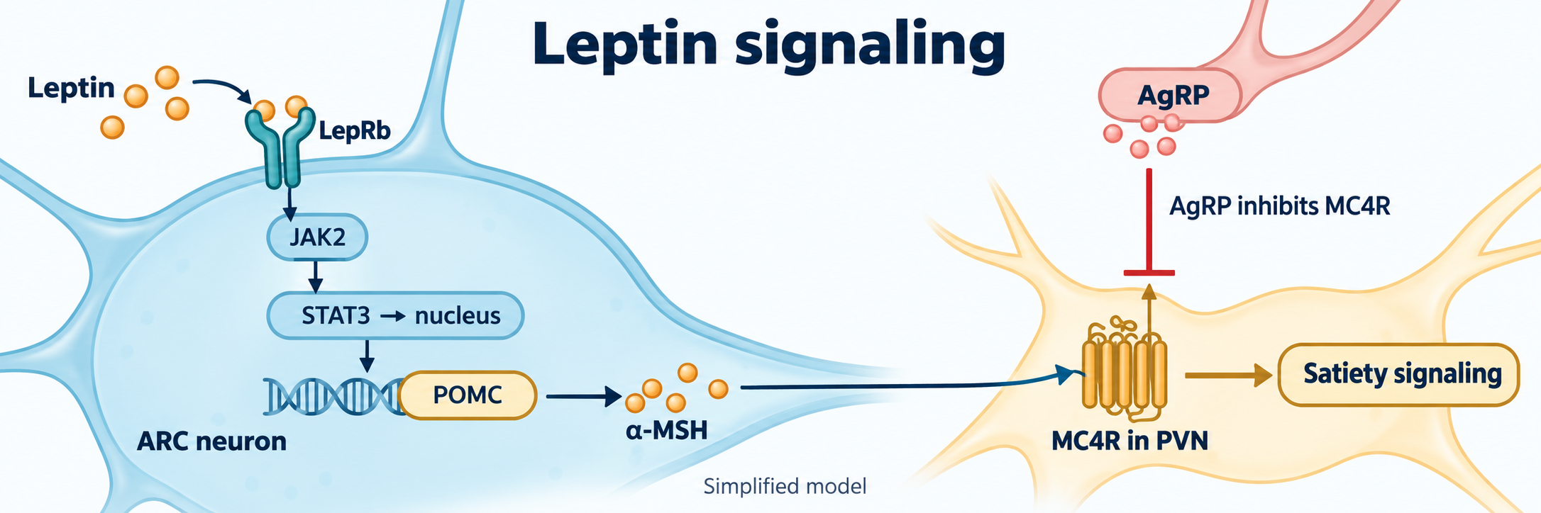
LepRb has no intrinsic tyrosine-kinase activity. Membrane-proximal motifs associate with JAK2. Ligand-dependent rearrangement promotes kinase activation and phosphorylation of activation-loop Tyr1007/Tyr1008, followed by receptor phosphotyrosine docking sites. In conventional LepRb numbering, Tyr1138 recruits STAT3, Tyr1077 is associated with STAT5, and Tyr985 with SHP2 and SOCS3. Residue numbers should be interpreted against the particular species and sequence.
A general phosphate-transfer reaction is:
Protein-Tyr-OH+ATPkinaseProtein-Tyr-OPO32−+ADP.
This is a reaction scheme, not a measured JAK2 rate or evidence for an unsupported fifteenfold fall in Km. STAT3 phosphorylation at Tyr705 enables dimer formation and transcriptional regulation, including Socs3 and energy-related neuronal programs. Receptor-site mutation in mice demonstrates the pathway's importance (Bates et al., 2003). These mice have severe energy-balance disturbance, but do not completely reproduce db/db: other pathways mediate some reproductive and additional leptin effects.
SHP2 connects the receptor to Ras–Raf–MEK–ERK; STAT5 provides another transcriptional branch. Their significance differs by cell population. A single linear pathway with identical output in all LepRb neurons is an inadequate model.
4. PI3K, FoxO1, PDE3B, and mTOR
Leptin and insulin signals converge on IRS/PI3K. PI3K converts PIP2 to PIP3, facilitating membrane recruitment of PDK1 and Akt. Activated Akt can alter FoxO1 localization, promoting 14-3-3 binding and nuclear export. Hypothalamic experiments link FoxO1 with energy status and Agrp/Pomc expression (Kitamura et al., 2006; Kim et al., 2006). STAT3 is a transcription factor, not the kinase directly phosphorylating FoxO1.
PDE3B hydrolyzes cyclic AMP and contributes to selected leptin-response models. Akt–TSC–Rheb–mTORC1 links signaling to S6K and 4E-BP1 and thus protein synthesis. Yet mTOR effects depend on duration and neuronal identity: persistent activation does not automatically improve sensitivity. Manipulating PI3K or mTOR in an animal brain does not establish a safe human intervention.
5. AMPK and hypothalamic lipid sensing
Leptin inhibits AMPK in experimental hypothalamic systems, whereas muscle responses can include AMPK activation (Minokoshi et al., 2004). Removing AMPK inhibition of acetyl-CoA carboxylase can increase malonyl-CoA production:
Acetyl-CoA+HCO3−+ATPACCMalonyl-CoA+ADP+Pi.
ACC means acetyl-CoA carboxylase and Pi inorganic phosphate. This links carbon metabolism to lipid sensing; it is not an appetite-calculation formula. Neuronal CPT1c is principally discussed as a malonyl-CoA sensor and should not be equated with conventional mitochondrial CPT1 acyl-group transport. Sympathetic outputs can affect UCP1-dependent thermogenesis, but their magnitude cannot be inferred from one leptin measurement.
6. Arcuate neurons and the melanocortin network
The arcuate nucleus, ARC, lies beside the median eminence and third ventricle. Fenestrated vessels and tanycyte barriers provide regulated sensory access rather than unrestricted entry of every plasma protein into every ARC neuron.
POMC and AgRP/NPY/GABA populations are important and heterogeneous. POMC/CART is convenient classical shorthand; cells do not all have identical receptors or peptide combinations. Leptin activates some POMC neurons directly and through their network (Cowley et al., 2001), while altering ionic currents and inhibitory inputs in other cells. KATP channels participate in selected responses; a fixed membrane-potential shift or eighty-percent inhibition cannot describe the entire population.
POMC is a precursor for multiple peptides. PC1/3, encoded by PCSK1, initiates cleavage producing ACTH; PC2 continues processing, carboxypeptidase E removes basic residues, and PAM participates in amidation. Subsequent acetylation alters α-MSH properties. PC1/3 therefore does not independently convert POMC into mature α-MSH. Nor is a universal thirty- to fortyfold affinity increase established for every acetylated form.
α-MSH activates MC4R in PVN and other regions. Canonical Gs–adenylyl cyclase–cAMP contributes, but MC4R cellular physiology exceeds a single PKA/TRPC5 pathway. AgRP is an antagonist and, in appropriate systems, an inverse agonist (Ollmann et al., 1997). NPY uses its own receptors and GABA inhibitory synapses. Inverse agonism reduces activity below the basal level; cAMP concentration never becomes negative.
Mouse Mc4r disruption and human POMC, PCSK1, and MC4R mutations demonstrate network importance (Huszar et al., 1997; Krude et al., 1998; Jackson et al., 1997; Farooqi et al., 2003). Rare monogenic conditions do not prove complete MC4R shutdown in everyone with common obesity.
7. SOCS3: feedback and inhibition
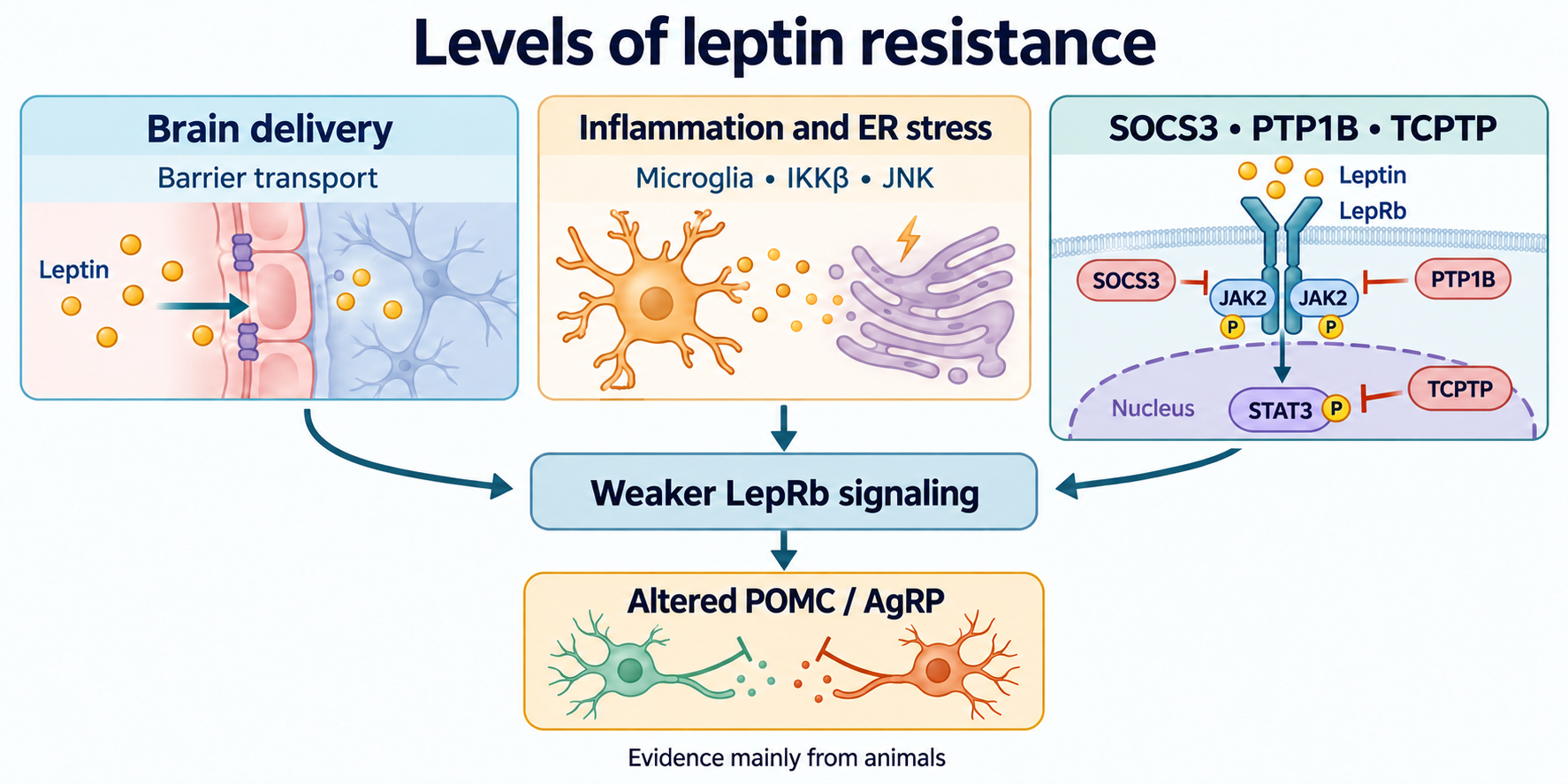
STAT3 induces SOCS3, providing negative feedback. Its SH2 domain recognizes phosphotyrosine motifs, the kinase-inhibitory region contributes to JAK inhibition, and the SOCS box recruits E3 ubiquitin-ligase machinery. These functions limit signal intensity and duration. E3 recruitment does not imply obligatory destruction of the entire LepRb/JAK2 complex after every response.
Kershaw et al. (2013) showed occlusion of JAK2's substrate-binding region. Structure 4GL9 contains SOCS3–JAK2–gp130 rather than LepRb. SOCS3 should not be described as an ATP-pocket plug (Babon et al., 2012).
Deleting Socs3 in POMC-expressing mouse cells increased leptin sensitivity and improved metabolic measures (Kievit et al., 2006). This is causal evidence within a model, not proof that general suppression is safe: SOCS3 also limits many immune cytokine signals.
8. PTP1B and TCPTP: distinct positions
PTP1B, encoded by PTPN1, is anchored to the cytoplasmic face of the endoplasmic reticulum. Its catalytic domain faces the cytosol; Cys215 forms a phosphoenzyme intermediate and Asp181 participates in acid–base catalysis. It can dephosphorylate JAK2 and restrict leptin signaling. Endosome–ER contacts help explain substrate access, but every contact does not mean receptor destruction. Neuronal deletion in mice affected weight, adiposity, and leptin action (Bence et al., 2006).
TCPTP, encoded by PTPN2, has several isoforms; nuclear TC45 can dephosphorylate STAT3. Loh et al. (2011) studied increased hypothalamic TCPTP in obesity and cooperation with PTP1B. One regulator acts near the receptor-associated kinase, the other on a transcriptional effector. Hyperleptinemia, inflammation, and phosphatase changes can sustain reduced responsiveness, but their order and relative contribution vary by model.
A correction to Loh et al. (2011) was published in 2022: the actin control blot in Figure 4A had been flipped and incorrectly reused as the tubulin control for p-JAK2. The authors supplied the correct control and stated that the scientific conclusions were unchanged (Loh et al., 2022). This matters when assessing that figure; the correction does not establish a human treatment effect.
Removing two phosphates consumes two water molecules and produces two Pi. This corrects the unbalanced original reaction.
9. Inflammation, glia, and ER stress
Thaler et al. (2012) reported early hypothalamic inflammation after high-fat feeding in rodents and an MRI association with human obesity. MRI does not measure SOCS3 or an individual POMC neuron's sensitivity. Glial responses can initially protect tissue; persistent changes do not make every instance of gliosis irreversible.
Milanski et al. (2009) linked saturated fatty acids with experimental hypothalamic TLR4-dependent inflammation. Palmitate should nevertheless not be called an established direct TLR4 ligand: Lancaster et al. (2018) distinguished receptor binding from TLR4-dependent metabolic inflammatory susceptibility. Their macrophage findings do not resolve every microglial question.
IKKβ phosphorylates IκB to release NF-κB. JNK links stress with AP-1 and IRS signaling changes. Zhang et al. (2008) demonstrated involvement of hypothalamic IKKβ/NF-κB in mouse energy imbalance. The meaning of a particular IRS1 serine depends on species and context; Ser307 is not a universal steric PI3K switch. Inflammatory pathways can induce SOCS3 independently of leptin stimulation.
The ER folds and modifies secreted and membrane proteins. Lipid composition, calcium homeostasis, and synthesis load can impair these functions. The unfolded-protein response includes PERK–eIF2α–ATF4, IRE1–XBP1, and ATF6. Initially adaptive, sustained stress can engage CHOP, JNK, and cell death. Ozcan et al. (2009) linked ER stress with mouse leptin resistance. Glycosylation, ER-associated degradation, and receptor-trafficking defects are potential components rather than the proven sole explanation of every phenotype.
Astrocytic contacts, microglial functions, synaptic organization, and POMC-cell survival extend the model beyond one receptor. Comparison with other neuroinflammatory pathways helps distinguish shared glial processes from leptin-specific signaling.
10. Delivery to the brain
Tight endothelial junctions restrict free leptin diffusion. Saturable transport, regional barriers, tanycytes, and receptor-related processes contribute to delivery. The CSF-to-blood ratio can fall with high peripheral concentration, but CSF is not ARC interstitial fluid. Universal Km values, plateau thresholds, or a diagnosis of brain leptin starvation are not established for all humans.
An illustrative saturable relationship is:
J=K1/2+CJmaxC.
J denotes transport flux, Jmax its limiting value in the same units, C concentration, and K1/2 the concentration giving half-maximal flux. It is a model, not a clinical calculator; parameters require measurement in a defined experimental system.
Banks et al. (2004) showed triglyceride inhibition of leptin transport experimentally. Banks et al. (2018) also investigated central leptin and insulin receptor effects. These findings do not establish physical endothelial clogging by VLDL or preservation of every insulin pathway. Clinically indicated triglyceride reduction does not equal proven restoration of brain signaling to a specified leptin threshold.
11. Humans: obesity, deficiency, and weight loss
High leptin in common obesity accompanies varied eating behavior; persistent uncontrollable hyperphagia is not compulsory. Heymsfield et al. (1999) found a dose-related weight response in some participants, so the trial was not wholly negative. Responses varied, and it did not establish universal treatment for common obesity.
Replacement in congenital deficiency is a different setting: Farooqi et al. (1999) described a marked effect in a child lacking leptin. Simply adding ligand cannot repair a defective receptor. Comparing these populations requires identifying the source of reduced signaling.
With energy restriction, leptin can decline faster than fat mass. After weight loss, this may accompany greater hunger, sympathetic and thyroid changes, and altered muscle efficiency. Rosenbaum et al. (2005) examined such adaptations and Rosenbaum et al. (2008) brain responses to food cues. “Leptin pit” is a metaphor, not an established syndrome with a universal threshold. fMRI changes do not demonstrate complete elimination of hunger or proven prevention of regain.
Low leptin during substantial energy deficit can contribute to reproductive suppression through intermediary neural circuits, not solely direct GnRH action. Thyroid, sympathetic, gonadal, and HPA responses depend on deficit duration and population. A universal expenditure fall of 300–500 kcal/day or endocrine collapse misrepresents individual variability. Regain is probabilistic and also depends on environment, support, behavior, and treatment; one hormone does not make it inevitable.
12. Experimental therapeutic targets
Chemical chaperones 4-PBA and TUDCA, ER-stress reduction, SOCS3 regulators, and phosphatases help investigate mechanism. Improved mouse responsiveness does not establish a human supplement effect.
Celastrol from Tripterygium wilfordii and withaferin A were studied as mouse leptin sensitizers (Liu et al., 2015; Lee et al., 2016). Their effects depend on the model and are not reducible to one direct target. Large changes in animal intake and weight do not define human doses; ashwagandha extract is not purified withaferin A. Ursolic acid is investigated as a metabolically active triterpene and enzyme inhibitor in cellular systems. A clinical leptin-resistance regimen of 150–300 mg several times daily is not established.
DHA contributes to membrane biology and precursors of inflammation-resolution mediators, including D-series resolvins and protectins. MFSD2A transports LPC-linked long-chain fatty acids (Nguyen et al., 2014). A triglyceride or ethyl-ester DHA capsule is not its direct substrate. Cintra et al. (2012) studied unsaturated fats in animal models, not restoration of human hypothalamic STAT3. M1/M2 labels oversimplify microglial states.
Curcumin has experimental anti-inflammatory effects. Greater bioavailability of selected preparations does not prove therapeutic concentrations in human ARC, IKKβ/JNK inhibition there, or resistance reversal. The same distinction applies to taurine: endogenous functions do not make an arbitrary dose a validated chemical chaperone for this condition.
13. Fructose, activity, and sleep
Hepatic fructose metabolism involves KHK and can promote lipogenesis and VLDL secretion under appropriate loading conditions. Shapiro et al. (2008) investigated fructose feeding and leptin responsiveness in rats. The work does not show that avoiding all fruit completely repairs the human barrier. Diet effects depend on energy intake, food form, and replacement. Time-restricted eating, activity, and triglyceride reduction may have metabolic aims, but 16/8, Zone 2, or a triglyceride target below 1.0 mmol/L is not a validated universal leptin-restoration protocol.
Sleep relates to appetite and endocrine regulation. Taheri et al. (2004) studied cohort associations, whereas Spiegel et al. (2004) examined brief restriction in healthy young men. These designs differ; hormone-change percentages do not apply automatically to everyone or prove receptor restoration. Daily fluctuations also depend on feeding. Fixed peak times, compulsory dinner four hours before sleep, and blue-light blocking are not established ways to eliminate SOCS3-mediated resistance. Circadian signaling and melatonin explain relevant rhythms without replacing leptin-response studies.
14. Comparing evidence levels
Level
What is studied
Translation limit
Molecule and structure
Leptin, LEPR, JAK2, SOCS3, phosphatases
A complex fragment does not describe an intact living neuron
Neuronal pathway
STAT3, PI3K/FoxO1, POMC processing, MC4R
Cell populations and stimulus duration differ
Glia and ER
IKKβ/NF-κB, JNK, UPR, synaptic changes
Causal experiments are predominantly rodent studies
Barrier
Saturable transport and lipid effects
No universal clinical flux threshold
Human weight loss
Hunger, expenditure, hormones, fMRI
No single syndrome or inevitable regain
Intervention
Deficiency replacement and sensitizer experiments
Different populations; mouse doses are not prescriptions
The mechanistic map frames questions: is hormone insufficient, delivery altered, intracellular responsiveness weakened, or network output changed? It does not diagnose the condition from leptin, HOMA-IR, or CRP. These markers do not measure local ARC inflammation or define a serum goal below 10–12 ng/mL. Assessment of obesity and associated disorders requires clinical context; the original nutraceutical algorithm lacks a confirmed status.
Leptin resistance brings together explanations for reduced neuronal responses despite adequate energy stores. Its strength is connecting receptor signaling, glial environment, delivery, and melanocortin circuitry. Its central limit is translation to an individual: attractive targets remain targets until clinical studies establish effects, safety, and the appropriate population.
FAQ
Can one blood test diagnose leptin resistance?
No. Serum leptin relates to fat mass and other factors, but does not directly measure ARC delivery or intracellular responsiveness. Clinical context is required.
Why is AgRP an inverse agonist?
In suitable experimental systems it reduces basal MC4R activity. This is a fall relative to baseline, not a negative cAMP concentration.
Does leptin help everyone with obesity?
Replacement in congenital deficiency differs from common obesity. The dose-ranging trial showed variable responses and did not establish a universal regimen.
Are DHA and curcumin proven treatments?
Their membrane and inflammatory mechanisms are of interest, but do not establish reversal of human hypothalamic resistance or a universal dose.
Does low leptin after weight loss make regain inevitable?
No. It can contribute to hunger and expenditure adaptation, but environment, support, behavior, and treatment also matter. A leptin pit is not one established clinical syndrome.