Does high urate necessarily cause hypertension?
No. Experimental mechanisms and small selected-adolescent trials support a possible contribution, but universal causality and treatment benefit for all adults are unestablished.

Purine degradation, KHK-C, ATP and Pi, XOR, vascular and renal hypotheses. What Feig, CARES, FAST and ALL-HEART show, and why asymptomatic urate does not establish a universal treatment regimen.
Uric acid is the terminal product of human purine degradation. At blood pH, approximately 7.4, it is predominantly present as the urate anion. Elevated serum urate without gout attacks is usually called asymptomatic hyperuricemia. Asymptomatic describes the clinical presentation; it does not establish that crystals are absent, because deposits can precede the first attack. Soluble urate, monosodium urate crystals and the activity of the enzyme producing urate are distinct biological exposures.
The approximate monosodium urate saturation concentration of 6.8 mg/dL under physiological conditions corresponds to about 404 µmol/L. This is a physicochemical value affected by temperature and the surrounding medium, rather than a precise threshold of vascular toxicity. Laboratory intervals vary; upper limits around 360 µmol/L for women and 420 µmol/L for men are often used. Randomized trials have not established a universal vascular safety threshold of 300–360 µmol/L. A treatment target for gout also does not automatically justify treating someone without gout (FitzGerald et al., 2020).
Fructose can connect hepatic energy metabolism to purine breakdown. Cell and animal experiments support plausible pathways involving oxidative stress, vascular remodeling and hypertension. An epidemiological association, however, does not demonstrate that urate causes every form of hypertension, chronic kidney disease or hepatic steatosis. These levels of evidence need to remain distinguishable.
In many mammals, urate oxidase, or uricase, initiates conversion of urate into 5-hydroxyisourate; subsequent reactions produce the more soluble allantoin. Apes, including humans, have lost functional uricase. Reconstruction of ancestral proteins suggests a progressive decline in activity rather than necessarily one abrupt event. The human UOX pseudogene contains stop codons at positions 33 and 187 and a splice-acceptor lesion. Different ape lineages have different histories of inactivation, so an identical mutation sequence cannot be assigned to all hominoids (Oda et al., 2002; Kratzer et al., 2014).
Proposed selective advantages include plasma antioxidant activity, maintenance of blood pressure with limited sodium and enhanced fat storage from seasonal fruit. Neel's thrifty-genotype concept and Johnson's fructose–urate hypothesis generate testable questions, but do not establish the historical cause of selection. A direct connection to human bipedalism is particularly uncertain: evolutionary chronology and reconstructed-cell findings do not prove it. These hypotheses cannot supply precise ancestral salt intakes, body heights or a mandatory modern treatment regimen.
Purine nucleotides are synthesized de novo from PRPP and other precursors or recycled through HPRT and APRT salvage pathways. AMP degradation has two branches. AMP deaminase produces IMP, followed by 5′-nucleotidase formation of inosine. Alternatively, AMP is dephosphorylated to adenosine, which adenosine deaminase converts to inosine. Purine nucleoside phosphorylase yields hypoxanthine and ribose-1-phosphate from inosine. Guanosine yields guanine, and guanine deaminase produces xanthine. Xanthine oxidoreductase (XOR) catalyzes hypoxanthine → xanthine → uric acid.
PDB structure 1FIQ is bovine milk xanthine oxidase, not the human protein. XOR is an approximately 290-kDa homodimer. Each monomer contains an N-terminal domain with two [2Fe–2S] clusters, a central FAD-binding domain and a large C-terminal molybdenum-cofactor domain. Molybdenum is coordinated by the molybdopterin dithiolene and oxo, hydroxo and sulfido ligands. Electrons travel from molybdenum through the iron–sulfur centers to FAD. Redox potentials, distances and residue numbers must be tied to the particular protein and experimental conditions (Enroth et al., 2000).
Aromatic and charged residues position the purine in the active site. Activated Mo–OH attacks substrate carbon, with coupled hydride transfer to the sulfido ligand and reduction of Mo(VI) to Mo(IV). Product release is followed by electron transfer to the final acceptor. Xanthine dehydrogenase (XDH) preferentially uses NAD⁺; xanthine oxidase (XO) uses oxygen. Thiol oxidation involving Cys535 and Cys992 in the studied bovine enzyme can reversibly change its conformation. Limited proteolysis produces irreversible conversion. This does not mean that every tissue stress produces the same transition, or that XDH cannot react with oxygen (Nishino et al., 2008).
Net reactions written using undissociated uric acid are:
XO can generate both superoxide and hydrogen peroxide. Their proportions depend on oxygen, pH and enzyme redox state; a fixed percentage cannot describe every tissue. XOR inhibition decreases both urate formation and enzyme-derived oxidants, so an allopurinol effect does not always distinguish these explanations.
Fructose enters enterocytes primarily through apical GLUT5, with basolateral GLUT2 participating in monosaccharide exit. GLUT2 also matters in hepatic transport. The intestine is not simply a passive conduit: isotope experiments in mice showed substantial conversion of small fructose doses into glucose and organic acids in the small intestine. High delivery rates can exceed intestinal metabolic capacity, increasing fructose delivery to the liver and colon. A universal hepatic extraction of 70–85%, independent of dose and conditions, is not justified (Jang et al., 2018).
Mutually exclusive splicing of KHK exon 3 produces A and C isoforms. KHK-C, prominent in liver, intestine and kidney, has greater fructose affinity than the widely expressed KHK-A. Approximate experimental Km values of 0.8 and 8 mmol/L illustrate this distinction, rather than dietary toxicity thresholds. In mice, deletion of KHK-A worsened adverse fructose effects, whereas deletion of both isoforms protected against the studied metabolic phenotype. That experiment does not prove that selective KHK-C blockade prevents all related diseases in humans (Diggle et al., 2009; Ishimoto et al., 2012).
Aldolase B cleaves fructose-1-phosphate (F1P) into dihydroxyacetone phosphate and glyceraldehyde; triokinase subsequently phosphorylates glyceraldehyde. Entry through this pathway bypasses the PFK-1-controlled step of glycolysis. Rapid phosphorylation can transiently deplete ATP and free inorganic phosphate, Pi, as phosphorylated intermediates accumulate. Nevertheless, ATP is itself a KHK substrate: describing the rate as completely uncontrolled is inaccurate. Depletion depends on dose, delivery rate and hepatic function; a 40–60% ATP fall within 15 minutes is not the ordinary response to every piece of fruit.
Adenylate kinase helps preserve ATP at the expense of AMP formation. Reduced Pi inhibition and AMP availability can increase AMPD2-mediated deamination:
Subsequent reactions generate inosine, hypoxanthine, xanthine and urate. This is degradation of previously synthesized nucleotides, not de novo purine synthesis. A transient serum-urate excursion after a fructose challenge is possible but does not equal persistent hyperuricemia.
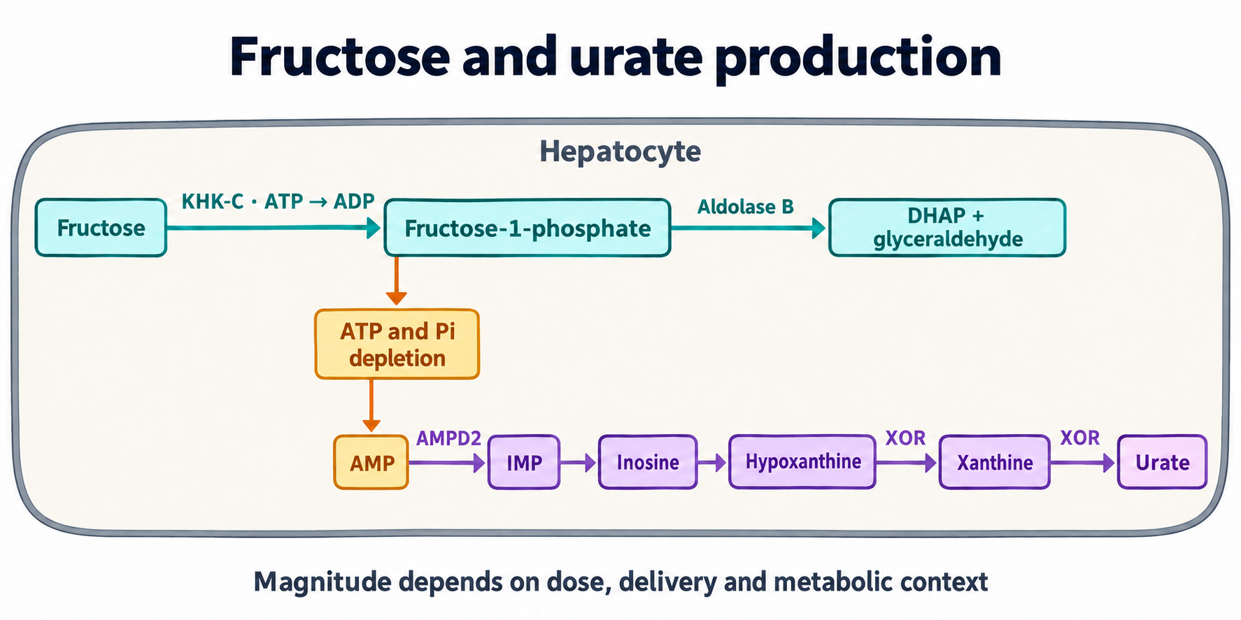
In cellular and animal models, intracellular urate is associated with mitochondrial oxidants, reduced aconitase activity and citrate accumulation. Citrate export through SLC25A1 and ATP-citrate lyase activity supply acetyl-CoA for lipogenesis:
Acetyl-CoA carboxylase generates malonyl-CoA, which inhibits CPT1; ChREBP and SREBP-1c regulate lipogenic genes including FASN and SCD1. These are potentially interacting pathways, not proof that the citric acid cycle or β-oxidation stops completely in every person with elevated urate (Lanaspa et al., 2012). The role of CPT1 is developed in the article on mitochondrial fatty-acid transport.
Monosodium urate crystals activate the NLRP3 inflammasome, caspase-1 and IL-1β maturation, driving inflammatory responses in gout (Martinon et al., 2006). This established mechanism cannot automatically be attributed to soluble urate.
Plasma urate can act as an antioxidant, whereas intracellular accumulation has prooxidant associations in some experimental systems. Total antioxidant capacity in an assay does not measure a person's overall protection from vascular disease. In kidneys, URAT1 mediates apical reabsorption, GLUT9 is a major transport component, OAT4 participates in organic-anion exchange and ABCG2 contributes to secretion. In vascular cells, transporter expression and function vary by model and cell type: renal functions do not prove identical roles in every endothelial cell. A probenecid effect also does not establish selective blockade of all these transporters.
Experimental pathways involve NADPH oxidases and mitochondrial reactive oxygen species. Enzyme specificity is crucial: p47phox-dependent assembly belongs to NOX2, whereas NOX4 generally does not require these cytosolic activators and releases predominantly H2O2 (Martyn et al., 2006). Superoxide from other sources can consume NO:
Peroxynitrite and other oxidants damage the tetrahydrobiopterin, BH4, system. Reduced BH4 availability relative to BH2 can uncouple eNOS: electron transfer no longer supports normal NO synthesis and can generate more superoxide. This is general vascular redox biology, not a hyperuricemia-specific diagnostic signature. Superoxide dismutase and superoxide clearance explain competing removal pathways.
Gersch et al. (2008) demonstrated a chemical reaction of uric acid with NO producing 6-aminouracil. The finding establishes chemical possibility, while its quantitative contribution to human vascular NO deficiency remains uncertain. A simplified “BH4 plus peroxynitrite yields BH2” expression is not a fully balanced unique reaction. Brachial flow-mediated dilation (FMD) assesses a conduit artery's response to altered flow; a universal 7% cutoff cannot diagnose urate toxicity or eNOS uncoupling.
Mazzali et al. (2001, 2002) inhibited rat uricase with oxonic acid. Moderately increased urate accompanied higher blood pressure and renal arteriolar changes without obligatory crystal injury. These findings support an early functional phase involving reduced NO and renin–angiotensin activation, followed by a structural phase involving preglomerular remodeling, interstitial inflammation and salt sensitivity.
PDGF, MAPK/ERK, COX-2 and MCP-1/CCL2 have been studied as proliferative and inflammatory signaling pathways in vascular smooth-muscle cells. Renal vascular narrowing and altered tubular transport may impair pressure natriuresis: higher pressure is required to excrete the same sodium load. NHE3, ENaC and local renin–angiotensin signaling matter, but this balance is not simply transmission of systemic pressure into glomeruli. Renal autoregulation and different nephron segments contribute.
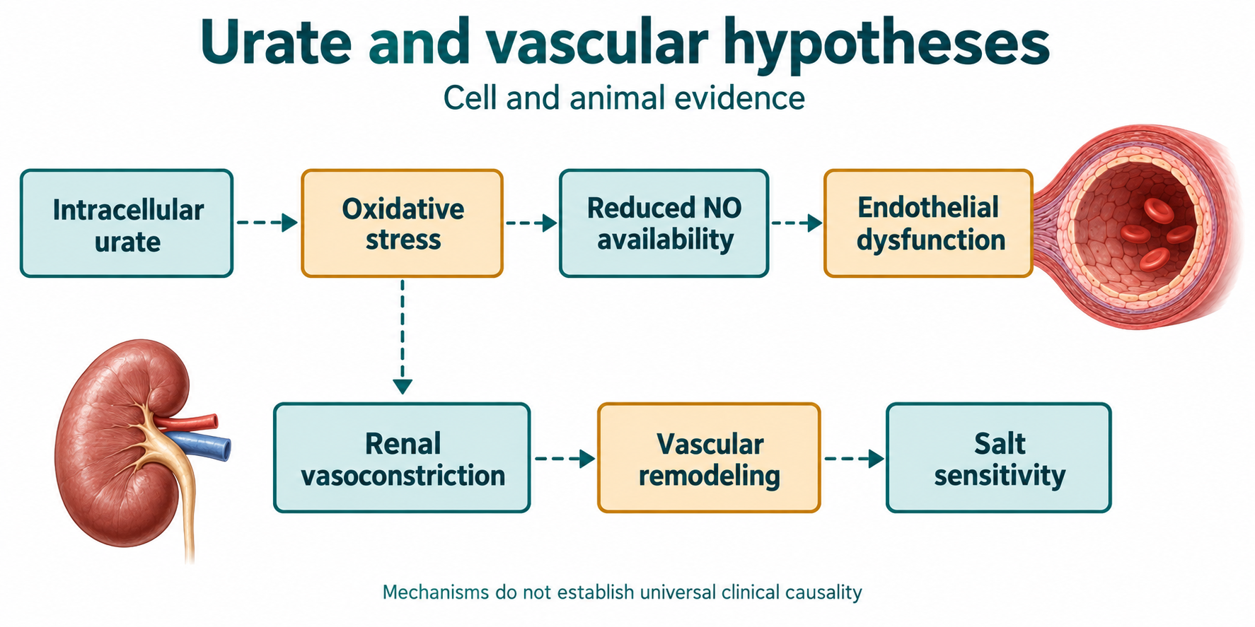
The two phases are a research model, not established stages shared by every patient. They do not demonstrate inevitable progression, absolute irreversibility or a narrow treatment window requiring urate-lowering drugs for everyone without gout. A neutral adult trial cannot be explained by renal fibrosis if that mechanism was not measured.
Higher urate predicts future hypertension, renal disease and cardiovascular events in observational studies. NHANES, PAMELA and PIUMA provide associations; adjustment for measured factors does not eliminate residual confounding or reverse causation. Reduced renal excretion itself raises urate. These cohorts cannot provide a universal mortality increase per mg/dL or an individually guaranteed eGFR decline.
Feig et al. (2008) enrolled 30 adolescents aged 11–17 with recent hypertension and urate ≥6 mg/dL in a crossover trial. Four weeks of allopurinol, 200 mg twice daily, lowered blood pressure. Both clinic and ambulatory criteria normalized in 20/30 receiving allopurinol versus 1/30 receiving placebo. The frequently cited 19/22, or 86%, refers to the subgroup achieving urate <5 mg/dL, not the full randomized sample. This small short trial did not measure long-term clinical events.
Soletsky and Feig (2012) compared allopurinol, probenecid and placebo in obese adolescents with prehypertension. In the combined urate-lowering groups, clinic systolic and diastolic pressure fell by 10.2 and 9.0 mmHg, versus increases of 1.7 and 1.6 with placebo. This supports a causal contribution in the selected population, but does not establish these drugs as routine therapy for all hypertension.
| Trial | Actual comparison | Main result | Interpretation limit |
|---|---|---|---|
| CARES, 2018; 6190 participants | Febuxostat versus allopurinol in gout with established cardiovascular disease | Composite endpoint HR 1.03; cardiovascular death HR 1.34, 95% CI 1.03–1.73 | Comparative drug safety, with substantial treatment discontinuation and follow-up loss |
| FAST, 2020; 6128 participants | People ≥60 with gout, already taking allopurinol, and an additional risk factor | On-treatment HR 0.85, 95% CI 0.70–1.03; p<0.0001 for noninferiority | Different eligibility and analysis; not a universal refutation of CARES |
| ALL-HEART, 2022; 5721 in analysis | Allopurinol plus usual care versus usual care in ischemic heart disease without gout | MI, stroke or cardiovascular death HR 1.04, 95% CI 0.89–1.21; p=0.65 | Adding allopurinol did not establish secondary-prevention benefit |
FAST excluded recent myocardial infarction or stroke, severe heart failure and severe renal impairment, which matters when comparing it with CARES. CKD-FIX and PERL (2020) also did not demonstrate the anticipated renal benefit of allopurinol in their populations. Together these findings oppose universal preventive prescribing based solely on serum urate.
SLC2A9 encodes GLUT9 and lies on chromosome 4's short arm. Variants rs16890979, rs12498742 and rs734553 are associated with urate; rs16890979 is a coding missense variant, not merely an intron-7 variant. Effects vary by population and sex, and association does not establish a separate cardiovascular risk for each allele. GLUT9 loss of function can cause renal hypouricemia type 2 and predispose to complications after intense exercise (Vitart et al., 2008; Matsuo et al., 2008).
ABCG2 is an ATP-dependent exporter involved in intestinal and renal excretion. The rs2231142 Q141K variant reduces protein function and is associated with gout risk. Its effect is not complete abolition of intestinal elimination or an established cause of resistant hypertension. Reduced extrarenal excretion can increase the renal urate load (Woodward et al., 2009).
URAT1/SLC22A12 supports reabsorption; the loss-of-function W258X variant causes renal hypouricemia type 1. Associations involving rs893006 do not establish an individual “super-reabsorber” phenotype. KHK and AMPD2 regulation is biologically interesting, but claims about common AMPD2 phosphate-pocket alleles producing guaranteed fructose hypersensitivity lack adequate support in the cited experiments. These ideas do not constitute a ready genetic prescribing algorithm.
Sucrose supplies glucose and fructose; HFCS and agave syrup can also deliver substantial fructose. Reducing sweetened drinks and excessive added sugars lowers rapidly delivered sugar loads and supports energy-balance management. Whole fruit, with fiber and different consumption rates, is not equivalent to a large free-fructose challenge. A universal 25–40% reduction in urate production after beverage removal has not been established.
Alcohol can increase urate production and reduce elimination. Ethanol oxidation raises NADH/NAD⁺ and can favor lactate accumulation; organic anions influence renal urate exchange. Acetate activation consumes ATP:
Beer additionally supplies purines. Effects depend on quantity and clinical context; emotive labeling as a metabolic poison adds no explanatory value.
| Intervention | Scientific signal | What is unestablished |
|---|---|---|
| Vitamin C | Small urate reductions in some trials and a meta-analysis; renal elimination may contribute | Universal direct URAT1/GLUT9 blockade, hypertension prevention or a standard gout-treatment dose |
| Cherries and anthocyanins | Small physiological experiments; Zhang et al. (2012) found an observational association with fewer recurrent gout attacks, OR 0.65 | Treatment of vascular “metabolic exacerbations”, equivalence to medicines or proven event prevention |
| Quercetin | Shi and Williamson (2016): 22 men, 500 mg/day for four weeks; urate change −26.5 µmol/L | Blood-pressure benefit, universal efficacy, long-term safety or replacement of urate-lowering medication |
| Luteolin and kaempferol | Laboratory XOR inhibition | Clinically established effective doses; IC50 depends on enzyme, substrate and assay |
Choi et al. (2009) studied gout risk, not asymptomatic hyperuricemia risk. An association between higher vitamin-C intake and lower gout risk does not justify prescribing 1500 mg/day to everyone. ACR 2020 conditionally recommends against adding vitamin C for gout. Ascorbate support of BH4 is mechanistically interesting but does not establish clinical-event prevention by such supplementation.
Losartan has a URAT1-related uricosuric effect; fenofibrate and SGLT2 inhibitors can also lower urate. For SGLT2 inhibitors, glycosuria and altered renal transport are implicated, but simple competition between glucose and urate at GLUT9b does not exhaust the mechanism (Hamada et al., 2008; Chino et al., 2014; Uetake et al., 2010). These effects are considered alongside the drugs' primary indications. They do not justify fenofibrate, an SGLT2 inhibitor or a supplement combination solely for presumed vascular protection against asymptomatic urate.
Repeated serum urate, renal function, medication exposure, alcohol, diet, blood pressure and signs of gout help establish context. Urate-lowering treatment follows clinical indications rather than “low FEUA → vitamin C; high FEUA → XOR blockade.” ACR 2020 conditionally recommends against initiating pharmacological therapy for asymptomatic hyperuricemia.
Fractional urate excretion uses concurrently measured urine, U, and plasma, P, urate and creatinine concentrations:
Urine and plasma concentrations of each analyte must use matching units. FEUA describes the fraction of filtered urate excreted in urine and depends on renal function, medicines and conditions. A high value alone does not prove hepatic overproduction; a low value does not diagnose a specific mutation.
| Measure | Appropriate interpretation |
|---|---|
| Serum urate | Balance of production and elimination; one concentration does not reveal intracellular urate or XOR activity |
| FEUA | Renal urate handling, rather than an independent classification of every hyperuricemia cause |
| FMD | Method-dependent endothelial-function research measure, not a specific urate-toxicity test |
| Ambulatory blood pressure | Pressure level and daily pattern; nondipping does not prove urate arteriolosclerosis |
| UACR and eGFR | Renal injury and function; confirmed UACR ≥30 mg/g is clinically meaningful but does not establish the cause |
Fructose–purine biochemistry explains how energy depletion can connect to urate generation. The clinical conclusion is narrower: elevated urate deserves assessment alongside cardiovascular and renal factors, while prevention of events by lowering it without gout needs evidence for the particular population and intervention.
No. Experimental mechanisms and small selected-adolescent trials support a possible contribution, but universal causality and treatment benefit for all adults are unestablished.
Whole fruit differs from a large rapidly delivered free-fructose load. Food form, added-sugar amount and overall energy balance matter.
No. It describes renal excretion relative to filtration and depends on renal function, medicines and measurement conditions.
Small trials and observational associations do not demonstrate this. Lowering a laboratory value does not establish prevention of gout or vascular events.