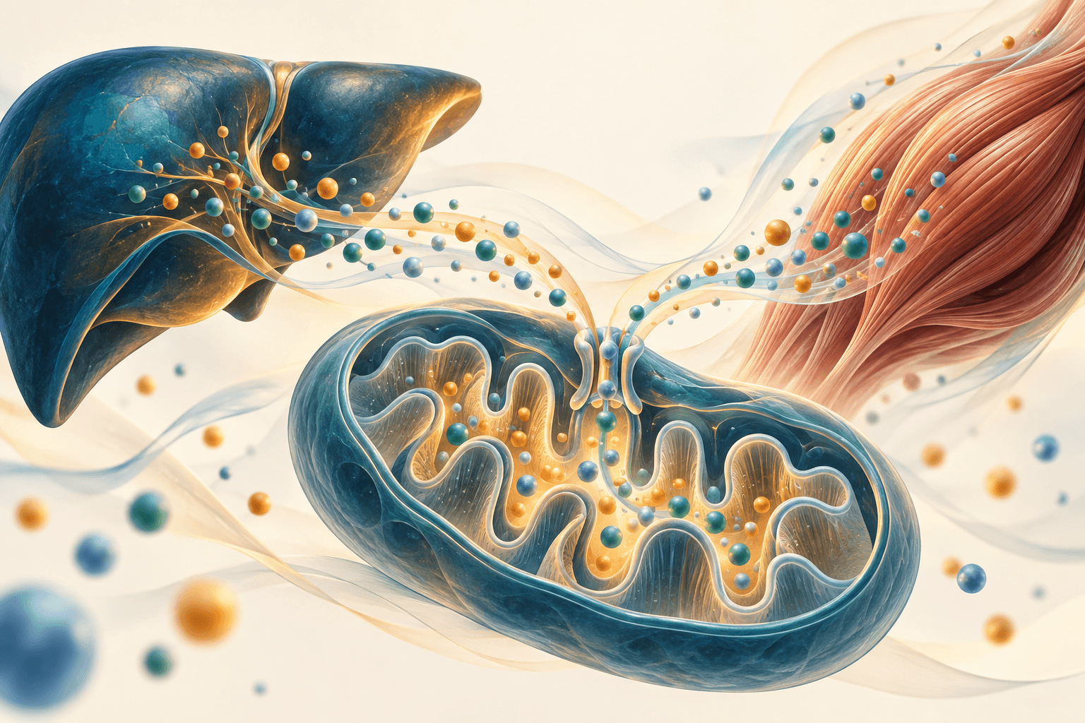
BCAA dysmetabolism: branched-chain ketoacids, BCKDH and insulin resistance
How leucine, isoleucine and valine metabolism connects BCKAs, BCKDH regulation and insulin signalling, with biochemical detail and clinical evidence limits.
Leucine, isoleucine and valine are essential branched-chain amino acids, or BCAAs. They support protein synthesis, nitrogen handling and adaptation to exercise. Their circulating concentrations often rise with obesity and predict subsequent type 2 diabetes in observational studies. Nevertheless, a blood concentration does not directly measure oxidation, BCKDH activity or the cause of insulin resistance. BCAAs, their branched-chain α-ketoacids (BCKAs), and metabolites formed further downstream are distinct molecules acting at different points in the pathway.
Molecular identity and exchange between organs
Their aliphatic side chains are hydrophobic, while the amino and carboxyl groups make free amino acids zwitterions at physiological pH. Side-chain hydrophobicity does not imply unrestricted passage of the whole molecule across membranes. Essential amino-acid requirements must be assessed alongside total-protein adequacy; these chemical properties cannot define a universal therapeutic BCAA dose.
Leucine is 2-amino-4-methylpentanoic acid; isoleucine is 2-amino-3-methylpentanoic acid; valine is 2-amino-3-methylbutanoic acid. Isoleucine has two chiral centres: natural L-isoleucine has the 2S,3S configuration. Sharing a branched side chain does not make the three interchangeable. Their oxidation products, transport interactions and signalling effects differ.
The plasma pool reflects dietary entry, protein breakdown, incorporation into new protein, exchange between organs and oxidation. Muscle, adipose tissue, kidney and brain participate in transamination. Liver has relatively low BCAT activity but processes ketoacids arriving from other tissues; BCAAs neither completely bypass liver nor undergo oxidation exclusively in muscle.
Cytosolic BCAT1 and mitochondrial BCAT2 transfer the amino group to α-ketoglutarate. Pyridoxal phosphate (PLP), derived from vitamin B6, supports the reaction through Schiff-base intermediates:
This step is reversible. Glutamate can transfer nitrogen to pyruvate through ALT, producing alanine. Alanine export to liver connects muscle nitrogen disposal with gluconeogenesis in the glucose–alanine cycle. Glutamine provides another nitrogen carrier. In brain, glutamate also contributes to GABA synthesis. These connections extend BCAA biology beyond muscle protein, but do not allow brain neurotransmitter levels to be inferred from one blood measurement.
LAT1 comprises the light chain SLC7A5 and heavy chain SLC3A2/CD98. It is a sodium-independent exchanger of large neutral amino acids. Nicklin et al. (2009) demonstrated intracellular glutamine exchange supporting essential amino-acid entry in their cellular models; glutamine is not thereby established as its exclusive substrate in every tissue. Yoneshiro et al. (2019) identified SLC25A44 as a mitochondrial BCAA transporter and linked brown-fat BCAA oxidation to energy homeostasis. That study does not identify it as a BCKA transporter. Evidence that monocarboxylate transporters carry some BCKAs across the plasma membrane similarly cannot establish a single universal inner-mitochondrial route for all three ketoacids.
Three ketoacids enter a common oxidative gate
Transamination produces α-ketoisocaproate (KIC) from leucine, α-keto-β-methylvalerate (KMV) from isoleucine, and α-ketoisovalerate (KIV) from valine. Their systematic names are 4-methyl-2-oxopentanoate, 3-methyl-2-oxopentanoate and 3-methyl-2-oxobutanoate, respectively. BCKAs have lost the original amino group; the abbreviation is not another name for BCAAs.
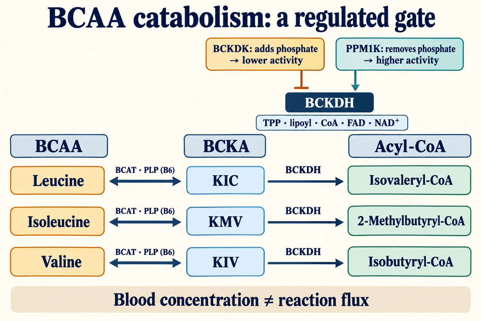
The diagram separates the three carbon routes from their common BCKDH gate. BCAT arrows are reversible, whereas BCKDH oxidative decarboxylation is effectively irreversible. Phosphorylation regulates the complex; circulating concentration remains a separate measurement.
BCKDH is a mitochondrial multienzyme complex related to pyruvate dehydrogenase (PDH) and α-ketoglutarate dehydrogenase. E1 contains BCKDHA and BCKDHB subunits in an α2β2 assembly. E2, encoded by DBT, forms a 24-subunit core; flexible lipoyl domains shuttle reaction intermediates between active sites. E3 is dihydrolipoamide dehydrogenase, DLD, shared with other dehydrogenase complexes. Overall mass and peripheral E1/E3 occupancy depend on assembly rather than a universally fixed stoichiometry.
E1 uses thiamine diphosphate, TPP/TDP, and Mg²⁺. The reactive thiazolium centre engages the ketoacid and permits CO₂ release. The reduced acyl intermediate is transferred to the E2 lipoyl group and then to CoA. E3 reoxidizes reduced lipoamide: its FAD is transiently reduced and subsequently transfers electrons to NAD⁺, generating NADH. The net reaction is:
| Component | Chemistry and cofactor | Limit of the nutritional inference |
|---|---|---|
| BCAT1/2 | PLP from B6; nitrogen transfer | Extra B6 does not necessarily increase flux when supply is adequate |
| E1 BCKDHA/BCKDHB | TPP from B1 and Mg²⁺; decarboxylation | Thiamine has not been shown to remove regulatory phosphorylation in ordinary insulin resistance |
| E2 DBT | Covalently attached lipoate and CoA, related to B5 | Supplemental free α-lipoic acid is not equivalent to enzyme lipoylation |
| E3 DLD | FAD from B2 and NAD⁺, related to B3 | Adequate cofactors do not guarantee a specified whole-body oxidation rate |
Lipoylation requires endogenous enzymes, including LIAS and lipoyl-transfer machinery. The shared cofactors help explain lipoic acid, PDH and redox metabolism, without making every complex equally sensitive to every metabolite.
| Amino acid | BCKA | Initial acyl-CoA | Carbon fate |
|---|---|---|---|
| Leucine | KIC | Isovaleryl-CoA | Acetyl-CoA and acetoacetate; ketogenic |
| Isoleucine | KMV | 2-Methylbutyryl-CoA | Acetyl-CoA plus propionyl-CoA, then succinyl-CoA; mixed |
| Valine | KIV | Isobutyryl-CoA | Through 3-HIB to propionyl-CoA and succinyl-CoA; glucogenic |
The leucine route includes isovaleryl-CoA dehydrogenase, biotin-dependent methylcrotonyl-CoA carboxylase and HMG-CoA. Methylmalonyl-CoA epimerase, MCEE, belongs to downstream propionyl metabolism from valine and isoleucine, rather than this leucine branch. Propionyl-CoA carboxylase uses biotin and ATP; methylmalonyl-CoA mutase requires adenosylcobalamin, a B12 cofactor, to form succinyl-CoA. The umbrella term BCAA therefore covers three different carbon outcomes.
BCKDK and PPM1K regulate activity, not every determinant of flux
BCKDK phosphorylates E1 and reduces BCKDH activity. Mitochondrial PPM1K, also called PP2Cm, removes phosphate and increases the active fraction. This reversible gate conserves essential amino acids under physiological conditions. KIC can inhibit BCKDK, providing substrate feedback: accumulation does not inevitably switch the complex off permanently.
Wynn et al. (2004) studied Ser292 in mature human E1α. Residue numbering changes with species and inclusion of the mitochondrial targeting sequence. Phosphorylation disorganizes a loop and interferes with E2 lipoyl-domain binding and reductive acylation. Decarboxylation itself was not severely impeded. Describing the phosphate as simply blocking ketoacid entry misrepresents this structural result.
Lu et al. (2009) established PP2Cm regulation in cultured cells and mice; deficient mice showed a phenotype resembling intermediate/intermittent maple syrup urine disease. This does not imply that every person with obesity has the same PPM1K reduction, or that a BCAA/BCKA ratio reports the percentage of phosphorylated enzyme. Expression, transport, substrate availability, NADH/NAD⁺, free CoA, nutritional state and tissue context all matter. Isotope tracing by Neinast et al. (2019) illustrates why enzyme activity, concentration and actual oxidation flux must be distinguished.
Leucine sensing and the limits of mTORC1 feedback
Leucine is part of normal nutrient signalling. Wolfson et al. (2016) identified its binding to Sestrin2. Binding releases GATOR2 from Sestrin2-mediated inhibition; GATOR2 promotes inhibition of GATOR1, comprising DEPDC5, NPRL2 and NPRL3. GATOR1 normally provides GAP activity toward RagA/B. Its inhibition permits the RagA/B–GTP, RagC/D–GDP state that recruits mTORC1 through Ragulator and Raptor to the cytosolic lysosomal surface (Sancak et al., 2008; 2010).
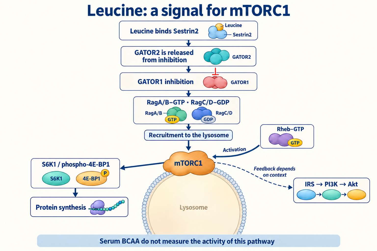
The diagram follows named states: inhibiting GATOR1 permits active Rag rather than directly inhibiting that active state. Rag provides localization; separate Rheb–GTP activates the kinase. Insulin signals through PI3K–Akt to restrain TSC1/2, a GAP for Rheb. Energy limitation and AMPK can constrain mTORC1 through TSC and Raptor. Leucine availability alone therefore does not ensure equal activation under every hormonal and energetic condition.
mTORC1 activates S6K1 and phosphorylates 4E-BP1, releasing its restraint on translation initiation. Sustained excessive signalling can feed back onto IRS and attenuate PI3K–Akt responses in specific models. However, IRS1 serine phosphorylation is not uniformly harmful. Copps et al. (2010) found that replacing mouse Ser307 with alanine worsened insulin resistance; the corresponding human residue is Ser312. Site, timing, tissue and parallel signals determine the effect.
In muscle, Akt2 regulates TBC1D4/AS160, Rab and GLUT4 trafficking. Disturbance can reduce glucose uptake without eliminating every Akt function. Biswas et al. (2020) examined BCKAs, mTOR and insulin signalling in myotubes and mouse models; KIC effects depended on exposure and differed from other ketoacids. This supports a possible mechanism, not a universal toxic serum threshold.
Downstream products require an operating pathway
Residual BCKDH flux produces branched acyl-CoAs and subsequent metabolites. Substrate overload can affect the acyl-CoA/free-CoA balance, reducing equivalents and oxidation of competing fuels. A complete BCKDH block cannot simultaneously generate unlimited downstream BCAA-derived acyl-CoAs. Partial defects, different organs and excess substrate create different situations.
Acylcarnitines can report acyl-group handling and help buffer free CoA. Elevated C3 or C5 does not automatically establish toxicity. Propionylcarnitine C3 also reflects odd-chain fatty acids, other amino acids, propionate and B12-related metabolism. A C5 signal can include isovaleryl-, 2-methylbutyryl- and pivaloylcarnitine, the last influenced by some medicines. C5-OH occurs in multiple metabolic disorders and is not a stand-alone biotin-deficiency test.
The valine product 3-hydroxyisobutyrate, 3-HIB, is particularly instructive. Jang et al. (2016) showed that muscle-derived 3-HIB promoted endothelial fatty-acid transport and insulin resistance in mouse models. Producing it requires downstream catabolism. This is a mechanism involving active flux, distinct from upstream BCKA accumulation or a complete BCKDH shutdown.
Acute leucine signalling, including glutamate-dehydrogenase activation, and KIC metabolism can stimulate β-cell insulin release through ATP production and ATP-sensitive potassium channels. An acute secretory response differs from chronic metabolic stress and dysfunction. Insulin stimulation is not inherently evidence of pancreatic injury.
Organ-specific evidence
Sun et al. (2016) linked impaired catabolic gene expression and KLF15 to human failing-heart samples and mouse pressure-overload models. BCKAs altered respiration and ROS in isolated mitochondria. Li et al. (2017) investigated BCAA accumulation, reduced hexosamine-pathway/O-GlcNAc activity, PDH suppression and mouse susceptibility to ischemia–reperfusion. In that study's direct comparison, BCAAs inhibited PDH whereas BCKAs did not. Combining these findings into a universal claim that BCKAs competitively inhibit PDH in all patients is unwarranted.
White et al. (2018) linked BCKDK/PPM1K to ATP-citrate lyase, ACLY, and lipogenesis in rats. Fructose-responsive ChREBPβ regulated this node; ACLY Ser454 numbering refers to the rodent protein studied. ACLY produces cytosolic acetyl-CoA from citrate, ACC produces malonyl-CoA, and FASN primarily forms palmitate, C16. Oleate, C18:1, requires elongation and desaturation rather than direct FASN production. Malonyl-CoA inhibits CPT1A, an acyltransferase controlling long-chain fatty-acid mitochondrial entry, rather than closing every oxidation “channel.” These links matter for steatosis/MASLD but do not establish BCKAs as the sole cause of fibrosis.
Zhenyukh et al. (2018) studied endothelial cells and high-BCAA-associated ROS/inflammatory signalling. This differs from the 2017 study in blood mononuclear cells. Cellular mTOR, NOX and NF-κB responses do not establish every intermediate or continuous exposure at equivalent concentrations in human vessels.
At the blood–brain barrier BCAAs compete with other large neutral amino acids, including tryptophan, tyrosine and phenylalanine. Precursor entry may influence serotonin and catecholamine synthesis, with free tryptophan, binding, transport affinities and local metabolism also relevant. Fernstrom's 2005 review does not establish a universal chain from high BCAAs to serotonin deficiency, depression or carbohydrate craving. Neurological toxicity in maple syrup urine disease cannot simply be transferred to ordinary protein intake.
Association, genetics and intervention answer different questions
Newgard et al. (2009) found a BCAA-related metabolic signature in obese versus lean people; the high-fat/BCAA supplementation experiments were in rodents. Wang et al. (2011) examined future diabetes: 201 cases developed over 12 years among 2422 initially eligible participants. Detailed metabolomic comparisons used selected groups. The three-amino-acid predictive score contained isoleucine, tyrosine and phenylalanine, not the sum of all three BCAAs. A large top-quartile relative risk in that analysis is neither a universal clinical cutoff nor proof of a fixed lead time over all conventional tests.
| Evidence | What it supports | Main limit |
|---|---|---|
| Newgard/Wang cohorts | BCAA-related concentrations track metabolic risk | Association and selection do not establish a single cause |
| Lotta (2016), Mahendran (2017), Jia (2025) genetic analyses | Relationships may run in both directions between BCAA metabolism and metabolic health | Instrument choice, pleiotropy and tissue effects constrain causal interpretation |
| Mouse genetics and cell models | BCAT/BCKDH/mTOR and individual metabolites can alter phenotype | Effects depend on the intervention point, dose and species |
| Karusheva (2019) crossover trial | Short-term BCAA reduction changed meal responses | No established long-term prevention or improved clamp sensitivity |
Lotta et al. (2016) used instruments including the PPM1K region to support a causal hypothesis of impaired BCAA metabolism. Mahendran et al. (2017) supported insulin resistance causing higher BCAA levels. Jia et al. (2025), a larger bidirectional analysis, linked BCAAs with lipids and also treated elevated BCAAs as a consequence of poor metabolic health. Feedback is plausible; “BCAAs caused this person's diabetes” is not established. Genetic findings for BCAA concentrations also do not automatically apply to BCKAs.
She et al. (2007) provide an important counterexample: mice lacking mitochondrial BCAT had high BCAAs yet increased energy expenditure and improved metabolic features. Blocking the first step differs from blocking BCKDH after ketoacid formation. Concentration cannot substitute for knowledge of the pathway.
Measurement and arithmetic without invented cutoffs
LC–MS/MS requires validated separation of amino-acid isomers. Leucine and isoleucine have the same molecular mass, approximately 131.17, and nominal protonated ion near 132.1. Mass alone cannot distinguish them. Alloisoleucine is relevant to suspected inherited defects. Interpretation depends on matrix, assay and preanalytics: diet, exercise, illness, renal function and sampling time should follow the specific laboratory's requirements rather than a universal fasting rule.
Olson et al. (2013) measured mouse tissue BCKAs with o-phenylenediamine derivatization and an isotope-labelled internal standard. This produces cyclic quinoxalinol derivatives. In contrast, 2,4-dinitrophenylhydrazine forms hydrazones, a different chemistry. Not every BCKA assay uses the same derivatization, and mouse tissue values are not human plasma reference intervals.
The BCAA sum retains concentration units:
For example, 130 + 65 + 220 = 415 µmol/L. This illustrates arithmetic, not an optimal value or diagnostic boundary. Summing KIC, KMV and KIV similarly describes the measured ketoacid pool. A BCAA/BCKA ratio is not a direct assay of BCAT activity or BCKDH phosphorylation.
| Marker | Measured quantity | Interpretation limit |
|---|---|---|
| Leu, Ile, Val and their sum | Amino-acid pool | Assay-specific references; no universal insulin-resistance threshold |
| KIC, KMV, KIV and their sum | Ketoacid pool | Plasma and tissue differ; concentration is not flux |
| C3 | Propionylcarnitine | Other substrates and B12 status contribute |
| C5 and C5-OH | Several acylcarnitines/isomers | Inherited disorders and medicines may contribute; identify the species |
| Fischer ratio | Usually BCAAs/(phenylalanine + tyrosine) | Historically used in liver disease; neither a brain neurotransmitter measure nor a universal diabetes test |
| HOMA1-IR | Fasting glucose–insulin model | Depends on units, insulin assay and β-cell function; does not measure BCKDH |
For HOMA1-IR (Matthews et al., 1985), with fasting insulin I₀ in µU/mL:
I₀ = 10 with G₀ = 5 mmol/L, approximately 90 mg/dL, gives about 2.22 in either form. This is not a universal cutoff. HOMA2 is a different model; low insulin secretion can produce a deceptively low index. Comparing HOMA-IR, TyG and QUICKI helps clarify their respective limits.
Intervention evidence and nutritional adequacy
BT2 is an experimental benzothiophene-carboxylate BCKDK inhibitor. Tso et al. (2014) reported an IC50 of about 3.19 µM in their assay, not a human dose. Zhou et al. (2019) found improvements in obese mouse models. Acevedo et al. (2024) showed an additional chemical-uncoupling action that lowers mitochondrial ROS and lipogenesis. BT2 effects therefore cannot be attributed entirely to selective BCAA lowering; these experiments do not provide an approved insulin-resistance treatment regimen. BCKDK binding by genistein or curcumin and clinical suppression by such supplements are not established here.
Karusheva et al. (2019) studied 12 people with type 2 diabetes in a small randomized crossover trial. An isocaloric diet and amino-acid mixture retained essential amino-acid adequacy. Short-term BCAA reduction lowered fasting levels by 17% and meal-related levels by 62%; meal-induced insulin secretion fell by 28%, while OGIS increased by 24%. Whole-body and hepatic sensitivity measured by hyperinsulinemic–euglycemic clamp did not differ. “Clamp sensitivity improved 28%” and “liver fat fell 22%” are not findings of this trial. Microbiome, FGF21 and adipose changes are interesting intermediate outcomes, not long-term clinical benefit.
Fontana et al. (2016) examined total-protein restriction in humans and selective BCAA restriction in mice; these are different interventions. Cummings et al. (2018) also studied diet-induced obese mice. None establishes a universal percentage reduction for every patient.
BCAAs remain essential. Protein needs depend on age, growth, pregnancy, muscle mass, exercise and disease, including kidney disorders. Isolated BCAA supplements differ from complete protein or whey; plant foods also contain BCAAs, and dietary quality depends on the whole amino-acid pattern. Physical activity and better metabolic health can change handling, without establishing one fixed “zone 2” exercise duration as a PPM1K activation protocol.
Thiamine-responsive maple syrup urine disease requires specialist management; a rare inherited condition does not justify high-dose B1/B6/magnesium for everyone with obesity. DLD deficiency can affect several complexes and cause lactic acidosis. Resveratrol–SIRT1, berberine–AMPK and sulforaphane–KEAP1/NRF2 are mechanistic research directions, not validated ways to dephosphorylate a patient's BCKDH. Glucoraphanin is a sulforaphane precursor; myrosinase and bioavailability alter exposure. Universal dose tables would imply unsupported precision. Metformin, SGLT2 inhibitors and incretin drugs have established indications, rather than demonstrated PPM1K-targeted regimens; tirzepatide is a dual GIP/GLP-1 agonist.
Clinical meaning and scope
High BCAAs may add information about metabolic risk but do not diagnose insulin resistance; normal values do not exclude it. Diabetes diagnosis relies on validated glucose and HbA1c measurements, sometimes glucose-tolerance testing, and clinical context (NIDDK). Blood pressure, lipids, body composition, diet, medicines and accompanying disease matter. A metabolomics panel is not mandatory for everyone. Markedly abnormal amino acids, alloisoleucine and neurological symptoms, particularly from childhood or during decompensation, warrant specialist investigation of inherited disease rather than self-directed protein restriction.
Prepared on 8 October 2026. This article uses targeted checks of primary studies and separately identified clinical/reference sources. It is not a systematic review, GRADE assessment or independent peer review. Some full texts were unavailable; accessible abstracts, metadata and passages were used, and exact inaccessible methods were not verified. Established biochemistry, cellular mechanisms, mouse phenotypes and clinical evidence carry different weight. Distinguishing the amino acid, ketoacid, downstream product and flux is necessary before turning an association into a diagnosis or treatment.
FAQ
Do high BCAAs prove insulin resistance?
No. They may mark risk, but diet, tissues and metabolism influence the result. Diagnosis requires clinical context and validated tests.
Does the BCAA/BCKA ratio measure BCKDH activity?
No. It compares concentrations affected by transport, production and clearance, rather than measuring enzyme phosphorylation.
Should BCAAs be removed from the diet?
BCAAs are essential. Experimental restriction does not establish a universal treatment diet; adequate protein and individual needs remain important.
Is BT2 a medicine for self-treatment?
No. It is an experimental compound with BCKDK inhibition and additional mitochondrial uncoupling effects, without an established treatment regimen here.
Sources
- Short-term dietary reduction of branched-chain amino acids reduces meal-induced insulin secretion and modifies microbiome composition in type 2 diabetes: a randomized controlled crossover trial (2019)
- The BCKDH Kinase and Phosphatase Integrate BCAA and Lipid Metabolism via Regulation of ATP-Citrate Lyase (2018)
- Bidirectional Mendelian Randomization Indicates Causal Relationships Between Circulating Branched-Chain Amino Acids and Metabolic Health (2025)
- The BCKDK inhibitor BT2 is a chemical uncoupler that lowers mitochondrial ROS production and de novo lipogenesis (2024)
- Irs1 serine 307 promotes insulin sensitivity in mice (2010)
- Molecular mechanism for regulation of the human mitochondrial branched-chain alpha-ketoacid dehydrogenase complex by phosphorylation (2004)
- Metabolite profiles and the risk of developing diabetes (2011)
- Protein phosphatase 2Cm is a critical regulator of branched-chain amino acid catabolism in mice and cultured cells (2009)
- Benzothiophene carboxylate derivatives as novel allosteric inhibitors of branched-chain α-ketoacid dehydrogenase kinase (2014)
- Quantitative Analysis of the Whole-Body Metabolic Fate of Branched-Chain Amino Acids (2019)
- BCAA catabolism in brown fat controls energy homeostasis through SLC25A44 (2019)
- Catabolic Defect of Branched-Chain Amino Acids Promotes Heart Failure (2016)
- Genetic evidence of a causal effect of insulin resistance on branched-chain amino acid levels (2017)
- Decreased Consumption of Branched-Chain Amino Acids Improves Metabolic Health (2016)
- Restoration of metabolic health by decreased consumption of branched-chain amino acids (2018)
- Sestrin2 is a leucine sensor for the mTORC1 pathway (2016)
- Branched-chain amino acids promote endothelial dysfunction through increased reactive oxygen species generation and inflammation (2018)
- A branched-chain amino acid metabolite drives vascular fatty acid transport and causes insulin resistance (2016)
- Defective Branched-Chain Amino Acid Catabolism Disrupts Glucose Metabolism and Sensitizes the Heart to Ischemia-Reperfusion Injury (2017)
- The Rag GTPases bind raptor and mediate amino acid signaling to mTORC1 (2008)
- Ragulator-Rag complex targets mTORC1 to the lysosomal surface and is necessary for its activation by amino acids (2010)
- Disruption of BCATm in mice leads to increased energy expenditure associated with the activation of a futile protein turnover cycle (2007)
- Targeting BCAA Catabolism to Treat Obesity-Associated Insulin Resistance (2019)
- Branched-chain amino acids and brain function (2005)
- Bidirectional transport of amino acids regulates mTOR and autophagy (2009)
- Branched-chain amino acid catabolism
- Maple Syrup Urine Disease (2020)
- Homeostasis model assessment: insulin resistance and beta-cell function from fasting plasma glucose and insulin concentrations in man (1985)
- Diabetes Tests & Diagnosis
- Quantification of branched-chain keto acids in tissue by ultra fast liquid chromatography-mass spectrometry (2013)
- Branched-chain ketoacid overload inhibits insulin action in the muscle (2020)
- Genetic Predisposition to an Impaired Metabolism of the Branched-Chain Amino Acids and Risk of Type 2 Diabetes: A Mendelian Randomisation Analysis (2016)
- A branched-chain amino acid-related metabolic signature that differentiates obese and lean humans and contributes to insulin resistance (2009)