Adiponectin connects adipose tissue with muscle, liver, vascular, and immune physiology. Its concentration often falls with visceral obesity, even though adipocytes are its principal source. This observation has stimulated research into adiponectin as both a biomarker and a therapeutic target. The underlying biology is substantial; an association with lower disease risk, however, does not establish that raising a blood concentration prevents disease.
From ADIPOQ to circulating multimers
Human ADIPOQ, at chromosome 3q27.3, encodes a 244-residue secreted protein, also called Acrp30, apM1, GBP28, or AdipoQ in earlier literature. Its architecture comprises a cleaved N-terminal signal peptide, a variable region, a collagen-like segment with Gly-X-Y repeats, and a C-terminal globular domain. The predicted precursor mass and apparent electrophoretic mass differ: post-translational modification and assay conditions help explain the familiar “30-kDa” designation. Precise domain boundaries depend on species and numbering conventions; they should not be transferred uncritically between mouse and human proteins.
The globular structure studied by Shapiro and Scherer (1998; PDB 1C28) reveals a C1q/TNF-related β-sandwich fold. Three globular heads associate through noncovalent contacts; the collagen-like chains form a triple-helical stalk. This structural relationship does not make adiponectin a pro-inflammatory TNF cytokine. Glycine at every third collagen-like position permits close chain packing, while hydroxylation and glycosylation influence assembly and secretion.
Circulating full-length adiponectin is chiefly oligomeric. Low-molecular-weight (LMW) trimers form the structural unit; middle-molecular-weight (MMW) hexamers contain two trimers; high-molecular-weight (HMW) assemblies contain several trimers, commonly described as 12–18 subunits. Apparent masses around 70, 140, and several hundred kDa are approximate analytical descriptions, not immutable physical constants. Relative fractions vary by sex, metabolic state, preparation, and method. LMW is biologically active in appropriate experimental systems and should not be called a passive reservoir simply because HMW predicts some metabolic outcomes better.
Proteolytically generated globular adiponectin, gAd, is widely used experimentally. Leukocyte elastase can cleave adiponectin, but a universal normal circulating gAd percentage and a precise physiological contribution are not established by these experiments. Results obtained with a recombinant globular fragment cannot automatically be assigned to the whole physiological hormone pool.
Modification and secretory quality control
A conserved N-terminal cysteine supports intermolecular disulfide bonds: Cys36 in the human precursor and Cys39 in mouse, rather than “Cys39 becoming Cys36 after signal-peptide cleavage.” Removing an 18-residue signal peptide changes numbering by 18, not three. Mutation experiments establish the importance of this cysteine for higher-order assembly, while noncovalent interactions still support trimers (Pajvani et al., 2003; Wang et al., 2007).
Conserved collagen-domain lysines undergo hydroxylation followed by glycosylation. Wang and colleagues' 2006 study describes positions 68, 71, 80, and 104 in its experimental sequence and shows that replacing the four lysines with arginine selectively disrupts HMW assembly. Species and precursor numbering must accompany residue lists. Hydroxylases use oxygen, iron, and 2-oxoglutarate; collagen-related glycosylation adds galactose and glucose to hydroxylysine. A simplified sequence is:
Lys⟶Hyl⟶Gal−O−Hyl⟶Glc−Gal−O−Hyl.
Here Lys is lysine and Hyl hydroxylysine. Proline hydroxylation helps collagen-like structural stability; this does not mean that proline and lysine undergo the same glycosylation reaction. Individual proline coordinates, exact enzyme assignments, and complete loss of biological activity need direct experimental support rather than extrapolation from collagen generally.
In the endoplasmic reticulum (ER), ERp44 retains adiponectin through a transient mixed disulfide. Ero1-Lα changes thiol-dependent retention and release, while DsbA-L supports multimerization and protects against ER stress (Wang et al., 2007; Liu et al., 2008). The designation DsbA-L reflects homology; it is not proof that the protein acts as a classical bacterial disulfide oxidase in every setting. Its human gene is GSTK1, not a newly defined DSBAL locus. Ero1 is an oxidizing ER enzyme: depicting its role simply as direct reduction of ERp44 obscures the coupled thiol-exchange system. Folding, retention, assembly, Golgi trafficking, and secretion are distinct stages.
What the HMW fraction measures
Pajvani and colleagues (2004) found that complex distribution related to thiazolidinedione-associated improvements in insulin sensitivity. Two dimensionless ratios can describe distribution:
SA=H+M+LH,RH=M+LH.
Here H, M, and L are HMW, MMW, and LMW concentrations measured in the same units and with a comparable analytical method. Provided the denominator is positive and the same fractions are counted,
RH=1−SASA,SA=1+RHRH.
A ratio of 0.5 in the first convention means 1.0 in the second. Confusing them creates a material interpretation error. Neither is a universal clinical “gold standard”; no single HMW percentage diagnoses insulin resistance or establishes metabolic health in every population. Total concentration, assay, kidney function, sex, age, and clinical context still matter.
Inflammation, hypertrophic adipocytes, hypoxia, oxidative stress, and ER stress can reduce adiponectin expression or secretion in obesity. TNF-α/IL-6, NF-κB/JNK-related pathways, impaired PPAR-γ activity, and the unfolded-protein response provide plausible experimental links. Chronic insulin resistance can also disturb secretion. A single cell diameter, obligatory ADIPOQ promoter methylation, or universal IRE1-mediated destruction of its transcript should not be presented as a demonstrated explanation in all patients. The obesity association and the high-adiponectin mortality paradox discussed below are different questions.
Receptors, adaptors, and sphingolipid metabolism
Yamauchi and colleagues (2003) identified AdipoR1 and AdipoR2. Both occur in multiple tissues; experimental emphasis on AdipoR1 in muscle and AdipoR2 in liver is useful but not an exclusive anatomical partition. Human AdipoR1 has 375 residues and AdipoR2 386; the latter is not a 311-residue protein. The source's ADIPOR1 locus “1p36.13–q41” combines incompatible chromosome coordinates and should not be retained.
The receptors belong to the PAQR family. Seven transmembrane helices do not make them conventional G-protein-coupled receptors: their N-terminus faces the cytoplasm and C-terminus the extracellular space. Tanabe and colleagues (2015) established structures with an internal zinc-containing cavity; nature14301 is a DOI suffix, not a valid PDB accession. Ligand recognition, membrane access to lipid substrates, and signaling are investigated experimentally; a completely resolved extracellular adiponectin–receptor binding geometry should not be inferred from an unliganded receptor structure.
Vasiliauskaité-Brooks and colleagues (2017) identified intrinsic basal ceramidase activity in purified AdipoR2, enhanced by adiponectin, and a structure with a bound fatty acid. This complements Holland and colleagues' 2011 receptor-associated ceramidase experiments. The zinc-containing pocket and proposed substrate pose support a catalytic model; every mechanistic detail and physiological substrate preference is not thereby settled. Sequence coordinates must be tied to the correct receptor and species rather than copied from the source's contradictory residue lists.
AdipoR-mediated hydrolysis does not directly produce sphingosine-1-phosphate (S1P); sphingosine kinases perform the subsequent phosphorylation. Ceramides can impair Akt signaling through phosphatase and kinase-dependent mechanisms and contribute to apoptosis. Altered sphingolipid balance may support survival in cardiomyocytes and pancreatic β-cells in experimental models, including AMPK-independent effects. Ceramide species and S1P actions are context-dependent: neither complete removal of “toxic lipids” nor universal S1P protection is a valid clinical inference.
T-cadherin, encoded by CDH13, is a GPI-anchored adiponectin-binding protein without a conventional intracellular tail. Hug and colleagues (2004) demonstrated binding to hexameric and HMW forms, unlike trimers and globular fragments. It supports tissue localization, particularly in cardiovascular tissues; Denzel and colleagues (2010) demonstrated its importance for cardioprotection in mice. It should not be reduced to a classical kinase receptor or a fully established obligatory AdipoR–integrin heterocomplex.
APPL1 has BAR, PH, and PTB domains that link membrane/endosomal interactions with receptor signaling. Mao and colleagues (2006) demonstrated interaction with AdipoRs and effects on lipid oxidation, glucose uptake, and GLUT4 translocation. The PTB domain can recognize the receptor's cytoplasmic region without implying that the receptor must first be tyrosine phosphorylated. APPL1 connects adiponectin and insulin pathways through interactions involving Akt, IRS proteins, and Rab trafficking. APPL2 can oppose signaling through receptor competition and APPL1 interactions. These regulatory experiments do not establish that every person with metabolic stress has complete APPL2 blockade. The source's 887-residue APPL1 size should not be used as a canonical human sequence value.
Muscle: access to the mitochondrial oxidation pathway
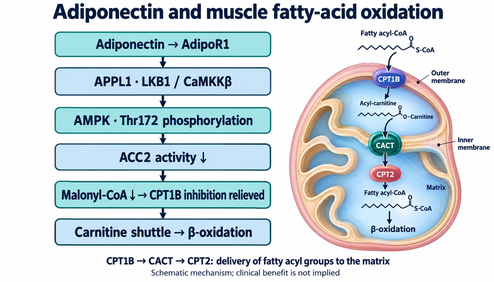
Adiponectin can activate AMPK by phosphorylation of Thr172 on its catalytic α-subunit, involving upstream LKB1 and calcium/CaMKKβ pathways. Iwabu and colleagues' muscle studies also connect AdipoR1 with calcium signaling, PGC-1α, and oxidative capacity. Activation magnitude depends on preparation; a universal 100–500-fold clinical response is inappropriate.
AMPK phosphorylates and inhibits acetyl-CoA carboxylase. ACC2-associated malonyl-CoA normally inhibits muscle CPT1B. Lower malonyl-CoA relieves this inhibition, increasing access of long-chain fatty-acyl groups to mitochondrial oxidation. The CPT1 and carnitine-shuttle article explains why this is transport regulation rather than β-oxidation itself. CPT1B forms acylcarnitine at the outer mitochondrial membrane; CACT exchanges carnitine/acylcarnitine across the inner membrane; CPT2 regenerates acyl-CoA on the matrix side. Omitting CPT2 or implying that cytosolic acyl-CoA crosses the inner membrane directly changes the mechanism.
AMPK-associated glucose uptake involves GLUT4-containing vesicles and Rab-GAP regulators TBC1D1 and TBC1D4/AS160, followed by Rab activation, docking, and SNARE-mediated fusion. Distinct phosphorylation sites and kinase contributions depend on species and stimulus. Insulin-independent signaling can coexist with improved insulin responsiveness; it does not guarantee normal glucose uptake in severe diabetes. DAG/PKC and ceramide/Akt mechanisms help explain lipid-associated insulin resistance, but DAG is not directly a β-oxidation substrate: its fatty-acyl components must first become available for metabolism.
PGC-1α coordinates longer-term mitochondrial adaptation. AMPK-dependent phosphorylation and NAD-dependent SIRT1 deacetylation can influence its activity, alongside NRFs, ERRα, and TFAM-mediated programs. NAMPT and NAD metabolism participate in this network; NAD is a cosubstrate for SIRT1, not merely an allosteric switch. These pathways are introduced further in the article on PGC-1α and mitochondrial biogenesis. Signaling does not make PGC-1α permanently hyperactive or ensure an obligatory conversion of all muscle fibers to type I.
Liver: oxidation, lipogenesis, and glucose production
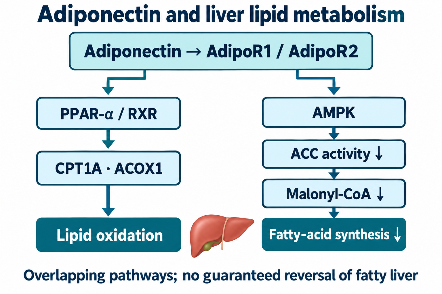
AdipoR2-associated PPAR-α activity promotes transcriptional programs for lipid handling. PPAR-α partners with RXR at response elements; targets include mitochondrial transport and peroxisomal oxidation genes such as CPT1A and ACOX1. APOA1 concerns lipoprotein biology, not a β-oxidation enzyme. Peroxisomes shorten selected very-long-chain fatty acids; their first oxidation step generates hydrogen peroxide rather than conserving electrons through the mitochondrial respiratory chain in the same way. AdipoR1 and AMPK pathways also operate in liver, so receptor functions overlap.
AMPK-dependent ACC inhibition can lower malonyl-CoA and de novo lipogenesis. SREBP-1c processing and transcriptional effects involve FASN and SCD1. Residue numbers often reported as ACC1 Ser79 or ACC2 Ser212/Ser221 differ across species and isoforms; they are not universal human coordinates. A diagram should emphasize inhibition and metabolic direction instead of unresolved residue assignments.
CREB–CRTC2 signaling regulates gluconeogenic genes PCK1 and G6PC. Phosphorylated CRTC2 can bind 14-3-3 and remain outside the nucleus. Nevertheless, adiponectin's suppression of glucose production is not reducible to an obligatory direct AMPK→CRTC2 Ser171 step: Miller and colleagues (2011) found effects persisting without the LKB1–AMPK pathway. Related kinases and other mechanisms matter. Nor does inhibition of a signaling node mean instant, complete disappearance of hepatic glucose production, a necessary physiological process. Mouse improvements in fatty liver cannot be described as established reversal of human MASLD or MASH by adiponectin treatment.
Vascular and immune effects: strong mechanisms, distinct endpoints
Endothelial experiments link adiponectin with AMPK/Akt, eNOS activation, NO availability, Hsp90, and angiogenic responses (Ouchi et al., 2004). NO activates soluble guanylate cyclase in vascular smooth muscle, increasing cGMP/PKG signaling and relaxation. Superoxide can consume NO and form peroxynitrite, connecting redox stress to endothelial function. A schematic arginine→citrulline+NO arrow is useful, but an unbalanced reaction should not be presented as exact eNOS stoichiometry. These experiments do not establish normalization of everyone's blood pressure.
Ouchi and colleagues (1999) showed modulation of TNF-induced endothelial adhesion molecules. NF-κB/IκB, VCAM-1, ICAM-1, and E-selectin connect inflammatory activation with monocyte recruitment. Macrophage experiments also support changes in inflammatory cytokines, IL-10/IL-1Ra, and lipid uptake/foam-cell formation. M1/M2 is a simplified experimental vocabulary for a much broader spectrum of macrophage states; adiponectin does not abolish all monocyte adhesion or all atherosclerosis.
Mouse ischemia–reperfusion studies implicate AMPK and COX-2-dependent protective mechanisms (Shibata et al., 2005), with T-cadherin-dependent localization and sphingolipid pathways providing complementary biology. Mouse pressure-overload studies support antihypertrophic effects (Shibata et al., 2004). The source's TSC2 direction requires correction: AMPK can activate TSC2-mediated restraint of mTORC1, not inhibit TSC2 to suppress mTOR. Experiment-specific infarct-size differences cannot be promised as a human 30–50% reduction.
Human associations and the adiponectin paradox
Lower adiponectin accompanies obesity, insulin resistance, type 2 diabetes, and fatty liver in many cohorts. Concentration ranges overlap and depend on assay and context; values such as 5–20 µg/mL are descriptive, not a universal reference interval, and low adiponectin is not pathognomonic. Sex differences and androgen effects are relevant but do not turn one concentration into a diagnostic rule. Experimental inhibition of hepatic stellate-cell activation provides antifibrotic plausibility; a blood measurement cannot independently grade MASH fibrosis.
Pischon and colleagues (2004) studied a nested case–control sample within 18,225 men in HPFS: 266 events and 532 controls over six years. Highest versus lowest adiponectin quintile gave RR 0.41, 95% CI 0.24–0.70, after broader adjustment; adding LDL/HDL cholesterol attenuated it to 0.56, 0.32–0.99. The source's RR 0.44 and “56% lower after lipid adjustment” are incorrect. With RR 0.56, the relative reduction is 44%, calculated as 100×(1−RR), not an absolute reduction in an individual's risk.
Spranger and colleagues (2003), using an EPIC-Potsdam nested case–control design, reported highest versus lowest quartile OR 0.3, 95% CI 0.2–0.7, P=.0051. This is an odds ratio, not the source's RR 0.37 with its stated interval. Neither study was an adiponectin-raising intervention, and neither establishes that HMW explains exactly 80% of protection.
In chronic heart failure, higher adiponectin can accompany higher mortality (Kistorp et al., 2005). Wasting, natriuretic peptides, altered secretion and clearance, reverse causation, and tissue resistance are possible contributors. Kidney disease changes interpretation, but the original Zoccali et al. 2002 study found lower adiponectin associated with more cardiovascular events in end-stage renal disease; it cannot support an undifferentiated claim that high adiponectin universally predicts death on dialysis. Renal handling is complex, not filtration of a normal pool of free monomers followed by complete cessation at one GFR. High concentration is therefore neither universal reassurance nor proof that adiponectin causes harm.
Swarbrick and colleagues (2006; corrected PMID 17019599) followed 19 women before, one month, and twelve months after Roux-en-Y gastric bypass. HMW increased and related to improved insulin sensitivity; this does not establish a universal 150–280% rise at 4–12 weeks, isolate adiponectin from weight loss and other changes, or prove the same sequence for every bariatric operation.
Genetics and interventions
ADIPOQ variants rs2241766 (+45T>G), rs1501299 (+276G>T), and rs266729 (−11377C>G) have been investigated for associations with adiponectin and metabolic outcomes. Hara and colleagues (2002) provide an early Japanese association study. Linkage disequilibrium, ancestry, sample size, diet, and confounding affect results. A synonymous variant does not automatically demonstrate altered splicing, a promoter allele does not universally reduce expression by 40%, and one SNP cannot prescribe carbohydrate intake. Candidate ADIPOR1/2 variants likewise do not establish personalized training intensity or reliable MASH prognosis.
AdipoRon, identified by Okada-Iwabu and colleagues (2013), is a small-molecule experimental agonist with receptor-dependent metabolic effects in mice. It improved glucose-related outcomes and survival under particular experimental conditions. Mouse oral activity does not establish an approved human drug, equivalence to exercise, or demonstrated human longevity. Manufacturing full-length adiponectin is challenging because assembly, modification, exposure, and delivery matter; “technologically impossible” and mandatory gram doses are unjustified absolutes.
Thiazolidinediones activate PPAR-γ and can increase adiponectin, particularly HMW. The Pajvani 2004 findings support a relationship with insulin-sensitivity changes, not a universal S_A shift from 0.25 to 0.65 or proof that all therapeutic benefit is mediated by adiponectin. Pioglitazone selection depends on the patient's disease, benefits, and risks; a low research biomarker is not an independent prescribing indication.
EPA/DHA and FFA4/GPR120, β-arrestin–TAB1–TAK1 inflammatory signaling, and PPAR-related effects are plausible experimental links. The Wu et al. 2013 fish-oil meta-analysis included 14 trials and 682 treated versus 641 control participants, rather than the source's 36 trials and 2,514 participants; its pooled increase was 0.37 µg/mL, with heterogeneity. Raising a surrogate does not establish prevention of diabetes or a universal 2.5–3.5-g daily prescription.
EGCG, curcumin, resveratrol, and astaxanthin have experimental anti-inflammatory or redox mechanisms and variable small human studies. Their formulations, doses, populations, and endpoints differ. A curcuminoid–piperine trial cannot prove an effect specific to a phytosomal formulation, and the source's asserted direct DNMT1/ADIPOQ mechanism or exact EGCG percentage is not established by its listed references. Astaxanthin-related membrane biology does not demonstrate normalization of LH3 kinetics in patients. These compounds should not be assembled into a mandatory adiponectin supplement stack.
Fermentable fibers, microbiota, short-chain fatty acids, intestinal barrier function, and metabolic endotoxemia connect nutrition with adipose inflammation. Cani and colleagues' 2007 mouse work supports a mechanistic research direction, not guaranteed barrier “sealing” in humans. Butyrate can affect histone deacetylases; H3K9ac denotes acetylation, and H3K4me3 methylation, not “hyperhydroxylation.” FFAR2/3 and GLP-1 effects add biological plausibility without proving a necessary ADIPOQ epigenetic chain in every individual.
Aerobic, interval, and resistance exercise benefit metabolic health through numerous pathways, including contraction-driven AMPK activation, mitochondrial adaptation, and altered body composition. Adiponectin responses vary with program, weight change, baseline health, and assay. Exercise reviews should not be converted into a universal 18–35% rise or obligatory two- to threefold receptor expression. A heart-rate percentage alone does not define everyone's physiological Zone 2 or FatMax; HIIT and resistance training are individualized independently of an unvalidated HMW target.
Exposure cannot be assumed equivalent to physiological full-length hormone
AdipoR1/R2
Inverted 7-TM topology; signaling and lipid-metabolic functions
Muscle/liver preferences do not imply exclusivity
T-cadherin
GPI anchoring and MMW/HMW tissue localization
Lacks a conventional intracellular signaling tail
Clinical evaluation addresses established questions: glycemic status, lipids, blood pressure, adiposity, liver disease when indicated, kidney function, symptoms, and treatment goals. HOMA-IR is an assay-dependent fasting surrogate; a universal 30% reduction is not a validated adiponectin-treatment endpoint. TG/HDL ratios require defined concentration units and cannot share a universal threshold across mg/dL and mmol/L. CAP and elastography address different aspects of liver assessment and require their own interpretation. A mandatory panel, S_A≥0.55 target, 12-week success algorithm, and fixed supplement/drug doses from the source are therefore replaced by context-dependent assessment.
This is a narrative scientific review with targeted verification of disputed source identifiers, molecular descriptions, and clinical results. Accessible primary abstracts, publisher materials, and structural publications were checked on 8 October 2026; it is not a systematic search, independent GRADE assessment, clinical protocol, or external peer review. Experimental mechanisms are retained with their model limitations, unsupported numerical rules are removed, and unresolved mechanisms are identified as such. Adiponectin is a productive research bridge between adipose biology and substrate metabolism, while clinical management remains anchored to demonstrated patient outcomes.
FAQ
Does high adiponectin always mean good health?
No. It can associate with better metabolic status in some populations and with severe illness, wasting or altered clearance in others.
Is HMW adiponectin a universal clinical target?
No. Assays and patient context differ; a fixed HMW ratio cannot independently diagnose insulin resistance or prescribe treatment.
Does AdipoR directly produce S1P?
No. Ceramidase hydrolysis produces sphingosine; sphingosine kinase then phosphorylates it to S1P.
Can AdipoRon replace exercise or diabetes treatment?
Its original effects were demonstrated in experimental models. Human efficacy and clinical equivalence cannot be inferred.
Should a low adiponectin result trigger a supplement stack?
No. Biomarker changes do not establish outcome benefits, and nutrition, activity and medicines require an individual clinical objective.