Secretory IgA (sIgA): polymeric immunoglobulin receptor (pIgR) transcytosis, immune exclusion of pathogens, and symbiosis with commensal microbiota
How secretory IgA is assembled and transported across the epithelium, how it can limit pathogens while supporting some commensals, and why stool sIgA alone is not a diagnosis or treatment target.
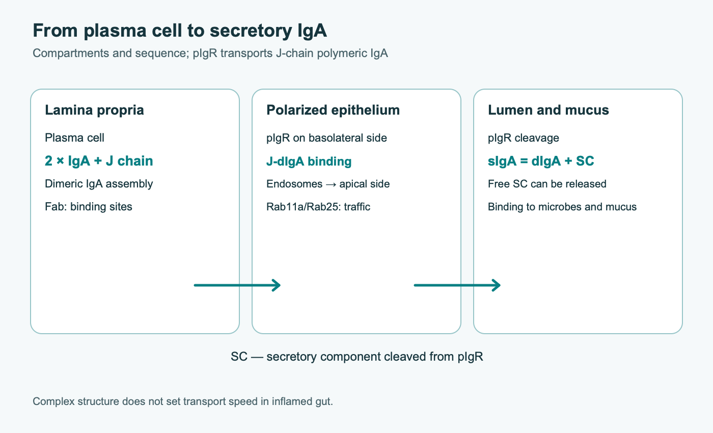
Secretory immunoglobulin A (sIgA) links plasma cells, intestinal epithelium, and luminal microbiota. Mostly polymeric IgA is made in the lamina propria; epithelial pIgR carries it to the apical surface and, after cleavage, leaves its secretory component attached to the antibody. The resulting molecule binds microorganisms and their products without necessarily initiating inflammation. sIgA does more than exclude pathogens: some commensals use IgA to maintain a stable position in mucus. A stool or saliva sIgA value alone neither describes the whole system nor shows that an “immune-boosting” supplement is needed.
1. Components of secretory IgA
An IgA monomer has two heavy α chains and two light chains, like other antibodies, but a secretory complex usually contains two monomers. Their Fc regions associate with the J chain, and the outside bears secretory component (SC), the cleaved extracellular portion of pIgR. A dimer has up to four Fab antigen-binding sites. Polymerization gives high avidity for repeated bacterial epitopes even when each Fab binds its antigen only moderately. Secretory IgM can also pass through pIgR because it carries a J chain; serum monomeric IgA is not the same molecule as luminal sIgA.
Humans have IgA1 and IgA2 subclasses. The longer O-glycosylated hinge of IgA1 gives Fab flexibility but can be targeted by some bacterial IgA1 proteases. The IgA2 hinge is shorter and therefore more resistant to those proteases specifically; it is not immune to all intestinal enzymes. Subclass proportions differ among serum, secretions, and intestinal regions and depend on sampling methods. Differences in IgA2 disulfide organization also mean that one diagram cannot represent every molecule.
The JCHAIN-encoded J chain is made by IgA- and IgM-secreting plasma cells. It directs the assembly of Fc tails into an asymmetric complex with interchain disulfide bonds. Cryo-electron microscopy structures from Kumar and colleagues and Wang and colleagues show that the J chain is more than a “staple”: it sets the geometry of the polymeric Fc core and the surface recognized by pIgR. These structures explain how dimeric and more highly organized IgA can assemble, but a structural model does not report the rate of secretion in a living inflamed intestine. Without direct verification of structure and residue numbering, specific Cys-partner lists from an earlier manuscript should not be treated as a universal map of human sIgA.
Covalent assembly occurs before contact with epithelium, in the plasma cell’s secretory apparatus. Oxidation of cysteine thiols forms disulfide bridges (2 R–SH → R–S–S–R); correct folding of heavy-chain and J-chain tailpieces determines whether the complex is recognized by the receptor. Finding JCHAIN in a biopsy transcriptome does not reveal how many mature dIgA molecules are present: protein and secreted-polymer measurements are needed. Structural descriptions should distinguish the stable principle—an asymmetric J-chain-assembled Fc core—from residue numbers that shift with signal-peptide counting, subclass, and recombinant construct.
The extracellular part of pIgR contains five immunoglobulin-like domains, D1–D5. D1 initiates recognition of J-chain-containing polymeric immunoglobulin; the remaining domains wrap around the complex. A disulfide bond between SC and some IgA molecules stabilizes the complex, but the proportions of covalent and noncovalent forms depend on the biological material and assay. After apical cleavage, the pIgR ectodomain remains on the antibody as SC. Some pIgR is transported without bound IgA and releases free SC, which can also interact with microbial surfaces.
IgA and SC glycans are functional parts of the molecule. N-glycans on heavy chains and SC, O-glycans on the IgA1 hinge, and their fucosylation and sialylation create a heterogeneous surface. This can affect proteolytic stability, interactions with mucin and microbial lectins, and act as a decoy target for adhesins. Glycosylation varies with the producing cell and tissue state. It is as incorrect to claim that every pathogen binds every sIgA through SC as it is to assume that Fab alone determines contact. For a particular microbe, intact antibody, Fab specificity, and glycoforms need to be compared.
SC can increase IgA stability in secretions under some conditions, but does not make it indestructible. Bacterial proteases, pH, digestive enzymes, and transit time affect antibody survival. A single structural study cannot establish “tens of hours” of persistence for every intestinal region and dose.
2. Directed route: plasma cell — pIgR — lumen
Lamina-propria plasma cells release J-chain-containing dimeric IgA beneath the epithelium. Epithelial cells synthesize pIgR, process it through the ER and Golgi, and place it on the basolateral surface. The receptor binds dIgA or polymeric IgM and is then internalized. Sorting signals in the pIgR cytoplasmic tail, adaptors, clathrin, and endosomal proteins regulate traffic. The complex moves from an early basolateral endosome through recycling endosomal compartments to the apical membrane. Rab11a and Rab25 participate in sorting and directed delivery in studied polarized cells, but no single GTPase is the universal “motor” for the whole pathway.
At the apical side, the pIgR extracellular portion is proteolytically released with dIgA as sIgA; some unloaded receptors yield free SC. Luminal sIgA therefore depends at least on plasma-cell number, dIgA synthesis, pIgR expression and polarity, vesicular traffic, proteolysis, and dilution of secretions. Increased PIGR mRNA does not guarantee increased functional transport, and a low concentration in diluted stool does not prove inadequate production.
Direction matters: pIgR is on the epithelium, and antibody reaches it from the tissue. Mixing apical and basolateral compartments in culture can make passive protein leakage look like transcytosis. A stronger model uses a polarized monolayer, adds dIgA below, measures sIgA and SC above, and checks junction integrity while blocking or genetically altering pIgR. If the monolayer is damaged, antibody can pass between cells, so IgA on the other side no longer demonstrates active receptor transport. Colocalization with Rab11a indicates a route but is not a quantitative measure of speed.
A “strictly fixed” pIgR-binding constant for every dIgA is misleading: affinity depends on whether the assay uses D1 or full SC, valency, glycans, and experimental conditions. The receptor favors J-chain-containing polymers; this does not prove the absolute absence of every other weak interaction. Nor is a single apical cleavage site and protease established for all epithelia. Structural binding, routing, and secretion are distinct experimental outcomes.
3. How B cells become a source of intestinal IgA
Peyer’s patches and isolated lymphoid follicles are sites where responses are induced. M cells in follicle-associated epithelium and antigen-presenting cells deliver and present parts of luminal antigens. After activation, B cells and plasmablasts migrate to the lamina propria, the effector site of secretion. Region, age, diet, infection, and prior antigen exposure change the relative contribution of pathways. The often-repeated estimate that “70% of all body lymphocytes are in GALT” is not needed to explain the mechanism and depends on how the tissue is defined.
In T-cell-dependent responses, CD40 on B cells interacts with CD40L on T follicular helper cells; IL-21 and other local signals support germinal centers, somatic hypermutation, and clone selection. Antigen specificity matters for neutralizing toxins and particular pathogens. In T-independent pathways, epithelial, dendritic, and stromal cells can support IgA class switching through BAFF and APRIL with a different pattern of T-cell help. This does not mean that most human IgA necessarily arises through one T-independent route or that every T-independent IgA is weak and polyreactive. The distribution depends on species, region, microbiota, and antigen.
Class switching is a chemical DNA rearrangement, not a simple change in protein secretion. TGF-β acting through SMAD promotes transcription of the Cα region. AID (AICDA) deaminates cytidines in transcribed switch regions (dC → dU). UNG removes uracil, and endonuclease and repair steps create breaks; end joining then brings VDJ next to Cα. AID activation and somatic hypermutation are related but occur in different parts of the immunoglobulin locus. IL-10, IL-21, BAFF/APRIL, and retinoic acid alter the response context; none alone turns any increase in stool IgA into high-quality protection.
Retinoic acid, produced from vitamin A by local cells, helps program gut homing. Integrin α4β7 interacts with endothelial MAdCAM-1; CCR9/CCL25 is particularly important for the small intestine, while CCR10/CCL28 helps distribute cells to other mucosal sites and intestinal regions. After leaving blood, plasmablasts mature into IgA-secreting cells. The homing axis is more complex than “vitamin A → IgA”: deficiency disrupts it, but extra high doses have not been shown to universally improve clinical protection in adequately nourished people.
4. Immune exclusion does not sterilize the microbiota
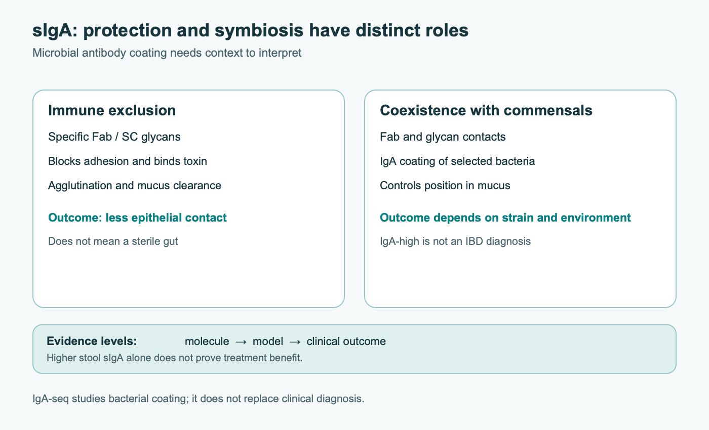
An antibody’s Fab can block a microbial adhesin or a toxin’s receptor-binding site. Multivalent sIgA binds repeated epitopes on neighboring cells and forms aggregates; Moor and colleagues also showed “enchaining” of dividing bacteria in a defined model. Clusters have more difficulty reaching the epithelial surface and may leave with mucus and intestinal flow. SC and IgA glycans can further retain some microbes in the mucus environment. Yet the organization, sterility, and renewal rate of inner and outer mucus differ between small and large intestine and change with inflammation. sIgA limits unwanted contact; it does not destroy every coated bacterium.
Antibodies to cholera toxin can prevent its B subunit from binding GM1; effects on other toxins depend on the specific epitope and local concentration. LPS recognition alone is not chemical detoxification of lipid A. sIgA may retain bacterial structures in the lumen and reduce tissue access, whereas LPS dephosphorylation is a different reaction, for example by intestinal alkaline phosphatase. The claim that any sIgA completely prevents LBP/CD14/TLR4 signaling is too strong.
Intracellular neutralization during transcytosis has been shown for particular antigen-specific IgA and viral models, including rotavirus. This does not show that every virus named in the earlier manuscript replicates in one endosomal compartment and is exported by this route. Antigen bound by dIgA on the lamina-propria side can also be carried toward the lumen, a mechanism called immune excretion. Its importance depends on antibody access to antigen and epithelial integrity; it is not a pump that reliably clears every allergen from blood.
IgA usually activates the classical complement pathway through C1q less strongly than IgG, which suits a persistently colonized mucosa. But “non-inflammatory” does not mean devoid of effector functions. Myeloid FcαRI/CD89 is found mainly on the tissue side of the barrier. Monomeric and multivalent IgA complexes can organize receptor-associated ITAM signals differently: in some models weak activation recruits SHP-1 and limits inflammation, whereas dense crosslinking activates Syk and effector cells. This is separate from luminal exclusion and is especially relevant when immune complexes enter tissue.
Binding and functional neutralization are also different. ELISA confirms antibody contact with a purified antigen, not blockade of infection in living epithelium. For a toxin, one must test whether antibody prevents receptor binding and toxic action; for bacteria, adhesion, invasion, and spread; for viruses, infectivity at the selected life-cycle stage. High avidity for purified LPS does not equal TLR4 deactivation after a complex enters tissue. Different infections can require different Fab specificities even when the transport route is shared.
5. Commensal coating: a contact marker, not a “pathogen” label
IgA coating can be measured by flow cytometry, after which fractions are sorted and characterized (IgA-seq). Palm and colleagues found that highly IgA-coated bacteria isolated from people with inflammatory bowel disease increased susceptibility to colitis after transfer to germ-free mice. This is a strong functional experiment, but not a diagnostic rule that every IgA-high taxon is “guilty” in every patient. The result depends on host state, microbial mixture, sorting method, and model. High coating can mean active immune control, proximity to mucus, or antigen exposure during inflammation, not uniform virulence of every member of a taxon.
Bunker and colleagues described polyreactive IgA that restricts pathobionts, while Donaldson and colleagues showed that some microbiota members use IgA for mucosal colonization. Thus, “less IgA on bacteria” is not always better, and “more” does not always indicate disease. The association can depend on Fab recognition of surface antigens, SC glycans, bacterial growth, and mucus properties. The contribution of each mechanism for Bacteroides or Akkermansia cannot be established without experiments on a defined strain and antibody. The earlier manuscript’s broad claim that IgA suppresses flagellin, invasins, and endotoxin synthesis across all named commensals is not supported by one series of experiments.
Retaining a microbe in outer mucus differs from uncontrolled growth of a dense biofilm. In a healthy colon, microbes interact with outer-layer MUC2, but inner-layer structure varies among species and is disrupted in colitis. The proportion of coated bacteria in stool does not describe their spatial distribution near the intestinal wall. The functional question is whether a particular antibody reduces invasion, pathological inflammation, or microbial spread in a given community.
6. IgA deficiency, inflammation, and disease: distinct clinical questions
Selective IgA deficiency is defined by persistently low serum IgA with normal IgG and IgM at an appropriate age; a commonly used criterion is below 0.07 g/L in people older than four years. Diagnosis requires clinical context and exclusion of secondary causes. It is not diagnosed from one stool sIgA result. Many people with this immune phenotype have no severe symptoms; some experience respiratory or gastrointestinal infections, autoimmune conditions, or allergic disease. J-chain-containing secretory IgM can compensate through pIgR, but this varies among patients. HLA and B-cell regulatory gene variants confer susceptibility, but a TACI defect alone does not describe all patients. When celiac disease is suspected in IgA deficiency, tests that do not rely solely on IgA are important. Adverse reactions to blood products can occur in some people, but anaphylaxis is not inevitable for everyone.
In inflammatory bowel disease, epithelial mass, pIgR expression, antibody composition and flow, permeability, and microbial load can all change. Cytokines can induce PIGR in some settings; severe epithelial injury may reduce effective transport in others. The claim that chronic IBD always suppresses pIgR by 60–80% and nearly stops sIgA is not generalizable to ulcerative colitis and Crohn’s disease. Biopsy cell composition and sampling location strongly affect mRNA levels. Increased IgA coating can coexist with a damaged barrier. Mechanistic studies need paired tissue, luminal, and clinical-activity measures, not one marker alone.
IgA helps limit microbial contact, but sIgA deficiency is neither a necessary nor sufficient cause of small intestinal bacterial overgrowth (SIBO). Motility, anatomy, acidity, medicines, bile, and diet affect small-intestinal bacterial growth. Aspirate counts and breath tests depend on method; a universal CFU threshold cannot be declared and used to infer “systemic endotoxemia” from low stool IgA. Bacterial translocation to mesenteric nodes is a separate phenomenon with different evidence, especially in severe injury models.
Interpreting sIgA requires attention to the sample matrix. Saliva, duodenal aspirate, stool, and tissue biopsy represent different organs, transit times, and dilution methods. Watery stool and inflammatory exudate change concentration per gram even if secretion is unchanged. Total IgA does not reveal antigen specificity, polymerization, SC composition, or binding to a particular strain. Longitudinal comparisons require the same laboratory method and attention to infection, medicines, and collection time. For recurrent severe infections, clinicians assess other immunoglobulins and functional responses too; a commercial “low sIgA” result should not replace that evaluation.
IgA nephropathy involves galactose-deficient IgA1, autoantibodies to its altered O-glycosylated hinge, and circulating complexes deposited in the mesangium. It is a systemic multistep disease; it cannot be explained by low stool sIgA or automatically attributed to faulty gut homing of plasma cells. Raising total sIgA with a supplement is not an established treatment. Associations among stress, sleep, diet, and immune measures also require attention to timing, baseline health, and sample type. Claims that cortisol invariably suppresses PIGR directly and reduces secretion by 50–70% in everyone are not established clinical rules.
Measurement
What it can tell you
What it does not prove
Serum IgA, IgG, and IgM
Helps assess a systemic immunoglobulin phenotype
Does not separately measure transport rate through pIgR
Stool or secretion sIgA
Antibody amount in a specific sample
Does not define a universal “weak barrier” diagnosis
PIGR mRNA or tissue staining
Expression at a selected site and time
Does not guarantee completed transcytosis
IgA-seq
Coating and composition of sorted bacteria
Does not establish causation of each taxon in a person
Infections, endoscopy, clinical course
Patient-relevant outcomes
Does not automatically identify which step of the sIgA axis changed
7. Diet, microbial preparations, and passive antibodies
Dietary fiber fermentation produces acetate, propionate, and butyrate. These short-chain fatty acids can act through FFAR2/FFAR3 and HCAR2 receptors, energy metabolism, and histone deacetylases. In mouse and cell experiments, acetate enhanced microbiota-directed IgA responses and butyrate affected mucosal IgA generation; pIgR expression was also studied separately in HT-29 epithelial cells. An acid’s effect on B cells and a fermentation mixture’s effect on epithelium are different experiments. These studies do not establish a fixed 2.5–4-fold increase in PIGR or a dose of inulin that treats IgA deficiency. Response depends on baseline microbiota, fiber tolerance, fermentation site, and disease.
Probiotic findings are also strain- and outcome-specific. Buts and colleagues found increased sIgA and SC secretion in the small intestine of rat pups after Saccharomyces boulardii; this was not a placebo-controlled study in adults with IBD and does not support a promise of a 40–65% rise in human stool sIgA after 7–10 days. Studies of Lactobacillus rhamnosus GG in children with rotavirus diarrhea and in Crohn’s disease involved different populations and immune outcomes. A measured antibody increase should be separated from reduced illness; findings from different strains cannot be combined into one “sIgA dose.” Safety of live products deserves particular attention in immunocompromised people. See the article on probiotic safety.
Vitamin A and its metabolite retinoic acid participate in homing and maturation of IgA responses; nutritional deficiency can impair mucosal immunity. Vitamin D and zinc affect many cells and barrier processes. But absence of deficiency does not mean that high doses improve infection protection. Claims of an obligatory human VDRE in PIGR, absolute dependence of every plasmablast on one receptor, or an “over 80%” fall in intestinal sIgA from vitamin A deficiency should not become universal recommendations. Correcting an established deficiency is a separate matter from treating immunodeficiency.
Bovine colostrum mainly contains cow immunoglobulins, especially IgG; its composition varies with time after calving, processing, and animal immunization. It is not human dIgA produced in the lamina propria, and oral intake does not correct a genetic or cellular IgA deficiency. In two double-blind placebo-controlled trials by Otto and colleagues, 90 healthy volunteers received tablets of hyperimmune colostrum from cows immunized against ETEC strains before experimental O78 ETEC challenge. One regimen provided 400 mg colostrum protein three times daily with a buffer; comparator groups differed in dose and buffer. Diarrhea protection was high in some groups, but this was a controlled challenge with one strain and specific antibodies. It does not show that ordinary colostrum at an arbitrary dose prevents all travelers’ diarrhea or treats IBD. Studies of childhood rotavirus diarrhea also used specifically immune material and a different endpoint. Dietary protein can be degraded and can cause adverse reactions, so these results cannot support mass “sIgA replacement therapy.”
8. Methods and limits of this review
This is a narrative review of structural, cellular, animal, and limited human evidence on intestinal sIgA. On October 9, 2026, primary records relevant to disputed claims about IgA structure, S. boulardii, and hyperimmune-colostrum trials were checked directly. Human source samples and microbiota transfer to mice in the IgA-coating study were also compared. This was not an exhaustive systematic search, meta-analysis, or formal risk-of-bias assessment of every cited publication.
Molecular structure explains a possible mechanism, cell and animal models test it under defined conditions, and human trials answer only the clinical question measured. Available studies do not establish a universal stool sIgA cutoff for diagnosing “leaky gut” or show that increasing one test result with a supplement improves IBD, SIBO, or IgA nephropathy. An individual result should be interpreted alongside symptoms, age, infections, treatment, and sample collection method.
9. Takeaway
The axis “plasma cell — J-chain-containing dIgA — epithelial pIgR — secretory component — lumen” explains why mucosal protection depends on several compartments. sIgA can exclude pathogens, neutralize some toxins, and help manage commensal location without obligatory inflammation. The same antibodies can become markers of active contact as microbiota and mucus change; by themselves they are not signs of health or disease. Clinical interpretation is more reliable when immune deficiency, inflammatory disease, and the outcome of a specific intervention are assessed separately instead of being reduced to one sIgA number.
FAQ
What is secretory IgA?
It is usually polymeric IgA that has passed through epithelial pIgR and retained its cleaved extracellular secretory component. It acts in mucosal secretions.
How does secretory IgA differ from serum IgA?
Serum IgA is often monomeric; sIgA usually includes a J chain and secretory component. Their concentrations and clinical meanings are not interchangeable.
Does low stool sIgA prove immunodeficiency?
No. Selective IgA deficiency is assessed with serum IgA, IgG, and IgM in light of age and possible causes. Stool results vary with collection, dilution, transit, and inflammation.
Why does IgA coat beneficial bacteria?
Coating can limit access to the epithelium and, in some systems, support mucosal colonization. It does not necessarily kill the bacterium or mean it is pathogenic.
Can a probiotic or fiber raise sIgA?
Some strains and metabolites changed immune measures in specific cell, animal, or limited human studies. No universal dose is established for treating a low stool sIgA result.
Does bovine colostrum replace human sIgA?
No. Trials of specific hyperimmune colostrum against ETEC studied passive antibodies in controlled challenge; ordinary colostrum does not correct human IgA deficiency.
Can IgA-seq diagnose IBD?
IgA coating helps study immune–microbiota interactions. It does not replace clinical diagnosis or automatically identify a culpable bacterial species in an individual patient.