Catalase: peroxisomes, H₂O₂ kinetics and mCAT — NutriFit
Catalase and peroxisomes: heme catalysis, H₂O₂ kinetics and limits of clinical translation
Catalase structure and NADPH, PEX5 import, ACOX1–H₂O₂ coupling, two catalytic routes, the GPx/Prx network, acatalasemia, rs1001179 and the limits of inference from mCAT mice.
Catalase solves an unusual redox problem: it uses two hydrogen peroxide molecules to produce water and oxygen without stoichiometric consumption of glutathione or thioredoxin. Yet the rate of a purified enzyme does not determine cellular protection by itself. The site of H₂O₂ production, substrate access, catalase distribution and the state of parallel peroxidases all matter. Connecting reaction chemistry with compartmentalization explains both peroxisomal function and the results obtained in mice expressing mitochondrially targeted catalase.
Hydrogen peroxide: signal, flux and damage
H₂O₂ is a reactive oxygen species, but not a free radical. It forms directly in oxidase reactions and through superoxide dismutation by superoxide dismutases. The neutral molecule moves between compartments; some aquaporins facilitate membrane transport. Its lifetime and concentration depend on local sources and sinks, so there is no single cellular half-life.
2O2∙−+2H+SODH2O2+O2.
Reversible oxidation of sensitive cysteines regulates phosphatases, kinases and transcription. Peroxiredoxin redox relays often transmit the signal, rather than H₂O₂ directly colliding with every target protein. Eustress and distress describe physiological outcomes, not universal bands of “1–10 nM” and “above 100 nM”. Interpreting a measured value requires the sensor, calibration, compartment and exposure time.
With labile iron present, H₂O₂ can generate highly reactive Fenton products:
Fe2++H2O2→Fe3++HO∙+OH−.
Damage then also depends on metal availability and proximity to lipids, proteins or DNA. Peroxynitrite primarily forms through the reaction of NO with superoxide; attributing it to a direct NO–H₂O₂ reaction is incorrect. This distinction matters when assessing the proposed selective effects of molecular hydrogen: different ROS are not interchangeable substrates.
Architecture of human catalase
Tetramer, domains and structural model
Human catalase, EC 1.11.1.6, usually forms a homotetramer with D₂ symmetry and a mass around 240 kDa. The canonical sequence contains 527 amino acids. An N-terminal arm contributes to subunit contacts, the central β-barrel forms the main fold, a wrapping region supports the heme environment, and a C-terminal helical domain contributes to surface organization and nucleotide binding. These domains describe architecture, not separate autonomous enzymes.
Putnam and colleagues studied active and inhibited structures. PDB entry 1DGB has a resolution of 2.20 Å; mentioning a 1.5 Å structure in the paper does not give 1DGB that resolution. The deposited crystal chain length and visible residue count may also differ from the full sequence. A segment missing from coordinates is not necessarily absent from the living protein.
Heme pocket and substrate selection
Each functional subunit contains heme b, an iron-bearing protoporphyrin IX. Resting iron is predominantly Fe(III). The proximal Tyr357 ligand, stabilized by an environment involving Arg353, distinguishes catalase from many heme proteins with a histidine ligand. On the distal side, His74 participates in acid–base catalysis, while Asn147 helps orient peroxide and hydrogen bonds.
The heme is buried deep within the protein. A channel roughly 25–30 Å long and its organized water molecules constrain access and substrate recognition. This is more than a diameter filter: a mutation enlarging the opening can reduce catalytic efficiency. Catalase acts mainly on H₂O₂ and certain small donors; it cannot replace GPx4 in reducing phospholipid hydroperoxides within membranes.
Bound NADPH and protective branches
The classic studies of Kirkman and Gaetani demonstrated tight NADPH binding to mammalian catalase, up to one molecule per subunit. The pocket lies outside the principal heme center, about two nanometers away. Bound nucleotide adopts an unusual fold compared with typical dehydrogenases.
NADPH is not an obligatory donor in every catalatic turnover. Its protective function involves suppressing accumulation of poorly active Compound II and other side states. Experimental kinetics allow more complex routes than obligatory direct reduction of already formed Compound II. Thus “NADPH donates an electron and G6PD regenerates the NADP radical” is not an established universal cycle: G6PD reduces NADP⁺ in the pentose phosphate pathway. No stoichiometric NADPH consumption in the main reaction does not mean complete independence from cellular redox status.
Peroxisomal targeting and proximity to oxidases
PTS1, PEX5 and the imported protein state
Peroxisomes have a single membrane and no genome of their own. Matrix proteins are synthesized on cytosolic ribosomes; membrane proteins use distinct delivery mechanisms and should not automatically be assigned the same pathway. The canonical C-terminal PTS1 is SKL. Human catalase has a weaker signal including terminal KANL. PEX5 recognizes it, but signal accessibility, other protein regions and receptor state also influence import. PTS2 uses PEX7 and the appropriate coreceptors.
PEX5 interacts with the PEX14/PEX13 docking system. After cargo delivery, receptor monoubiquitination is linked to extraction by the AAA ATPases PEX1/PEX6 and return to the cytosol; the apparatus includes PEX2/PEX10/PEX12 and membrane anchors. The ability to import folded proteins and some oligomers does not show that catalase invariably enters as a completed tetramer. Freitas and colleagues demonstrated PEX5 binding to monomeric catalase, inhibition of tetramerization and release upon interaction with PEX14. Other systems support tetramer import; the route depends on experimental conditions. Including yeast PEX17 as an obligatory component of human docking is also incorrect.
PEX defects cause a spectrum of peroxisome biogenesis disorders, including Zellweger syndrome. Catalase mislocalization is one sign, but disease severity reflects multiple lipid metabolic disturbances, not just loss of a “catalase barrier”.
ACOX1, VLCFA and product transfer
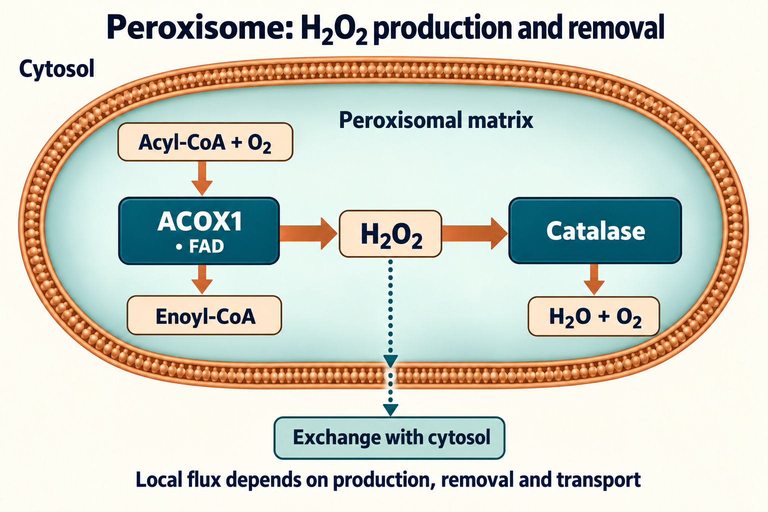
Peroxisomal β-oxidation is especially important for very long-chain fatty acids, including C₂₄–C₂₆ species. ABCD proteins participate in substrate delivery; ABCD1 is associated with X-linked adrenoleukodystrophy. ACOX1 contains FAD and transfers electrons to oxygen, generating H₂O₂, unlike mitochondrial acyl-CoA dehydrogenases linked to ETF and the respiratory chain.
Acyl−CoA+O2ACOX1/FADtrans−2−Enoyl−CoA+H2O2.
One H₂O₂ molecule corresponds to one oxidase step, not complete degradation of one fatty acid. Subsequent hydration, NAD⁺-dependent oxidation and thiolysis shorten the chain; products can continue metabolism in mitochondria. Branched fatty acids need additional pathways: phytanic acid first undergoes α-oxidation, and PHYH is an Fe(II)/2-oxoglutarate-dependent hydroxylase, not simply another H₂O₂-producing flavin oxidase. D-amino acid oxidase can produce H₂O₂, whereas humans lack active urate oxidase.
Catalase lowers local peroxide burden, but the membrane does not completely isolate H₂O₂. Changes in import, oxidase abundance and transport alter cytosolic signaling and mitochondrial interactions. Universal percentages for oxygen consumption or catalase abundance in human liver cannot be inferred from a rodent experiment. A flux balance is more realistic:
This is a qualitative model; quantitative use requires all fluxes to refer to the same volume and use compatible units.
Two routes through Compound I
Catalatic cycle and side states
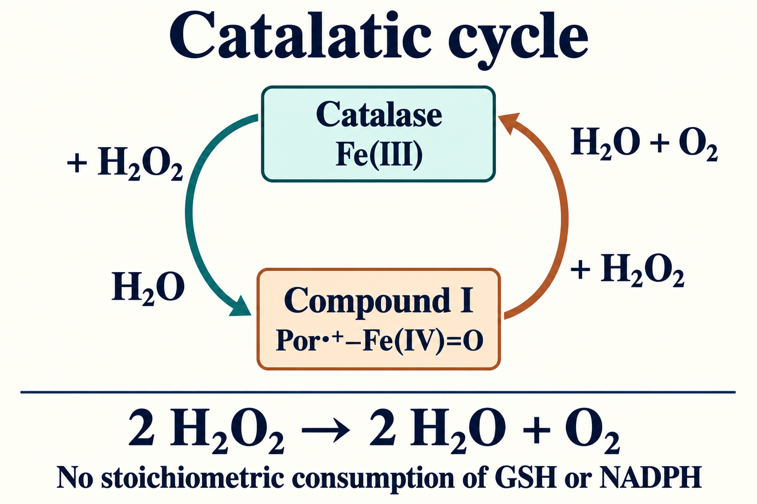
The first H₂O₂ molecule oxidizes the heme center by two equivalents, producing ferryl Fe(IV)=O and a porphyrin cation radical, together called Compound I. The distal environment facilitates heterolytic O–O cleavage and proton transfer. A second H₂O₂ molecule reduces Compound I to resting Fe(III) while becoming O₂. The same substrate therefore acts as oxidant and reductant. Net two-electron accounting does not establish simultaneous transfer of an electron pair: Alfonso-Prieto and colleagues’ QM/MM models of Helicobacter pylori and Penicillium vitale catalases proposed sequential one-electron transfers through a Compound II-like state, with His involvement or direct hydrogen atom transfer. These are models of particular enzymes, not direct observations of the atomic route in human CAT.
One-electron reduction of Compound I can produce Compound II, a ferryl state without the porphyrin radical, which does not support the usual cycle as efficiently. Compound III is another oxygen-bound state; its formation does not imply obligatory immediate irreversible heme destruction. Accumulation, protein damage and recovery depend on substrate, donors, NADPH and exposure duration. An intermediate’s name is not a fixed measure of irreversibility.
What does an “extremely high rate” measure?
Bimolecular step constants have units M⁻¹·s⁻¹, whereas turnover numbers have units s⁻¹. Numerical resemblance does not make them interchangeable. Forty to fifty million molecules per minute corresponds to approximately 0.67–0.83 million per second, retaining the same normalization per active site or tetramer. This does not establish kcat>107s−1 without a different experimental basis.
For an idealized two-step cycle, constant substrate concentration and no side pathways, H₂O₂ disappearance can be written as:
vH2O2=k1+k22k1k2[E]active[H2O2].
The factor two reflects consumption of two peroxide molecules per cycle; O₂ evolution is half as fast. The model gives linear substrate dependence in its applicable regime, not a universal quadratic dependence at low concentrations. Its limits include steady-state intermediate occupancy, substrate availability and absence of inactivation.
Apparent Km depends on the model. Abe and colleagues obtained about 0.025 M for purified beef liver catalase at pH 7.0, with measurements at 2 °C. This describes a preparation and method, not the concentration at which human catalase switches on. High peroxide can produce kinetic deviations, side intermediates and inactivation; “unlimited capacity without saturation up to 1 M” is too strong.
Peroxidatic branch: alcohols and toxicology
Compound I can oxidize a suitable small donor instead of a second H₂O₂ molecule. For a primary alcohol, the net reaction is:
R−CH2OH+H2O2→R−CHO+2H2O.
Relative fluxes depend on competing reaction rates and both substrate concentrations; there is no strict switch at 1 µM. Ethanol produces acetaldehyde and methanol produces formaldehyde. Formate or nitrite oxidation in enzyme systems likewise does not establish their physiological contribution in every tissue.
In human liver, alcohol dehydrogenase and, depending on conditions, CYP2E1 remain the major ethanol pathways; a fixed 10–20% catalase share is unsupported by this base. Rodent brain and 3-amino-1,2,4-triazole experiments support catalase involvement in local acetaldehyde formation and some behavioral effects, but do not measure a universal human “60–70%”. Oxidizing a toxic alcohol is not necessarily detoxification: formaldehyde and formate are themselves toxic, and species differences are particularly relevant to methanol.
CAT, GPx and Prx: a network rather than three thresholds
Property
Catalase
Glutathione peroxidases
Peroxiredoxins
Catalytic group
Heme b
Selenocysteine in GPx1/GPx4
Reactive cysteine
Main chemistry
H₂O₂ disproportionation
Donor-dependent peroxide reduction
Catalytic thiol oxidation
Net reductant
Second H₂O₂
Usually 2 GSH
Often thioredoxin; isoform-dependent
Regeneration
Main cycle without GSH/NADPH consumption
Glutathione reductase reduces GSSG using NADPH
Thioredoxin reductase uses NADPH
Compartments
Mainly peroxisomes; other pools also exist
Isoform-dependent; cytosol, mitochondria, lipids
Cytosol, nucleus, mitochondria and elsewhere
Particular role
Large local peroxide fluxes
GPx4 protects phospholipids and limits ferroptosis
H₂O₂ interception, redox relays and signaling
Constraint
Substrate delivery, active enzyme abundance, inactivation
The latter represents the net thioredoxin-dependent system, not direct H₂O₂ reaction with thioredoxin. Prx6 and some other contexts use different donor systems. Ordinary GSSG regeneration requires NADPH; this is the metabolic cost of maintaining the pool, not ATP expenditure at every peroxidase step.
Rapid Prx reaction with H₂O₂ is compatible with slower complete turnover because regeneration can limit the cycle. Hyperoxidation of some 2-Cys Prx to sulfinic forms temporarily changes peroxide interception and signaling; sulfiredoxin can restore particular forms in an ATP-dependent reaction. The floodgate model is useful in some conditions, not a universal threshold for every Prx. Each sink’s contribution depends on abundance, localization and regeneration. CAT can influence low fluxes in its compartment, and placing GPx4 in a middle “H₂O₂ tier” ignores its lipid substrates.
CAT genetics, acatalasemia and a common promoter variant
Human CAT is located at 11p13 and contains 13 exons. Its GC-rich promoter interacts with several transcriptional regulators; protein abundance, heme loading and distribution jointly determine activity. CAT involvement in WAGR-region deletions depends on individual deletion boundaries; a fixed 50% reduction is not obligatory in every case.
Acatalasemia is a rare, usually autosomal recessive condition caused by pathogenic CAT variants. Takahara’s historical observations linked poor oxygen evolution from peroxide-exposed blood with oral necrotic lesions. Hemoglobin oxidation and bacterial H₂O₂ production may promote local injury. However, many people with marked deficiency lack these lesions: hygiene, infection burden and other defenses modify the phenotype.
Splicing defects, frameshifts and altered protein stability have been described across populations. Interpretation requires the precise variant and transcript; a geographic label is not a molecular diagnosis. Observational reports of diabetes in catalase deficiency are interesting, but small selected groups do not establish a universal “4.5–5.2-fold” risk or one mechanism of β-cell death. Erythrocyte activity measurement requires a method, normalization to hemoglobin or protein and sample context; it is not a standalone test of the “rate of aging”.
The −262C>T polymorphism, rs1001179, differs from rare causes of acatalasemia. Forsberg studied factor binding, reporter transcription and blood catalase levels, not established hypertension. The T variant gave about 1.5-fold higher reporter transcription in HepG2 and K562; among 29 donors, T carriers had higher erythrocyte catalase levels than CC donors (p < 0.03). Direction and magnitude cannot automatically transfer to every tissue or disease. Associations with vitiligo, tumors and hypertension depend on population and design; genotype does not justify prescribing “catalase activators” or prove each outcome’s causal pathway.
mCAT: what changing the compartment demonstrated
In 2005, Schriner and colleagues compared transgenic human catalase targeted to mouse peroxisomes, nuclei or mitochondria. Mitochondrial targeting produced the most pronounced lifespan increase; the paper reported median and maximum lifespan extensions of about five months. This was a genetically modified animal experiment, not a supplement trial. It supports a causal role for that intervention in the model and the importance of localization, but does not make every ROS the sole cause of aging.
Dai and colleagues described reduced age-associated cardiac hypertrophy and fibrosis, preserved diastolic function and less of certain mitochondrial damage in mCAT mice. This is more accurate than complete prevention of dilated cardiomyopathy and all heart failure. Oxidative stress can influence the mitochondrial permeability transition pore, mPTP, but the protective phenotype alone does not prove obligatory direct cyclophilin D oxidation. Changes in mtDNA, cataracts, muscle or insulin sensitivity belong to specific models and endpoints; they cannot be combined into one numerical effect without separate evidence.
Evidence level
Supported conclusion
What it does not show
Purified enzyme
Mechanism, kinetics and inhibition under specified conditions
Cellular distribution or clinical benefit
Cellular import systems
CAT–PEX5 interaction and assembly dependence
A single route in all tissues
mCAT mice
Genetic targeting effects on lifespan and selected phenotypes
Human lifespan extension by oral catalase
Acatalasemia and rs1001179
Genetic mechanisms and limited associations
Universal risks or preventive treatment protocols
Regulation, inhibitors and enzyme delivery
FoxO factors connect stress defense with Akt signals, energy status and protein modifications; SIRT1 can alter FoxO transcriptional responses. Nrf2/Keap1 coordinates a cytoprotective network, but a change in CAT activity after stimulation does not prove direct Nrf2 promoter binding in every tissue. PPARα regulates lipid metabolism and peroxisomal enzymes; oxidase and clearance-system induction may differ, so greater β-oxidation does not guarantee proportional catalase induction.
Resveratrol, sulforaphane, EGCG, quercetin and omega-3 fatty acids have been studied in cells and animals. These studies do not provide a proven treatment regimen for catalase deficiency or human aging. Even increased blood activity remains a surrogate endpoint without demonstrated tissue targeting and clinical benefit. The details of resveratrol, SIRT1 and bioavailability matter especially: a laboratory effect does not ensure direct enzyme activation at achievable exposure.
Iron and heme synthesis are necessary for a functional hemoprotein; ALAS and ferrochelatase participate in that pathway. This does not imply additional iron is needed without confirmed deficiency. Excess labile iron promotes Fenton chemistry. Selenium is required by some GPx enzymes, not as CAT’s prosthetic group. One antioxidant measurement does not describe the whole network.
3-Amino-1,2,4-triazole is used as a mechanism-dependent catalase inhibitor associated with its peroxide cycle; structural data show interaction in the active region. Azide and cyanide bind heme centers and affect other enzymes. Their effects, particularly behavior after systemic administration, are not perfectly specific evidence for CAT without additional controls.
Delivering a large protein is constrained by distribution, proteolysis, immune reactions and reaching the appropriate compartment. PEGylation, liposomes, extracellular vesicles and cellular carriers are under study. Batrakova’s 2007 work concerns macrophage-mediated catalase delivery; it is not a clinical trial of intranasal exosomes. In ischemia–reperfusion, neuroinflammation and wound models, carrier, exposure, timing and tissue endpoints matter; one percentage reduction in infarct size cannot transfer to all formulations.
CeO₂, MnO₂ and Fe₃O₄ are studied as materials with enzyme-like activity. They differ from protein catalase: activity, ion release, distribution and possible prooxidant effects depend on surface properties, pH and environment. Resistance to proteolysis does not establish absolute safety or identical kinetics. This evidence base contains no human randomized trial supporting their prescription for lifespan extension, CAT replacement or diabetic foot treatment.
Reading the evidence base
This is a narrative review of the supplied manuscript and its literature base, with targeted checks of disputed structural, bibliographic and mechanistic claims on October 8, 2026. The previous editor’s saved research notes were absent from the working package; confirmed corrections supplied at handoff were used during recovery. No systematic search, formal GRADE assessment or external peer review was performed. Coverage of recent publications is not guaranteed; precise values without adequate support were not carried over.
Catalase protects cells through heme chemistry, peroxide access and correct localization. It saves reducing equivalents in the main cycle but operates in a network with GPx and Prx, rather than possessing infinite autonomous capacity. Genetic models and enzyme delivery let researchers test particular compartments; clinical decisions additionally require human exposure, efficacy and adverse-effect data.
FAQ
Why does the main reaction not consume glutathione?
The second H₂O₂ molecule reduces Compound I. Two peroxide molecules become two waters and O₂, without obligatory GSH consumption per turnover.
Is catalase always imported as a tetramer?
No. Evidence includes PEX5 binding to monomers and tetramer import systems; cargo state depends on conditions.
Can rs1001179 guide supplement selection?
These data do not support that use. Functional effects and associations depend on the experimental system and population.
Does mCAT establish human lifespan extension?
No. It is a genetic intervention in mice. Human delivery, efficacy and adverse effects require separate evidence.