Betaine HCl: gastric acid, pepsin, B12, and evidence — NutriFit
Betaine HCl and hypochlorhydria: proton secretion, pepsin, and absorption
Gastric proton-pump physiology, pepsin activation, mineral and B12 absorption, betaine HCl chemistry and human pH studies. Diagnosis, interactions, regulatory status, and the limits of acid replacement.
Betaine hydrochloride can add acid to the gastric lumen. Small human experiments show that it can temporarily lower gastric pH when acid secretion has been suppressed by rabeprazole. They do not establish that it restores parietal cells, replaces intrinsic factor, cures dyspepsia, corrects iron deficiency, or prevents small intestinal bacterial overgrowth. The distinction between a measurable chemical effect and an effective treatment is central to interpreting this compound.
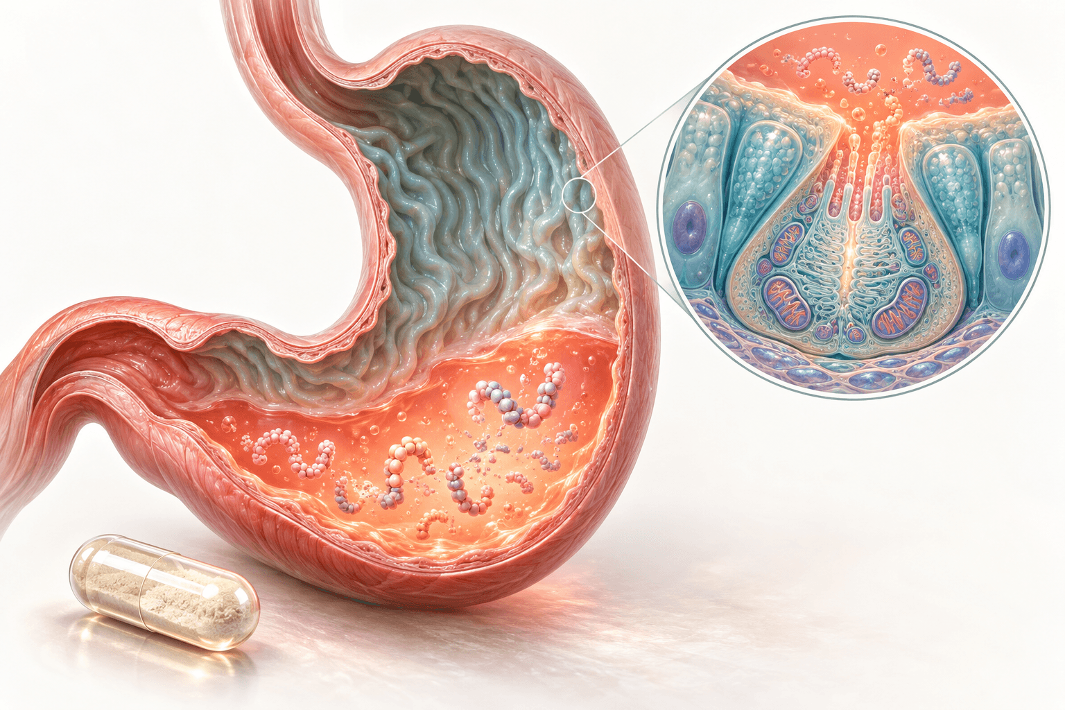
The stomach is also more than an acid reservoir. Its glands coordinate ion transport, enzyme secretion, endocrine feedback, mucosal protection, and preparation of food for intestinal absorption. Understanding these compartments explains both why an acidifier may alter digestion and why acid alone cannot correct every consequence of atrophic gastritis.
How this review uses the evidence
This is a narrative scientific review and a targeted correction of the source material. Molecular experiments, human absorption studies, and official clinical or regulatory guidance were checked where they could resolve consequential claims. It is not a systematic review, a formal grading of evidence, or a validated treatment protocol. Some full-text pages were inaccessible; for those publications, conclusions are restricted to accessible primary abstracts, indexed primary passages, or official publication records. Exact pharmacopoeial specifications could not be checked against a current authenticated monograph.
The strongest direct betaine HCl evidence discussed here concerns short-term intragastric pH and drug exposure in healthy volunteers with experimentally induced hypochlorhydria. Clinical outcome evidence for people with established gastric atrophy is a separate question. Biochemical plausibility is identified as such rather than presented as proof of benefit.
1. Acid secretion is a coordinated cellular process
Oxyntic glands and mucosal protection
The oxyntic glands of the gastric body and fundus contain parietal cells that secrete acid and intrinsic factor, chief cells that release pepsinogens and gastric lipase, mucous neck cells, and endocrine cell populations. Enterochromaffin-like cells, or ECL cells, are epithelial endocrine cells in these glands. They release histamine close to parietal cells; they are not a separate cell population located in the lamina propria.
Surface mucus is enriched in MUC5AC, whereas glandular and neck mucus prominently contains MUC6. Together with bicarbonate secretion, epithelial junctions, blood flow, and restitution, mucus helps keep the epithelial surface less acidic than the lumen. Acid does not inevitably injure a healthy gastric lining, but failure of these defenses can expose tissue to acid and proteolytic enzymes. A capsule does not make externally supplied acid harmless to an injured mucosa.
Parietal cells have abundant mitochondria because proton transport requires ATP. At rest, much of the gastric proton pump resides in tubulovesicular membranes. Stimulation recruits pump-containing membrane to the apical secretory canalicular system. The resulting increase in exposed transport capacity connects receptor signaling to acid secretion; it is not new synthesis of all the pump molecules with each meal.
ATP4A, ATP4B, and the transport cycle
The gastric H+/K+-ATPase is a P-type ATPase composed of the catalytic alpha subunit, ATP4A, and the glycosylated beta subunit, ATP4B. ATP4A contains ten transmembrane helices and cytoplasmic nucleotide-binding, phosphorylation, and actuator domains. ATP4B is a type II membrane protein with a large extracellular domain that supports assembly, trafficking, and stability of the complex (Abe et al., 2018).
ATP-dependent phosphorylation of a conserved aspartate, conventionally numbered Asp385 in structural work, drives transitions between E1 and E2 conformations. The pump exports hydrogen ions from the cytoplasmic side and imports potassium from the canaliculus. Potassium binding promotes dephosphorylation and return of the transport cycle. A structural study found one occluded potassium site, supporting an electroneutral 1H+/1K+ exchange per ATP in the examined state (Yamamoto et al., 2019). Older proposals of a pH-dependent change in stoichiometry should not be confused with a directly measured universal constant under every experimental condition.
A lumen near pH 1 and a cytoplasm near pH 7 imply a proton activity ratio of approximately one million:
Here aH+ is hydrogen-ion activity; pH is its negative base-10 logarithm. Plasma pH around 7.4 should not be substituted automatically for intracellular pH. The relation describes a gradient, not the amount of acid secreted in an hour. Secretory output, gastric volume, and luminal pH are distinct measurements.
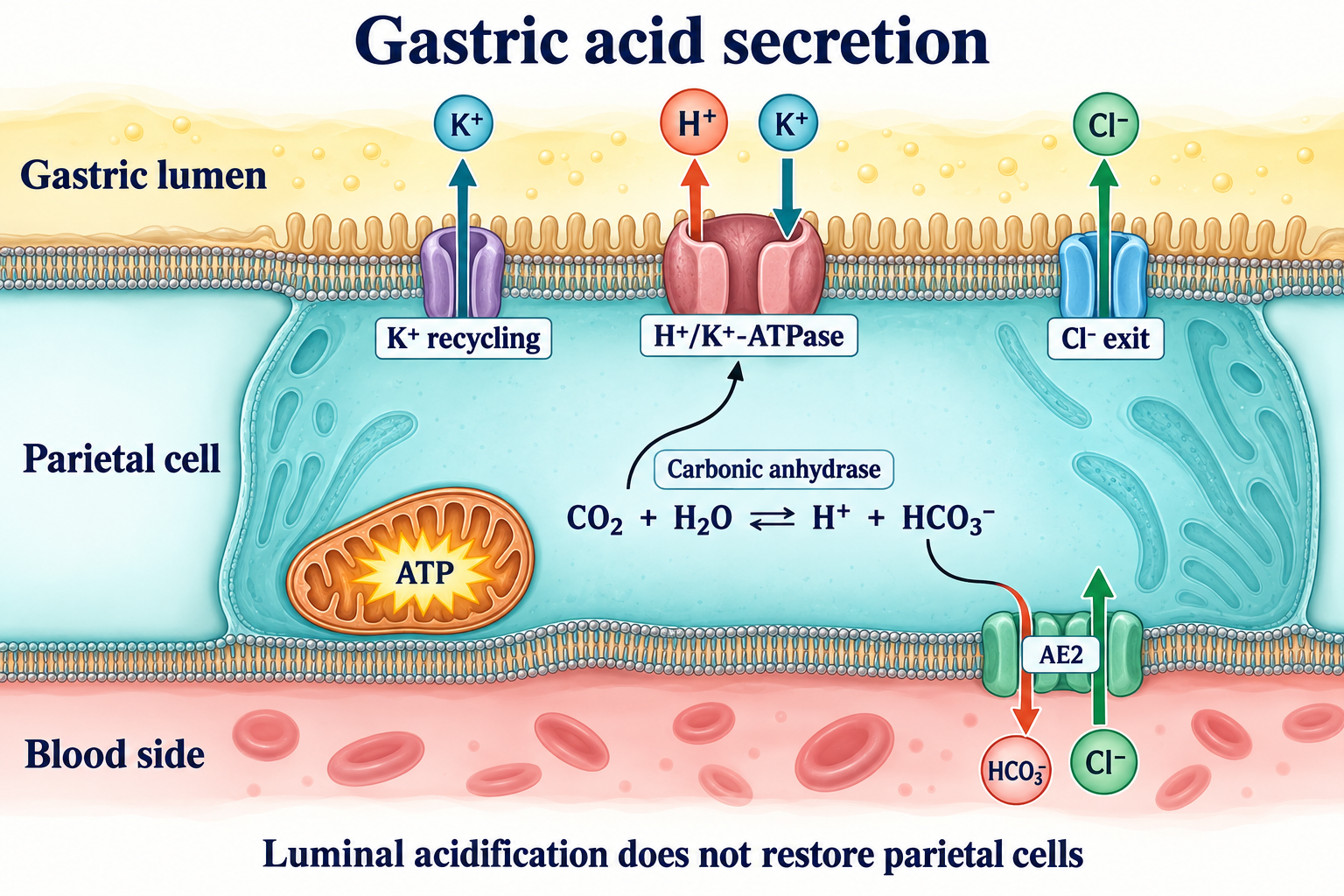
Carbonic anhydrase accelerates the supply of hydrogen ions and bicarbonate from carbon dioxide and water:
CO2+H2O⇌H2CO3⇌H++HCO3−.
Apical potassium recycling, prominently through the KCNQ1/KCNE2 channel system, supplies luminal potassium for continued pump activity. Chloride exits through apical transport pathways, while the basolateral AE2 exchanger, SLC4A2, brings chloride into the cell in exchange for bicarbonate. Chloride pathways include experimentally implicated proteins such as SLC26A9, but their contributions should not be reduced to a single universally exclusive channel. Basolateral sodium/potassium ATPase maintains supporting ionic gradients. Bicarbonate entering gastric venous blood contributes to the postprandial alkaline tide. Hydrogen and chloride ions coexist in the aqueous lumen; secretion does not require assembly of undissociated HCl molecules inside the cell.
The diagram separates the lumen, parietal cell, and blood side. An orally administered acidifier changes luminal chemistry without reproducing the pump's membrane recruitment or basolateral bicarbonate transport.
Gastrin, histamine, acetylcholine, and somatostatin
Acetylcholine released by enteric neurons stimulates parietal M3 receptors, engaging Gq, phospholipase C, IP3/DAG signaling, and intracellular calcium. Histamine from ECL cells activates H2 receptors coupled to Gs, adenylyl cyclase, cAMP, and protein kinase A. These signaling systems potentiate one another; an exact fold of synergy depends on the preparation and stimulation used.
Gastrin, produced mainly by antral G cells, acts through CCK2 receptors. Much of its acid-stimulating action is mediated by ECL histamine release, with direct and trophic effects also contributing. Peptides, amino acids, gastric distension, and vagal pathways participate in meal responses; vagal stimulation of G cells involves gastrin-releasing peptide rather than a single acetylcholine pathway for every cell.
Somatostatin from D cells restrains G cells, ECL cells, and parietal secretion. Falling antral pH supports this negative feedback. Consequently, substantial loss of acid secretion in corpus atrophy can produce hypergastrinemia. It is increased gastrin, not hypogastrinemia, that contributes to ECL hyperplasia and type 1 gastric neuroendocrine tumor risk in autoimmune atrophic gastritis (Shah et al., 2021).
2. Pepsin activation and the acid–peptic barrier
Pepsinogen families and proteolytic activation
Chief cells secrete inactive precursors rather than exposing their own secretory apparatus to fully active proteases. Pepsinogen I comprises group A pepsinogens associated mainly with oxyntic glands; human gene nomenclature includes PGA3, PGA4, and PGA5. Pepsinogen II is associated with PGC and progastricsin, with a broader gastric distribution. Historical electrophoretic pepsinogen fractions are not interchangeable with a list of five distinct human genes named PGA1 through PGA5.
The N-terminal prosegment occludes the catalytic cleft and stabilizes the zymogen through interactions with the protease domain. Acidification changes ionization and disrupts these contacts. Exposure of the active site permits proteolytic removal of the prosegment, followed by autocatalytic amplification. Structural work on porcine pepsinogen establishes this general principle, but prosegment length, cleavage sites, activation rate, and isoenzyme behavior should not be assigned one exact value for all human gastric proteases (James and Sielecki, 1986).
Pepsin is an aspartic endopeptidase. Its catalytic aspartates, conventionally Asp32 and Asp215 in mature pepsin A, support water-mediated peptide-bond hydrolysis. Acid helps unfold many dietary proteins and exposes susceptible bonds; pepsin then preferentially cleaves particular hydrophobic or aromatic sequence environments. Gastric acid and enzymatic proteolysis are complementary processes, not the same chemical reaction. Pancreatic proteases and intestinal peptidases subsequently perform much of the digestion and absorption of dietary protein.
Activity is different from irreversible denaturation
A pH optimum is specific to an enzyme, substrate, and assay. Human pepsin preparations often have high activity near pH 2, declining as pH rises. This does not justify a claim that all activity disappears at pH 3.5 or that every pepsin molecule is irreversibly destroyed at pH 6.5.
Johnston and colleagues (2007) found that tested human pepsin was inactive at pH 6.5 or above but could remain structurally stable and regain activity after reacidification. The result matters in reflux as well as digestion: reduced current activity is not proof that retained pepsin can never become active again. It also prevents an unjustified claim that a transient rise in gastric pH permanently eliminates all gastric proteolysis.
Acid defense has limits
Low gastric pH restricts survival of many swallowed organisms. Susceptibility nevertheless differs across organisms and depends on exposure time, food buffering, inoculum, and acid-resistance mechanisms. The stomach is not a sterile compartment, and acid does not uniformly eliminate viruses, spores, or preformed toxins. The acid barrier operates alongside mucus, motility, immune defenses, bile, and pancreatic secretion.
Acid therefore has three related roles: preparing proteins for pepsin, maintaining chemical conditions for certain nutrients and drugs, and limiting microbial passage. Demonstrating temporary acidification does not demonstrate restoration of all three roles to a clinically effective level.
3. What hypochlorhydria means and why its cause matters
Hypochlorhydria denotes reduced acid secretion; achlorhydria denotes absent or profoundly deficient acid output, including failure to respond adequately to stimulation in classical testing. A high gastric pH supports low acidity only in a specified setting. Recent food, antacids, acid-suppressing drugs, sampling site, and observation duration all affect interpretation. A single post-meal pH above 4 cannot independently establish permanent gland failure.
Autoimmune gastritis involves immune injury directed against oxyntic structures, including the proton-pump complex. Progressive loss of parietal cells reduces both acid and intrinsic factor. Chief-cell function and gland architecture may also deteriorate. Antiparietal-cell and anti-intrinsic-factor antibodies assist evaluation, but neither one positive antibody nor one negative antibody replaces histology and clinical interpretation. Pernicious anemia is a later manifestation of B12 deficiency in this setting, while iron deficiency may appear earlier (Shah et al., 2021).
The effect of Helicobacter pylori depends on the distribution and stage of gastritis. Antral-predominant disease may coexist with increased acid secretion, whereas corpus-predominant inflammation and atrophy can reduce secretion. Bacterial virulence factors and host inflammation are relevant, but infection does not imply that every patient needs an acidifier. Confirmed infection requires appropriate eradication and confirmation of success.
Older age increases the likelihood of accumulated disease and medication exposure. It does not necessarily cause acid-secretory failure by itself. In the primary study by Katelaris and colleagues (1993), acid secretion was preserved with aging in healthy men without atrophy. A population-specific prevalence of low acidity should not be recast as an inevitable physiological fate of everyone over 60.
Proton-pump inhibitors inhibit stimulated acid secretion; H2 antagonists act at a different regulatory step. Gastric pH during treatment varies with agent, schedule, adherence, and the individual. Medication-induced acid suppression is not identical to autoimmune gland destruction. Many patients have a sound indication for suppression, such as ulcer healing, reflux complications, or bleeding-risk reduction. Adding acid to oppose a prescribed treatment requires assessment of that indication, rather than assuming that all suppression is harmful.
4. Nutrients: chemical preparation is only one step in absorption
Iron: solubility, reduction, and intestinal regulation
Acidity helps keep many nonheme iron species soluble. Ascorbate can support reduction of ferric iron to ferrous iron. DCYTB is a ferric reductase at the duodenal brush border, and DMT1 transports divalent iron across the apical enterocyte membrane. DCYTB should not be placed in the gastric juice, and acid alone should not be said to reduce all ferric iron (McKie et al., 2001).
Absorption also depends on dietary ligands, hepcidin, inflammation, enterocyte export, and the balance between intake and losses. Heme iron follows a different initial pathway. Atrophic gastritis can contribute to iron deficiency and an inadequate oral-iron response, but not every oral iron preparation becomes completely unabsorbable. Annibale and colleagues (2001) identified gastrointestinal causes of refractory deficiency even in patients without gastrointestinal symptoms; the lesson is to investigate the cause, not to presume acid deficiency from a poor response.
Calcium: the meal changes the answer
Acid can dissolve calcium carbonate:
CaCO3+2HCl⟶CaCl2+H2O+CO2.
Dissolution precedes intestinal uptake, which includes transcellular pathways such as TRPV6 and paracellular transport and is influenced by vitamin D and the overall calcium load. Calcium citrate behaves differently from carbonate. In Recker's human absorption experiment (1985), fasting carbonate absorption was impaired in achlorhydria, but taking carbonate with a normal breakfast restored absorption. That finding contradicts a universal claim of an 80% loss of dietary calcium absorption whenever gastric acid is low.
Associations between long-term PPI use and fractures do not prove that acid suppression alone produces osteoporosis through carbonate malabsorption. Bone risk depends on age, falls, nutrition, glucocorticoids, endocrine disease, and other factors. Neither the dissolution equation nor a short pH experiment establishes fracture prevention by betaine HCl.
Magnesium and zinc: different transport problems
Severe hypomagnesemia can occur in susceptible long-term PPI users, including cases resolving after the drug is withdrawn (Mackay and Bladon, 2010). Intestinal magnesium absorption includes paracellular flux and regulated TRPM6/TRPM7-related pathways. Changes in luminal chemistry and channel regulation are proposed contributors. This adverse effect does not establish that all hypochlorhydria directly blocks both channels, that gastric acid deficiency is the leading cause of hypomagnesemia, or that betaine HCl corrects it. Renal losses, diuretics, gastrointestinal losses, and other causes still require assessment.
Zinc uptake through ZIP4 depends on the availability of zinc and the diet. Phytate can reduce availability, but physiological gastric acid is not a phytase that reliably hydrolyzes every phytate complex. Reduced absorption is also not equivalent to genetic or acquired absence of ZIP4. A small primary comparison found a smaller plasma zinc response to supplementation in PPI users (Farrell et al., 2011). Its size and nonrandomized comparison limit generalization, and it did not test betaine HCl as a corrective treatment.
Nutrient
Acid-related step
What also matters
What acidification alone has not established
Nonheme iron
Solubility and access to reduction pathways
Diet, hepcidin, inflammation, losses, intestinal transport
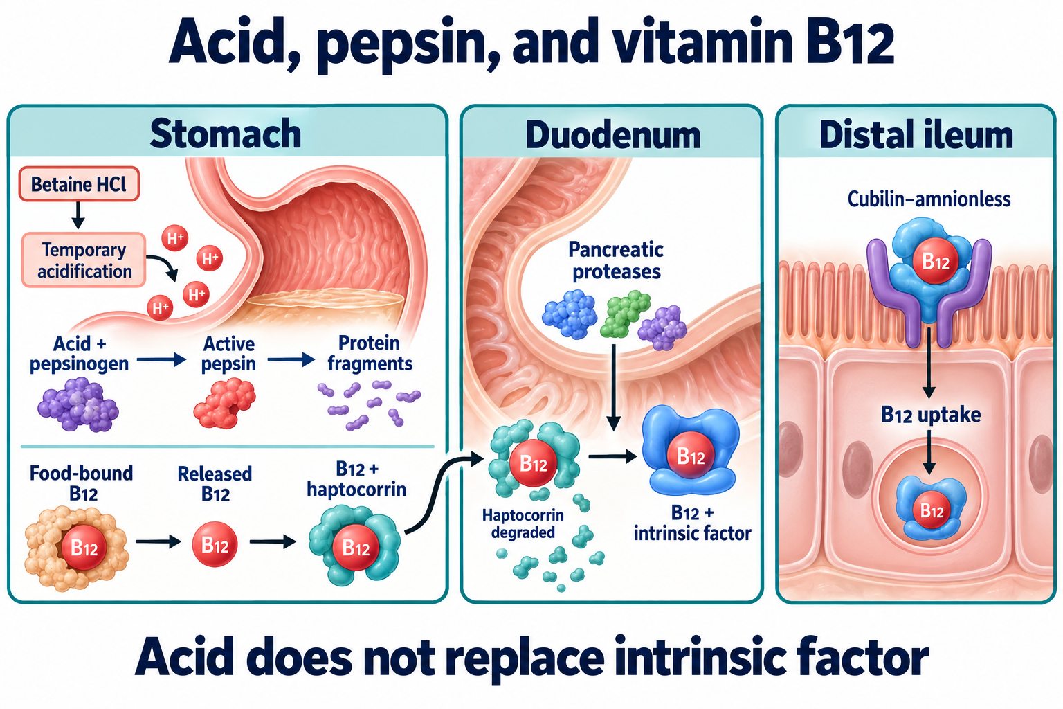
Acid and pepsin help release food-bound cobalamin. In the stomach, the released vitamin associates predominantly with haptocorrin, also called R protein. After the contents enter the duodenum, pancreatic proteases digest haptocorrin, permitting transfer of B12 to intrinsic factor. The intrinsic-factor–B12 complex is taken up in the distal ileum through the cubilin–amnionless receptor system. These are different anatomical and biochemical stages.
Food-cobalamin malabsorption can occur with reduced acid despite preserved intrinsic factor. Marcuard and colleagues (1994) demonstrated reduced absorption of protein-bound B12 during omeprazole exposure. Free crystalline B12 does not require release from dietary proteins, although its uptake still depends on dose and the available absorption pathways. Autoimmune destruction may additionally cause intrinsic-factor deficiency; adding acid does not supply the missing binding protein or repair an ileal disorder.
B12 deficiency may cause anemia, neuropathy, and spinal cord injury, sometimes without a typical macrocytic blood count. Iron deficiency can mask macrocytosis. Long-term stores explain why onset may be delayed, but a fixed three-to-five-year countdown is inappropriate for every patient. Neurological symptoms require prompt assessment and B12 replacement; a trial of betaine HCl should not delay this treatment.
The diagram shows where acid-dependent release ends and intrinsic-factor-dependent absorption begins. The arrow from betaine HCl concerns temporary luminal acidification, not replacement of the later steps.
Folate and the intestinal acid microclimate
PCFT, encoded by SLC46A1, mediates proton-coupled uptake of monoglutamyl folates. Identification of the transporter and loss-of-function variants in hereditary folate malabsorption established its physiological importance (Qiu et al., 2006). Folate transport experiments demonstrate pH dependence, with strong activity in acidic conditions.
However, gastric pH is not the same as pH at the intestinal brush border. Bicarbonate neutralization, mucus, epithelial ion transport, and the local surface microclimate all intervene. A PCFT assay near pH 5.5 does not demonstrate that gastric hypochlorhydria automatically causes folate deficiency or that betaine HCl restores folate status. Dietary folate deconjugation and the patient's nutritional and intestinal context must also be considered.
5. Microbial ecology and pancreatic–biliary responses
Reduced gastric acidity can permit greater survival of swallowed organisms and change gastric and intestinal microbial composition. A primary study found more positive glucose breath tests in PPI users than in its comparison groups (Lombardo et al., 2010). Breath tests, selected populations, motility, and medication indications affect interpretation. There is no universal SIBO prevalence for all people with achlorhydria, and the study does not show that betaine HCl prevents recurrence.
The distinction between altered microbial composition and symptomatic overgrowth is developed further in the article on SIBO, intestinal motility, and breath-test interpretation. Gastric acidity is one part of this system; a transient pH change does not restore migrating motor complexes or resolve every anatomical cause of overgrowth.
Nitrite formation and N-nitroso chemistry are plausible concerns in an atrophic stomach, but cancer risk also reflects inflammation, metaplasia, bacterial factors, and host susceptibility. Acidification has not been shown here to prevent gastric adenocarcinoma or neuroendocrine tumors. Surveillance decisions therefore remain based on the underlying gastric disease.
Observational associations of PPIs with infection or pneumonia also need causal restraint. In a large randomized pantoprazole trial, enteric infections were modestly more frequent, whereas most other assessed safety outcomes did not differ significantly over approximately three years (Moayyedi et al., 2019). Those findings neither remove every possible long-term risk nor justify an assertion that acid suppression routinely causes severe infections.
Acid entering the duodenum stimulates secretin release and pancreatic and biliary bicarbonate secretion. Nutrient products, particularly fats and proteins, stimulate CCK and pancreatic enzyme or gallbladder responses. Neural and other hormonal signals provide additional control. Low gastric acidity may alter these stimuli, but this does not establish obligatory secretin deficiency, pancreatic exocrine failure, or biliary sludge. The specific sensing proteins, magnitude of hormone changes, and clinical consequences require evidence in the relevant human condition. Steatorrhea or malnutrition should be investigated on its own merits.
6. Betaine HCl chemistry and acid capacity
A hydrochloride salt is not anhydrous betaine
Betaine, or trimethylglycine, is a quaternary ammonium compound. At physiological pH it largely exists as a zwitterion with a permanently positive nitrogen and a negatively charged carboxylate. In betaine hydrochloride, the carboxylate is protonated and chloride balances the positive charge. The formula is C5H12ClNO2 and the molar mass is approximately 153.61gmol−1 (NIST Chemistry WebBook).
Writing BH+ for protonated betaine and B for the zwitterion, dissolution and acid dissociation can be represented as:
BHCl(s)⟶BH+(aq)+Cl−(aq),BH++H2O⇌B+H3O+.
Chloride is a spectator counterion in the acid dissociation. This is not hydrolysis of a peptide or a mechanism that guarantees complete release of a fixed concentration of free protons at every pH. The dissociation constant of protonated betaine must not be confused with the dissociation constant of free hydrochloric acid. For the acid–base pair, an ideal dilute-solution approximation is:
pH=pKa+log10([BH+][B]).
The approximation requires appropriate activities and equilibria; a meal adds a large and variable buffering system. Exact solution pH cannot be predicted from capsule mass alone.
Stoichiometry is not a dosing protocol
For pure, anhydrous betaine HCl, one mole supplies one mole of titratable acid equivalent. If m is the mass in grams:
nacid(mmol)=153.61(gmol−1)1000m(g).
Thus 1.00 g corresponds to approximately 6.51 mmol of acid equivalents, and 1.30 g to approximately 8.46 mmol. Expressed as component masses, a gram contains approximately 0.763 g betaine equivalent and 0.237 g HCl equivalent. These are composition calculations, not proof that the stomach contains 6.51 mmol of free hydrogen ions afterward. Proton binding to food, gastric volume, endogenous secretion, and emptying determine the actual pH trajectory.
Equating 1.30 g with exactly 20–30 minutes of normal secretion would require the individual's measured acid-output rate. Similarly, a primarily luminal effect does not establish that every dose is systemically risk-free. Betaine can subsequently be absorbed and participate in metabolism; quantitative human intestinal uptake should not be assigned to a single proposed transporter without evidence.
Product quality and pepsin combinations
A useful quality assessment distinguishes identity, betaine HCl content, water content, contaminants, disintegration, and batch consistency. Pepsin products require an activity assay with a defined substrate, pH, and unit system. A mass of pepsin or a historical ratio such as 1:10,000 is not universally comparable with another enzyme-unit claim.
Coformulation has a biochemical rationale: acid can favor pepsin activity when the preparation and gastric conditions are suitable. That rationale does not establish clinical superiority over acid alone or prevention of dietary-protein allergy. A certificate of analysis or pharmacopoeial designation concerns the tested substance and method; it does not demonstrate efficacy of an entire supplement. Exact purity ranges, a trimethylamine limit, and storage characteristics cannot be asserted as universal current USP or European Pharmacopoeia requirements without the relevant monograph.
7. What the human acidification studies actually measured
Study
Design and setting
Main finding
Boundary of interpretation
Yago, 2013
Six healthy volunteers; single-period fasting pilot after rabeprazole-induced hypochlorhydria; Heidelberg capsule
With 1500 mg betaine HCl, mean pH over the following 30-minute interval fell from 5.2 ± 0.5 to 0.6 ± 0.2; mean time to pH below 3 was 6.3 ± 4.3 minutes
Temporary luminal effect; not a randomized nutrient-outcome trial
Yago, 2014
Randomized three-way crossover comparing dasatinib alone, rabeprazole exposure, and rabeprazole plus betaine HCl
Dasatinib exposure was restored under the studied fasting conditions
A drug-specific pharmacokinetic experiment in healthy volunteers
Faber, 2017
Crossover experiment with ritonavir-boosted atazanavir and a light meal
Betaine HCl did not adequately reverse the reduction in atazanavir exposure
Food buffering, formulation, and administration conditions affect translation
In the 2013 experiment, pH below 3 lasted 73±33 minutes and pH below 4 lasted 77±30 minutes. These are reported means with standard deviations, not guaranteed treatment windows. The 30-minute mean pH after administration is not a minimum pH measurement. The study used a Heidelberg capsule; attribution of these particular data to SmartPill is incorrect. Short-term tolerance in six subjects does not establish long-term safety in older adults with mucosal disease.
In the dasatinib experiment, rabeprazole reduced Cmax and AUC0−∞ by approximately 92% and 78%; adding betaine HCl restored them to approximately 105% and 121% of control exposure, respectively (Yago et al., 2014). Cmax denotes peak plasma concentration; AUC denotes the time-integrated concentration. These ratios do not establish equivalence or an appropriate regimen for every patient. The original attribution to an itraconazole study was incorrect.
Under fed conditions, the atazanavir study recovered only a small fraction of exposure lost after rabeprazole (Faber et al., 2017). A temporary fall in pH may occur too late, last too briefly, or interact with meal and formulation effects. Consequently, the dose used in these experiments is not a universal dose for ordinary meals or atrophic gastritis.
8. Diagnosis, treatment boundaries, and safety
Confirm the disease rather than use symptoms as an acid test
Bloating, early satiety, belching, and epigastric discomfort are nonspecific. They can accompany functional dyspepsia, reflux, ulcer disease, delayed gastric emptying, biliary disease, medication effects, or intestinal disorders. Improvement or burning after a capsule does not diagnose low acidity. A baking-soda belch test and escalating acid until a sensation of warmth appears do not provide validated diagnostic thresholds.
Evaluation follows the clinical question. Endoscopy with appropriately located biopsies establishes atrophy and metaplasia when indicated; body and antrum/incisura samples should be distinguished. Sydney sampling and OLGA/OLGIM staging inform distribution and cancer-risk stratification, not the number of acidifier capsules needed. Pepsinogen I, the pepsinogen I/II ratio, gastrin, and antibodies may support assessment, but cutoffs vary by assay and population. Gastrin is affected by PPIs and other conditions; a low pepsinogen ratio does not directly measure luminal pH (Shah et al., 2021).
Direct intragastric pH recording measures acidity in its observed conditions. Classical stimulated acid-output testing answers a related but different question. Neither test justifies a universal pH definition detached from timing and medication exposure. H. pylori antibody positivity can persist after eradication; it is not the preferred test of cure.
Treat established deficiencies and causes directly
Atrophic gastritis requires assessment of iron and B12 status, management of H. pylori when present, and appropriate consideration of endoscopic surveillance. B12 replacement is needed when deficient; intrinsic-factor loss may require a replacement strategy that bypasses or compensates for normal absorption. Acid does not substitute for that strategy.
For iron deficiency, assess bleeding, diet, inflammation, celiac disease, treatment adherence, and other causes of malabsorption. An iron-bisglycinate–betaine HCl–vitamin C cocktail has not been established here as a superior universal regimen. AGA guidance allows intravenous iron when oral iron is not tolerated, stores do not improve, or absorption is unlikely to be adequate (DeLoughery et al., 2024). Fixed supplement doses and a promised 2.5–3-fold ferritin increase would exceed the verified evidence.
Betaine HCl also has no established role here in tapering PPIs, preventing SIBO recurrence, reversing sarcopenia, or treating persistent dyspepsia. Rebound acid symptoms after stopping a PPI are a reason to reassess the treatment plan; they are not evidence that acid should be added. If an acidifier is considered clinically, the objective, formulation, medication context, mucosal status, and monitoring need to be specified individually. No validated general maximum dose or burn-based titration schedule follows from the volunteer studies.
Regulatory status and mucosal risk
The United States regulation for OTC drugs offered as stomach acidifiers explicitly states that adequate evidence has not established general recognition of safety and effectiveness for that use (21 CFR §310.540). This contradicts a blanket claim that betaine HCl is FDA-recognized as safe and effective treatment of hypochlorhydria. Availability as a supplement is a different issue, and it does not imply approval of disease-treatment claims. Regulatory provisions also differ by country.
Acid exposure is a concern in active ulceration, erosive disease, significant reflux, swallowing difficulty, or mucosal injury. Aspirin, NSAIDs, and other treatments that increase bleeding or injury risk warrant particular review. The exact risk of an acidifier combined with each drug has not been quantified adequately; plausible risk should not be turned into invented universal contraindication statistics.
New burning, chest pain, severe abdominal pain, vomiting, or bleeding should prompt stopping the suspected product and clinical assessment as appropriate. Hematemesis, black stool, progressive dysphagia, weight loss, and persistent vomiting require timely medical evaluation. Routine home neutralization with spoonfuls of bicarbonate is not an evidence-based rescue protocol and can introduce additional risks or delay care.
Drug interactions can be clinically consequential
Changing gastric pH can change dissolution and exposure of acid-dependent medicines, including particular antifungal, antiviral, and anticancer formulations. An observed restoration of dasatinib exposure is also a warning that a supposedly minor supplement can change systemic drug concentrations. Effects cannot be generalized across itraconazole formulations, different kinase inhibitors, or all enteric-coated products.
An acidifier opposes part of the intended luminal effect of an antacid or acid-suppressing drug. Timing alone may not remove this interaction. Decisions involving oncology or antiviral treatment require the prescribing team's assessment and the specific product information; supplement self-experimentation is inappropriate.
9. Monitoring and the other biological roles of betaine
Monitor the problem being treated
A falling or rising symptom score is not a measure of parietal-cell recovery. When iron deficiency is treated, blood count, hemoglobin response, ferritin, and transferrin saturation are interpreted with inflammation and the underlying disease. Soluble transferrin receptor can sometimes clarify an ambiguous result, but no single ferritin or transferrin-saturation target applies to every patient.
B12 assessment can include total or active B12 and, when indicated, methylmalonic acid. Homocysteine is less specific because folate status and other factors affect it; renal impairment can also elevate methylmalonic acid. Mixed deficiencies and neurological findings matter more than reliance on a single reference cutoff. Magnesium, calcium, vitamin D, zinc, or bone-density assessment follows individual risks and findings. Fixed two-month blood tests or routine yearly DXA for every PPI user are not a betaine HCl monitoring standard.
Improvement after combined deficiency treatment cannot be attributed automatically to acidification. The nutrient replacement, dietary change, drug review, and underlying-disease treatment may account for it. Relevant outcomes include functional recovery and safety, rather than merely a lower gastric pH.
BHMT and homocysteine
Absorbed betaine can donate a methyl group through betaine–homocysteine methyltransferase, BHMT, principally in liver and kidney:
This pathway is distinct from B12-dependent methionine synthase. Methionine can contribute to S-adenosylmethionine synthesis; dimethylglycine undergoes further metabolism. The relationship to methyl donation is explained in the article on S-adenosylmethionine and one-carbon metabolism.
Trials of betaine supplementation have lowered plasma homocysteine (Olthof et al., 2003). A biochemical reduction does not prove prevention of thrombosis or cardiovascular events, and it does not correct B12-dependent neurological dysfunction. In another primary analysis, higher-dose betaine increased LDL cholesterol and triglycerides (Olthof et al., 2005). These studies of betaine cannot be used interchangeably with studies of acidifying betaine HCl products.
Phosphatidylcholine synthesis and hepatic methyl balance provide a rationale for studying fatty liver. They do not establish that betaine HCl activates PEMT or prevents metabolic dysfunction-associated steatotic liver disease. A randomized high-dose betaine trial in NASH had important limitations and did not establish broad improvement of the proposed disease mechanisms (Abdelmalek et al., 2009).
Osmoprotection remains a separate line of evidence
Betaine is a compatible organic osmolyte. Its accumulation can help cells respond to hyperosmotic stress without the disruptive effects of accumulating high concentrations of inorganic ions. Transporter expression, barrier measurements, and inflammatory responses have been studied in experimental intestinal models.
A primary 2024 study in porcine IPEC-J2 cells found osmolyte-transporter responses to hyperosmotic stress and protective effects of betaine on barrier function (De Angelis et al., 2024). This is a useful cellular observation, not proof that oral betaine HCl repairs human intestinal permeability. Tight-junction expression, electrical resistance, and clinical intestinal disease are different outcomes, as discussed in the article on intestinal permeability and epithelial junctions. Gastric acidification and cellular osmoprotection should be evaluated separately.
10. Alternatives and persistent symptoms
Approach
Mechanistic distinction
Evidence boundary
Betaine HCl
Protonated betaine salt with quantifiable titratable acid capacity
Temporary pH effects and selected drug-exposure findings; no universal outcome regimen
Anhydrous betaine, TMG
Zwitterionic betaine used as an osmolyte and methyl donor
Does not supply the same hydrochloride acid load
Vinegar or citric acid
Organic acids with concentration-dependent dissociation and buffering
Cannot be assigned a universal final gastric pH or proven replacement role
Pepsin preparation
Adds an enzyme whose activity depends on conditions
Does not replace acid secretion, intrinsic factor, or pancreatic function
Nutrient replacement and cause-directed treatment
Targets the demonstrated deficiency or gastric disorder
Appropriate intervention depends on diagnosis and clinical guidance
The pKa of a weak acid does not create an absolute lower bound below which no solution can go. Concentration, acid capacity, competing buffers, and mixing matter. Conversely, an acidic beverage's measured pH does not show that it will overcome a meal's buffering capacity. Claims that vinegar can never lower pH below 3.8, or that betaine HCl is the only scientifically possible acidifier, are chemically unjustified. Acidic drinks may also contact teeth and the esophagus; a swallowed capsule can have its own transit and mucosal risks.
Persistent symptoms should lead to reassessment of the diagnosis, rather than repeated dose escalation. Delayed gastric emptying requires appropriate evaluation after exclusion of obstruction; fecal elastase or other pancreatic testing follows a compatible presentation and has interpretive limitations. Suspected bile reflux, SIBO, celiac disease, or medication intolerance needs its own assessment. Neither one symptom pattern nor a low pH selects a universal prokinetic, antibiotic, bile-acid drug, or enzyme regimen.
The defensible place of betaine HCl is therefore a limited one: it is a chemically understandable, experimentally demonstrated temporary acidifier with important questions about formulation, meals, interactions, and clinical outcomes. Established atrophic gastritis and nutrient deficiencies require diagnosis and cause-directed care. Future trials would need to evaluate those patients, measure meaningful outcomes, and monitor mucosal and drug-related safety before replacing that care with a general acid-supplementation protocol.
FAQ
Does bloating mean I have too little stomach acid?
No. Bloating, belching, and early satiety have many possible causes. Symptoms and the response to an acid capsule do not establish hypochlorhydria.
Is 1500 mg a proven dose for every meal?
No. That amount was studied in a small fasting pharmacological model. Meal buffering, formulation, disease, and medications prevent treating it as a universal regimen.
Can betaine HCl replace B12 treatment in autoimmune gastritis?
No. Acid does not replace intrinsic factor or reverse neurological B12 deficiency. Confirmed deficiency requires an appropriate B12 replacement strategy.
Should I increase capsules until I feel warmth or burning?
No. This is not a validated diagnostic or dosing method. Burning can signal irritation and should not be used as a therapeutic target.
Can acidification improve absorption of medicines?
It can change exposure of particular pH-dependent formulations, sometimes substantially. The prescribing team must assess this interaction; results from one drug cannot be generalized to another.
Is anhydrous betaine the same as betaine HCl?
No. Anhydrous betaine is used as a methyl donor and osmolyte. The hydrochloride salt additionally supplies titratable acid and has a different immediate gastric effect.
Does betaine HCl prevent SIBO or osteoporosis?
The cited primary studies do not establish those benefits. Mechanistic links between acidity, bacteria, and nutrient chemistry are not clinical outcome evidence.