
Apigenin: GABA Receptors, CD38, NAD⁺ and Sleep
How apigenin is absorbed and metabolized, why its GABA-receptor effects vary, and what CD38 and chamomile studies actually establish about sleep, anxiety, NAD⁺ and safety.
How apigenin is absorbed and metabolized, why its GABA-receptor effects vary, and what CD38 and chamomile studies actually establish about sleep, anxiety, NAD⁺ and safety.
Many people encounter apigenin before learning its name: it is found in chamomile, parsley and celery. Yet a cup of infusion, plant food, a standardized extract and a capsule of purified flavone expose the body to different preparations. Dissolution, intestinal and hepatic metabolism, distribution among tissues and access to a specific target all lie between the amount on a label and an effect in the brain. Following that route helps explain why interesting receptor and enzyme findings have not yet established a treatment for insomnia or ageing.
Apigenin has several experimentally investigated actions: interactions with GABA receptors, inhibition of the NAD⁺-consuming enzyme CD38 and changes in inflammatory signalling. These actions involve important physiological processes, but each requires its own evidence in humans. Lower enzyme activity in a test tube does not mean that an ordinary oral dose will reproduce the necessary concentration inside a cell. Below, molecular mechanisms are discussed alongside the conditions of the experiments, while human chamomile studies are considered in terms of their clinical results and limitations.
Apigenin is 4′,5,7-trihydroxyflavone, with the formula C₁₅H₁₀O₅ and a molar mass of 270.24 g/mol. Its C₆–C₃–C₆ backbone comprises two aromatic rings, A and B, and the oxygen-containing ring C that connects them. Ring C contains the C2=C3 double bond and a carbonyl group at C4; hydroxyl groups are located at C5, C7 and C4′. This structure determines electron distribution, hydrogen bonding and the potential to interact with proteins, but it does not itself establish high affinity for CD38 or a particular receptor subtype.
The conjugated system makes the chromone core relatively rigid, although rotation of ring B and the geometry of a bound complex depend on the surroundings. Molecular selectivity therefore cannot be inferred from a claim of an unchanging “perfect plane”. The phenolic OH groups participate in interactions with solvents and also provide sites for metabolic conjugation. The chemical capacity for binding or antioxidant reactions is likewise not equivalent to a clinical antioxidant effect: concentration, environment and competing reactions matter.
Low water solubility is a substantial issue for the purified aglycone. In an early physicochemical study, the octanol–water partition coefficient corresponded to log K = 2.87, while solubility varied considerably across the solvents and surfactant media examined. These findings explain the interest in delivery systems, but they do not establish a universal solubility for every product or assign every preparation to one BCS class. Biopharmaceutics classification requires assessment of solubility under defined conditions and of permeability; the notation “II/IV” cannot replace that assessment.
In plants, apigenin is often attached to sugar residues. Apigetrin is apigenin-7-O-glucoside, whereas apiin contains an apiosylglucose group at the same position. The difference between these glycosides affects their accessibility to hydrolases and where the aglycone is released in the intestine. Consequently, even an identical amount of the apigenin backbone in parsley, chamomile extract and a purified supplement does not guarantee the same timing or magnitude of systemic exposure.
Before a plant molecule becomes available to tissues, it must first be released from the food matrix and, for many O-glycosides, separated from its sugar group. Brush-border hydrolases, including lactase-phlorizin hydrolase, and the intestinal microbiota can contribute to this process. Cytosolic β-glucosidase can cleave suitable substrates inside a cell, but the presence of that enzyme does not explain how a particular glycoside crossed the membrane. The additional sugar residue in apiin makes hydrolysis more complex, shifting some transformations towards distal intestinal regions.
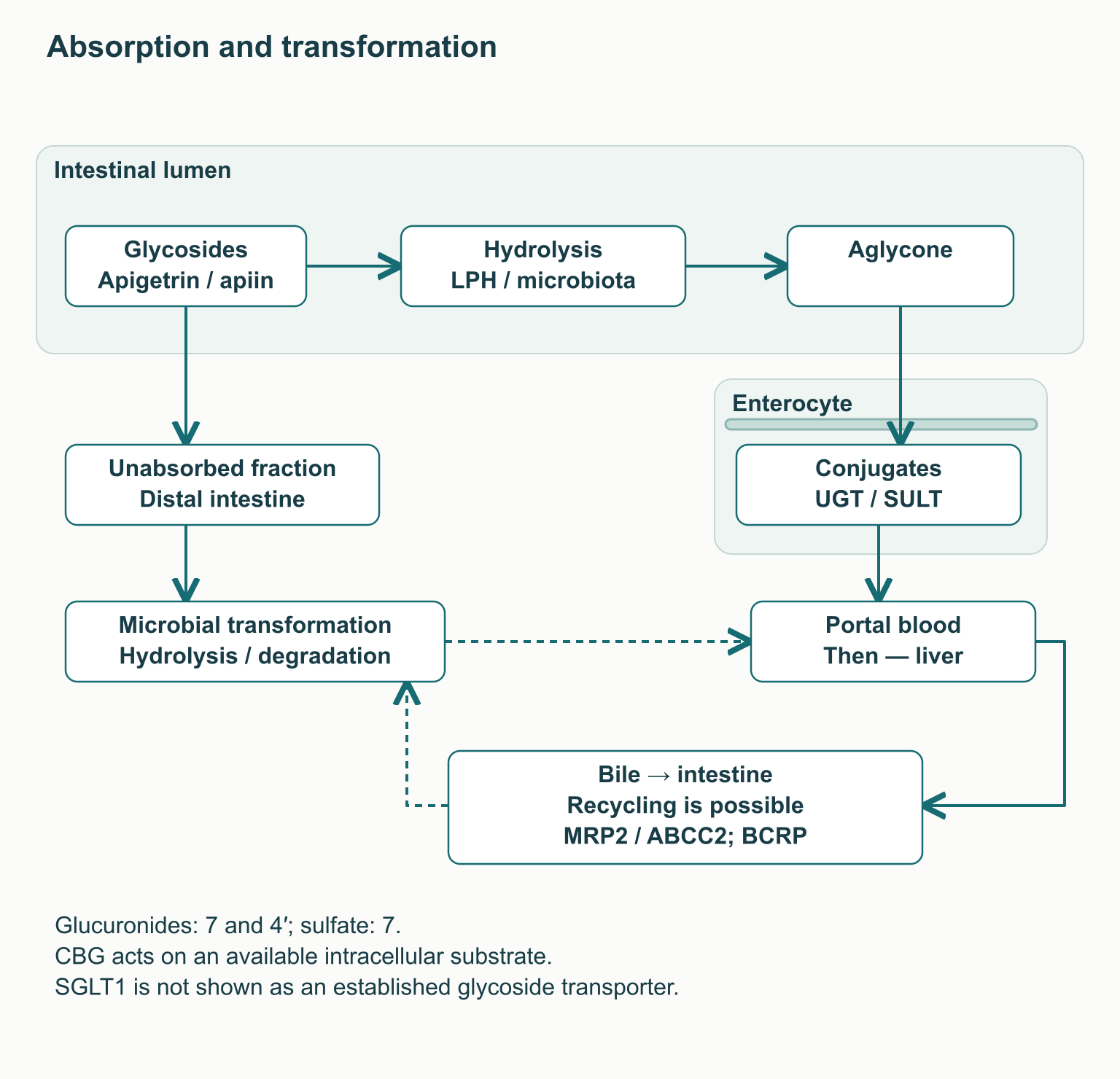
The popular diagram in which SGLT1 transports flavonoid glycosides alongside glucose is too definitive. In experiments with human SGLT1 expressed in oocytes, the flavonoid glycosides examined did not produce a transport current, although some inhibited glucose uptake. This is a transport model rather than an absorption study in a living person, but it illustrates why a direct “SGLT1 → apigenin” arrow cannot be treated as an established route. The most defensible account describes absorption as dependent on glycoside structure, hydrolysis and the properties of the intestinal environment.
Once it enters an enterocyte, the aglycone undergoes glucuronidation and sulfation, followed by further metabolism in the liver. A study of healthy men identified apigenin-4′-glucuronide, apigenin-7-glucuronide and apigenin-7-sulfate. UGT and SULT denote the relevant enzyme families; the contribution of individual isoforms depends on tissue and experimental conditions. A conjugated metabolite cannot automatically be regarded as pharmacologically equivalent to free apigenin, because membrane permeability, protein binding and access to targets change.
Some conjugates are removed by intestinal and hepatic transport systems; biliary excretion and subsequent bacterial hydrolysis can allow reabsorption. Efflux proteins, including MRP2/ABCC2 and BCRP, participate in such routes, but their relative contributions cannot be transferred from one cellular model to every supplement formulation. Microbial transformations can also break down the flavone backbone into simpler phenolic compounds. Thus, prolonged detection of a label or metabolites does not demonstrate equally prolonged persistence of the active aglycone; a universal human half-life of 12–24 hours has not been established for all products.
An informative food experiment involved 11 healthy adults given blanched parsley at 2 g/kg body weight. The mean load was 65.8 μmol of apigenin, approximately 18 mg, and the measured plasma peak reached 127 ± 81 nmol/L after 7.2 ± 1.3 hours. Urinary recovery over 24 hours was 0.22 ± 0.16% of the administered amount; this proportion is not absolute bioavailability, since it does not capture all metabolites and elimination routes. The large food quantity was used to investigate kinetics, not as a practical recommendation to eat kilograms of parsley or as a model for any capsule.
A separate metabolism study in men likewise showed that the chemical form and food source change the profile of circulating compounds. Its initial part included four participants, while subsequent repeated feedings examined variability; the number of feedings must not be mistaken for the number of independent individuals. These studies provide human support for discussing absorption, but they do not measure free apigenin concentrations in the brain. When comparing them with micromolar cell experiments, total analytically measured exposure, the unbound fraction and local tissue concentrations must be distinguished.
GABA is the main transmitter of fast inhibition in many neuronal networks, and the GABA_A receptor is a pentameric ion channel. Common synaptic variants contain two α, two β and one γ subunit, although their composition varies between brain regions. GABA binds at the extracellular β⁺/α⁻ interface, whereas the classical benzodiazepine site lies at the α⁺/γ⁻ interface. These are different sites on one protein complex: an allosteric ligand can change the channel’s response to GABA without replacing GABA at its principal binding site.
The open channel conducts primarily anions, and the effect of GABA is determined by the electrochemical gradient rather than simply by the presence of chloride. In mature neurons, the response usually supports inhibition through hyperpolarization and/or shunting of excitatory current. With a different intracellular Cl⁻ concentration, for example at particular developmental stages or in pathological states, the direction of the membrane-potential change can differ. Thus, Cl⁻ entry and hyperpolarization do not form an inevitable sequence in every cell.
In an early study by Viola, apigenin displaced a ligand of the central benzodiazepine site with a Ki of about 4 μmol/L. At certain doses, mice showed behaviour interpreted as anxiolytic without pronounced motor changes; a tenfold higher dose produced signs of sedation. This supports interest in interaction with the receptor system, but it does not establish an effective concentration in the human brain or the absence of adverse effects. Ki describes binding in a particular experiment and is not an oral dose.
Later functional studies do not provide a uniform picture of positive modulation. In Avallone’s experiments, apigenin reduced GABA-activated currents in neuronal cultures; Losi likewise reported reduced currents through recombinant α1β2γ2 receptors and changes in inhibitory transmission in cortical cultures. Losi also observed inhibition of NMDA responses with an IC₅₀ of about 10 μmol/L, with much less effect on AMPA responses. These findings concern cellular systems and indicate multiple targets, rather than clinical treatment of anxiety through selective enhancement of GABA.
In another recombinant experiment, apigenin at 1 μmol/L enhanced diazepam’s positive modulation of the GABA response, with the effect depending on the combination of substances examined. Enhancing a benzodiazepine already present is not equivalent to an independent positive effect of apigenin. Differences among experiments may reflect receptor composition, concentration, expression system and baseline activation. In practice, this argues against combining it with sedative medicines at home in pursuit of “synergy”.
| α-subunit subtype | What is known about receptor physiology | What cannot be claimed about apigenin |
|---|---|---|
| α1 | Contributes to several sedative and other benzodiazepine effects | That weak α1 activity reliably excludes drowsiness |
| α2 | Linked to components of anxiolysis in experimental pharmacology | That apigenin is clinically selective for α2 |
| α3 | Contributes to inhibitory transmission and the effects of some modulators | That a distinct beneficial “moderate” activity is established for apigenin |
| α5 | Important in certain networks, including hippocampal processes | That lack of α5 activity guarantees preserved memory |
Separating receptor-subtype functions is useful in drug development, but it does not turn a plant molecule into a selective medicine. There are insufficient data to describe apigenin as an established α2-preferring partial positive allosteric modulator. Even less can that label support claims of no amnesia, muscle relaxation, tolerance or withdrawal in humans. These clinical properties require separate studies with a known formulation, dose and duration.
Falling asleep depends on the interaction of circadian signals, accumulated sleep pressure and wake-promoting systems. GABA-containing neurons in preoptic regions help inhibit several wake centres, but a substance binding a receptor does not demonstrate selective action on these networks. Delta activity at approximately 0.5–4 Hz characterizes slow-wave processes; the theta range at about 4–8 Hz occurs in several states and is not, by itself, a marker of restorative sleep. High-quality human trials have not shown that apigenin increases N3, preserves REM in a specified proportion or improves memory by changing these rhythms.
In a sleep-disturbance experiment in rats, chamomile extract at 300 mg/kg shortened sleep latency but did not change total wakefulness, NREM or REM time. An extract contains several substances, and the amount given to an animal cannot be directly converted into recommended milligrams of pure apigenin for a person. This finding does not establish an increase in deep N3 sleep or “normalization of sleep architecture” in humans. Discussions of glymphatic processes and nocturnal hormone secretion also do not establish that an infusion or supplement improves brain clearance and tissue recovery.
The most direct placebo-controlled trial in chronic primary insomnia included 34 adults. They received standardized chamomile extract at 270 mg twice daily for 28 days. No significant advantage was found for total sleep duration, sleep efficiency or the principal nocturnal measures. The pilot sample size leaves uncertainty, but the result does not support a reliable hypnotic effect; still less does it test a separate apigenin capsule.
Anxiety has a different clinical research history. A small eight-week study compared chamomile extract and placebo in 57 people with generalized anxiety disorder; the extract dose was increased within the protocol rather than prescribed as an amount of pure flavone. The study found a signal of possible improvement in anxiety symptoms, but the mixture studied cannot identify which substance produced the result. Anxiety symptoms and sleep measures are different outcomes, so even a positive result on one scale cannot be transferred to sleep stages or relapse prevention.
A longer study first enrolled 179 patients in an open-label, 12-week phase using 1500 mg of chamomile extract daily. Subsequently, 93 responders were randomized to continue the extract or receive placebo for 26 weeks. Relapse occurred in 7 of 46 participants continuing treatment and 12 of 47 receiving placebo; the hazard ratio was 0.52, but its 95% interval of 0.20–1.33 included no effect. The primary result was not statistically significant, although differences emerged for some symptom measures; selecting previous responders limits generalization to everyone with anxiety.
| Study | Population, preparation and duration | Practical interpretation |
|---|---|---|
| Insomnia pilot | 34 adults; chamomile extract 270 mg twice daily, 28 days | No demonstrated advantage for principal sleep measures |
| Initial anxiety trial | 57 patients; titrated chamomile extract, 8 weeks | Possible symptomatic effect of a mixture; not a pure-apigenin trial |
| Continued anxiety treatment | 93 responders after an open-label phase; 1500 mg extract/day for another 26 weeks | The primary relapse-prevention result did not reach statistical significance |
Milligrams of chamomile extract must not be read as milligrams of apigenin. Moreover, standardization does not make different mixtures interchangeable: composition, extraction method and analytical control matter. In chronic insomnia, the initial task remains assessment of its causes and selection of treatment, and multicomponent cognitive behavioural therapy has a strong recommendation in the relevant guideline. An infusion can remain part of an evening routine, but it should not postpone care for persistent insomnia, marked daytime sleepiness or suspected sleep-related breathing problems.
NAD⁺ has two fundamentally different roles. In redox reactions it accepts electrons and becomes NADH, which is subsequently oxidized again; in reactions involving sirtuins, PARP and several other enzymes, the molecule is consumed as a substrate. The total NAD⁺ pool, the NAD⁺/NADH ratio and consumption rate are therefore related but distinct measures. Measuring them in a tissue also does not automatically reveal the concentration in the nucleus, cytosol or mitochondrial matrix.
CD38 has NAD⁺ glycohydrolase and ADP-ribosyl cyclase activities. Hydrolysis can be written as NAD⁺ + H₂O → nicotinamide + ADP-ribose; cyclization produces nicotinamide and cyclic ADP-ribose, cADPR. cADPR participates in calcium signalling, so CD38 is not simply a “harmful destroyer of energy”. The product ratio depends on reaction conditions: a claim that one hundred NAD⁺ molecules are invariably consumed for each cADPR molecule is not a universal stoichiometry.
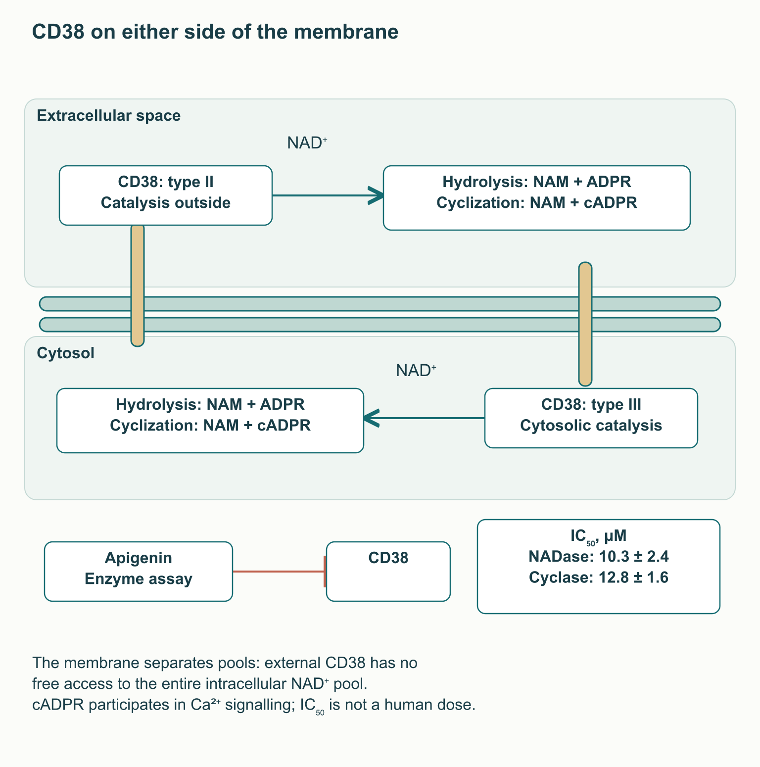
Enzyme orientation matters when interpreting the cellular result. In the common type II form of CD38, the catalytic region faces outside the cell or towards the lumen of the relevant membrane compartment; in type III it faces the cytosol. An extracellular enzyme therefore cannot be depicted freely consuming the entire intracellular NAD⁺ pool across an impermeable membrane. Changes in extracellular precursor availability, intracellular forms of the protein and tissue organization between cells help explain effects, but require their own evidence.
In mouse models, CD38 made a substantial contribution to age-related NAD⁺ decline and mitochondrial changes involving SIRT3. Other experiments showed that signals from senescent cells can increase the number and activity of CD38-positive macrophages in tissues. This links the inflammatory environment to NAD⁺ consumption rather than establishing one mechanism for ageing throughout the organism. The size of the change depends on tissue, model and measurement method; a universal 50–80% NAD⁺ decline in every ageing person has not been established.
Apigenin was indeed investigated as a CD38 inhibitor, but its activity was micromolar. In Escande’s study, IC₅₀ was 10.3 ± 2.4 μmol/L for NADase activity and 12.8 ± 1.6 μmol/L for cyclase activity. This is roughly two orders of magnitude above the 100–300 nmol/L range claimed in popular descriptions of the original material. Moreover, IC₅₀ depends on substrate concentration and assay conditions; it is not a binding constant, a blood concentration or a safe supplement dose.
After the cell experiments, the authors studied 12 mice fed a high-fat diet, divided into two groups. Apigenin was administered intraperitoneally at 100 mg/kg for seven days, with changes observed in hepatic NAD⁺, protein acetylation and metabolic measures. A short intervention bypassing ordinary intestinal absorption does not establish an oral protocol for humans or demonstrate prevention of age-related neurodegeneration. Findings concerning a more potent experimental CD38 inhibitor designated 78c involve a different molecule; its results cannot be attributed to apigenin.
SIRT1 and SIRT3 are NAD⁺-dependent deacetylases, but operate in different cellular contexts. SIRT1 acts mainly in nuclear and cytosolic processes, whereas SIRT3 regulates several mitochondrial-matrix proteins. Under certain conditions, increased NAD⁺ availability can support their reactions, but activity also depends on enzyme abundance, substrate state, reaction products and other regulators. Thus, the sequence “more NAD⁺ → all sirtuins switch on → the cell becomes younger” loses important qualifications.
Regulatory connections are easier to interpret alongside the specific proteins and reactions. The table lists steps that explain cell biology; it does not establish an effect of taking apigenin in humans. Neither enhancement of one reaction nor the availability of NAD⁺ alone guarantees clinical improvement.
| Step | Reaction or targets | Meaning and limitation |
|---|---|---|
| SIRT1 and autophagy | FOXO, p53; Atg5, Atg7, Atg8 | Changes in acetylation and stress-response programs depend on the cell and conditions |
| SIRT3 and IDH2 | Isocitrate + NADP⁺ → α-ketoglutarate + CO₂ + NADPH + H⁺ | A source of mitochondrial NADPH, not an established supplement effect |
| Glutathione reductase | GSSG + NADPH + H⁺ → 2 GSH + NADP⁺ | Glutathione regeneration; H₂O₂ removal requires the relevant enzymes |
| SIRT3 and the respiratory chain | Complex I, including NDUFA9; energy metabolism | Regulation of mitochondrial proteins does not guarantee an increase in ATP in humans |
A simple kinetic approximation helps explain substrate dependence: v = Vmax × [NAD⁺] / (Km + [NAD⁺]). Here v is reaction velocity, Vmax the limit at saturation and Km a parameter of a particular enzyme–substrate system. The equation shows gradual saturation rather than a switch with a single “normal” NAD⁺ range. Actual sirtuin reactions are more complex, so one Km range cannot serve as a universal test of NAD⁺ deficiency in a patient.
One established SIRT1 partner is the coactivator PGC-1α, which links energy-state signals with transcription of metabolic genes. Depending on the tissue, its regulation is associated with substrate oxidation, gluconeogenesis or mitochondrial-biogenesis programmes. PGC-1α interacts with the nuclear respiratory factors NRF1 and GABP, historically called NRF2, and with regulation of TFAM, which is required for mitochondrial DNA maintenance. This NRF2 must be distinguished from the antioxidant factor NFE2L2; a shared abbreviation does not make them the same protein.
SIRT1 also changes acetylation of FOXO factors, p53, autophagy components and the p65 subunit of NF-κB. These changes can redistribute stress responses, but their outcome depends on the cell and the task: suppressing apoptosis, for example, is not beneficial in every situation. For p65, deacetylation of Lys310 and changes in phosphorylation of Ser536 are different chemical events. They cannot be combined into an unlabelled “apigenin switches off inflammation” arrow, because the pathways and evidential bases differ.
In mitochondria, SIRT3 can regulate MnSOD/SOD2, IDH2 and respiratory-chain components, including proteins associated with complex I. SOD2 converts superoxide according to 2 O₂•⁻ + 2 H⁺ → H₂O₂ + O₂: the resulting hydrogen peroxide must then be removed by other systems. IDH2 produces NADPH during conversion of isocitrate to α-ketoglutarate; NADPH is subsequently used by glutathione reductase to restore oxidized glutathione. This is a specific chain supporting redox balance, not the literal disappearance of all reactive oxygen species.
SIRT3-knockout and calorie-restriction studies help establish these functions in cells and animals. Changes in the respiratory chain, mitochondrial-membrane permeability and cytochrome c release matter in injury and apoptosis, but do not demonstrate that an apigenin capsule prevents these events in people. Clinical translation requires measurements of exposure, pathway activity and a meaningful outcome within the same population. Until these links are connected, the mechanistic map is useful for explaining biology rather than promising higher ATP production or a longer life.
NF-κB is a family of transcriptional regulators through which cells coordinate many immune and stress responses. In the classical pathway, the IKK complex phosphorylates the inhibitor IκBα, promoting its degradation and allowing NF-κB, often as p50/p65, to influence transcription in the nucleus. This sequence describes a general pathway, not the complete mechanism of every inflammatory disease. In different models, apigenin affected individual steps in this system, and the findings did not always agree.
In experiments with LPS-activated mouse macrophages, apigenin reduced COX-2 and iNOS expression and associated transcriptional responses. COX-2 contributes to formation of prostanoids, including PGE₂, whereas iNOS/NOS2 supports inducible NO production. Concurrent excess NO and superoxide can permit NO• + O₂•⁻ → ONOO⁻, although NO also has normal signalling functions. Reduced production in an inflammatory culture does not mean beneficial suppression of all these substances throughout the body.
In another primary study involving human monocytes and mouse macrophages, reduced proinflammatory responses accompanied lower phosphorylation of p65 at Ser536. Yet IκBα degradation continued, and NF-κB DNA binding did not disappear. A separate study of related flavonoids under TNF stimulation likewise reported no blockade of several early steps. Apigenin therefore cannot be presented as a universal switch simultaneously disabling IKK, nuclear transport and DNA binding.
Reduced COX-2 expression in cells also does not establish drug-level selectivity or prove an absence of gastrointestinal risk compared with anti-inflammatory medicines. Protection in a mouse model of LPS-induced lethality is not equivalent to treating human sepsis. Specialists need to distinguish three levels: a change in a signalling protein, reduced mediator production and an improved disease outcome. These may be related, but each transition requires its own investigation.
For microglia, one experimental study found that apigenin and luteolin reduced IFN-γ-induced CD40 expression by affecting STAT1. The researchers examined a cell line and primary microglial cultures, as well as inflammatory mediators including IL-6 and TNF. This is not evidence of clinical conversion of microglia from “M1” to “M2”, nor a study of a single universal STAT3 pathway. Actual microglial states are more diverse than two poles, so the practical conclusion is a promising research target whose therapeutic applicability remains unestablished.
Poor aglycone dissolution makes formulation important, but improving one step does not remove the others. Particle dissolution is often described using the Noyes–Whitney relationship: dm/dt = D × A × (Cs − C) / h. Here dm/dt is the rate of transfer into solution, D the diffusion coefficient, A the available particle surface, Cs the saturation concentration, C the current concentration and h the effective diffusion-layer thickness. Micronization can increase A, but particle aggregation and the properties of the medium can alter the expected result.
Phospholipid complexes and liposomal systems use a different approach: they change how the substance interacts with aqueous and lipid environments. In an experiment with the apigenin–phospholipid complex APLC, solubility increased more than 36-fold relative to the free substance examined; subsequent pharmacokinetics and antioxidant measures were investigated in animals. This is a result for a specific formulation, not a guaranteed advantage of every bottle labelled “phytosome”. Liposomes also depend on size, stability, the proportion encapsulated and their behaviour after oral administration, rather than one assumed nanometre range.
Even an increase in concentration–time area under the curve does not necessarily improve a useful clinical outcome. One needs to know whether the original molecule or conjugates were measured, in which population, and how safety and target-tissue access changed. An oral lipid formulation does not automatically “bypass the liver”, and an exposure multiplier in rats cannot be transferred to humans without a study. Claims of a 3.5–5.2-fold increase in human bioavailability or attainment of a therapeutic brain concentration therefore require data for the stated product.
In practice, assessment begins with the identity of the formulation and the intended use. A food preparation, chamomile extract and purified apigenin should be considered separately; the label should make clear which component is present and in what quantity. For a specialist, the next question is whether a human study of that formulation measured the relevant outcome, rather than laboratory solubility alone. Without such a study, delivery technology remains a pharmaceutical property rather than a confirmed treatment protocol.
Apigenin is often combined with magnesium, L-theanine or glycine in a “sleep stack”. However, describing molecules that act on the nervous system together does not establish the efficacy or safety of a finished mixture. A combination requires specific forms, doses, timing and a clinical outcome; the experiments discussed do not provide a sufficient basis for prescribing such a stack. When several supplements are changed simultaneously, subjective improvement or an adverse effect is difficult to attribute to one component.
Magnesium participates in physiological NMDA-channel regulation, including voltage-dependent block, but taking a magnesium salt is not equivalent to selectively increasing that block in the brain. L-theanine cannot be described as an established selective NMDA antagonist, while glycine is an NMDA-receptor coagonist and separately acts through glycine receptors. A simple drawing in which all three substances identically “quench excitation” is therefore biologically inaccurate. Individual studies of another component also do not establish its combined effect specifically with apigenin.
For NAD⁺, the logic of combinations centres on the balance between synthesis and consumption. Nicotinamide, NAM, is converted by NAMPT, using PRPP, into NMN; nicotinamide riboside, NR, can be phosphorylated by NMRK to NMN. NMNAT then uses ATP to form NAD⁺ and pyrophosphate: NMN + ATP → NAD⁺ + PPi. These pathways explain precursor biochemistry, but do not mean that any oral molecule directly and equally replenishes every tissue pool.
The complete early-step reactions are NAM + PRPP → NMN + PPi for NAMPT and NR + ATP → NMN + ADP for NMRK. Sirtuins and PARP, like CD38, consume NAD⁺, but they perform different functions and do not form a single switch for cellular state. Precursor-derived synthesis, distribution across compartments, and the activity of each consumer therefore need to be considered separately.
Some nicotinamide can be methylated by NNMT: NAM + SAM → 1-methylnicotinamide + SAH. The methyl-group donor here is S-adenosylmethionine, SAM, not trimethylglycine. Betaine, or TMG, participates in a different reaction catalysed by BHMT, mainly in the liver and kidneys: betaine + homocysteine → dimethylglycine + methionine. A connection through methionine metabolism does not make TMG a direct NNMT cofactor or establish an obligatory need to take it with NAD⁺ precursors.
Potentially limiting NAD⁺ consumption with apigenin while increasing synthesis through precursors is a testable hypothesis, not established therapeutic synergy. The same caution applies to resveratrol: studies of direct SIRT1 activation depended on the substrate and assay design. Experiments using unmodified substrates and findings supporting context-dependent allosteric activation cannot be reduced to “resveratrol switches on SIRT1”. These data do not create a clinically established apigenin–NMN/NR–resveratrol–TMG combination for extending life or preventing neurodegeneration.
No generally accepted human therapeutic range for purified apigenin has been established for insomnia, anxiety or NAD⁺ preservation. Doses of 50–100 mg “for sleep” and 250–500 mg “for CD38” therefore cannot be presented as tested clinical protocols. Taking it 45–60 minutes before bedtime also cannot be inferred from food kinetics with a delayed plasma peak or from an intraperitoneal mouse experiment. Different formulations can change exposure, but do not yet justify calculating a therapeutic window independently from IC₅₀.
| Goal | What is actually available | What remains insufficient |
|---|---|---|
| Improving sleep | A pilot study of a specific chamomile extract | Reproducible benefit and a pure-apigenin regimen |
| Reducing anxiety | Small extract studies in patients with GAD | Equivalence of extract and apigenin; a universal prescription |
| Maintaining NAD⁺ | Enzyme, cell and mouse findings | A clinical outcome and a safe, effective oral dose |
| Reducing neuroinflammation | Altered signalling in experimental models | Demonstrated treatment of a neurological disease |
In a laboratory study, apigenin inhibited CYP2C9-mediated losartan metabolism; IC₅₀ in human liver microsomes was 4.10 μmol/L. A different assay with diclofenac showed competitive CYP2C9 inhibition with an IC₅₀ of 9.51 μmol/L. This is not the measured size of an interaction in a patient taking a capsule, but it is an important signal when considering concentrated supplements and medicines with a narrow safety margin. Studies of apigenin metabolism by CYP1A2 and CYP3A4 also cannot automatically be read as clinical evidence of equivalent inhibition of all these enzymes.
Official chamomile information warns of possible interactions with warfarin, some medicines metabolized by the liver and sedatives. An extract and a pure flavone are not identical, so the degree of risk for each product remains a separate question. A person taking an anticoagulant, hypnotic, anti-anxiety medicine or multiple medicines should discuss concentrated apigenin with a physician or clinical pharmacologist. Neither stopping prescribed treatment independently nor deriving a “safe interaction threshold” of 300–500 mg from laboratory results is justified.
Allergic reactions to chamomile, including rare severe reactions, have been reported, particularly in people sensitive to related plants in the Asteraceae family. Nausea or dizziness can also occur; ordinary food amounts are not equivalent to a concentrated preparation. There are insufficient data to prescribe purified apigenin during pregnancy, breastfeeding or childhood. Liver and kidney diseases affecting metabolism and excretion further complicate the transfer of standard dose assumptions.
Low acute toxicity in a particular animal experiment does not establish the safety of use for months in humans. The long-term chamomile trial examined 1500 mg of extract, not a thousand milligrams of apigenin, and involved selected patients with an anxiety disorder. A lack of established dependence is also not equivalent to proof of no tolerance, withdrawal or cognitive effects for every formulation and dose. These questions should remain open rather than turning limited observation into an advertising advantage.
In practice, apigenin is currently most useful as an example of the distance between molecular plausibility and readiness for clinical use. Flavone-containing foods can form part of a varied diet without trying to reach a laboratory concentration of an individual compound. When choosing a supplement, it is sensible first to define the problem, review medicines and seek human data for the specific preparation; persistent insomnia or anxiety still calls for proper clinical care. Research on CD38, sirtuins and inflammation provides meaningful directions for future work, but does not yet turn apigenin into a universal hypnotic, anxiolytic or anti-ageing treatment.
No. Binding at a receptor site does not determine the entire pharmacological profile. Functional experiments gave variable results, and human evidence does not establish a selective anxiolytic without sedative or other risks.
There is no generally accepted regimen for purified apigenin. A chamomile-extract pilot trial did not show significant benefit on principal sleep measures. Milligrams of extract cannot be converted into a prescription for the same dose of pure flavone.
There are no high-quality human data establishing increased N3 or enhanced glymphatic processes with apigenin. Findings on rat sleep latency and general sleep physiology do not prove these effects.
It is the concentration at which activity fell by half in a defined assay. For apigenin it was approximately 10–13 μmol/L, not 100–300 nmol/L. This value does not establish a supplement dose or show that this tissue concentration is reached in humans.
The NAD⁺ pool reflects molecular availability, whereas NAD⁺/NADH describes part of the redox state. Both depend on compartment and measurement method. Changing one does not guarantee a change in the other or clinical benefit.
There is no established clinical basis for making that combination obligatory. NAD⁺ synthesis and consumption pathways explain a hypothesis, but do not prove a mixture's efficacy. TMG participates in the BHMT reaction and is not a direct methyl-group donor for NNMT.
No. A specific experimental phospholipid complex improved solubility, and its pharmacokinetics was investigated in animals. Clinical superiority requires a study of that product in humans measuring the relevant outcome.
People taking anticoagulants, sedatives or other medicines, those with liver or kidney disease, and pregnant or breastfeeding people need individual assessment. Children must not be given a dose calculated from an adult or animal model. Chamomile can also cause allergy in people sensitive to Asteraceae.