TMAO: microbiota, FMO3, kidneys, and evidence on risk — NutriFit
TMAO: microbial TMA, hepatic FMO3, renal clearance, and cardiovascular evidence
The complete pathway from choline and carnitine through CutC/CutD, CntA/B, and FMO3 to TMAO elimination; mechanistic models, human feeding studies, observational associations, fish, and CKD.
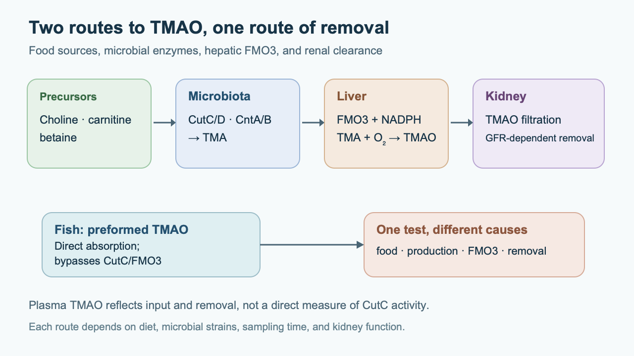
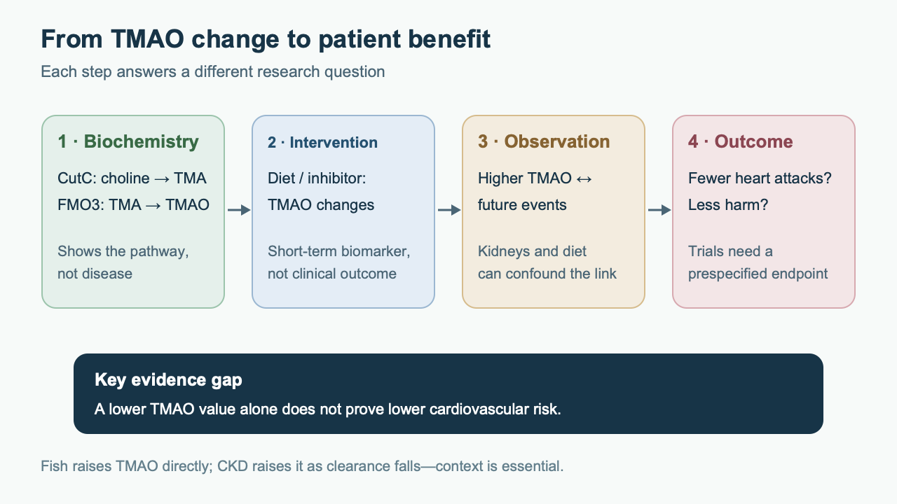
The plasma concentration of trimethylamine N-oxide (TMAO) brings together several processes that are easy to conflate. Some dietary compounds are first converted by gut bacteria into trimethylamine (TMA), hepatic FMO3 oxidizes TMA, and the kidneys eliminate the resulting TMAO. Fish can supply preformed TMAO, bypassing the microbial step. A high test result therefore does not by itself show which step has changed, much less establish that TMAO itself caused cardiovascular disease. The case of a patient with reduced kidney function is especially instructive: metabolite production may be unchanged while its concentration rises because removal has slowed.
This article follows the chemistry of CutC/CutD and carnitine pathways, the FMO3 cycle, clearance, cellular mechanisms, observational cohort findings, and the limits of dietary and drug interventions. It is a narrative review of the source bibliography with targeted checking of disputed claims on 9 October 2026; no full systematic search or formal certainty assessment was performed. Population, source of TMAO, kidney function, and a prespecified clinical outcome matter more than one metabolite value.
From food to microbial TMA
Choline is an essential component of phosphatidylcholine and sphingomyelin and a precursor of acetylcholine and betaine. Eggs, meat, liver, legumes, and other foods contribute it in different forms. Phosphatidylcholine is digested through several pancreatic and intestinal pathways; some choline is absorbed in the small intestine, while some is available to microbes in the distal gut. It is not valid to assume that a fixed 30–40% of every meal reaches the cecum: the fraction depends on food composition, dose, absorption, and transit. Betaine also comes from foods, including grains and beets, and from oxidation of choline in host tissues. It supports homocysteine remethylation through betaine-homocysteine methyltransferase (BHMT); some microbes can use its trimethyl group. A “TMA precursor” is therefore not synonymous with a “dietary toxin.”
L-carnitine carries long-chain acyl groups into the mitochondrial matrix through the carnitine shuttle; it is obtained from meat and synthesized endogenously. Absorption and renal reabsorption involve, among other proteins, OCTN2/SLC22A5. At large single doses, fractional absorption may fall, leaving more substrate for microbes, but a “2 g” threshold and “below 15–20%” bioavailability cannot be applied to everyone without a measurement protocol. Repeated carnitine exposure also changes the community’s capacity to produce TMA. Human feeding experiments found different responses in people with different habitual diets, but the label “vegetarian” alone does not predict an individual’s TMAO.
CutC is a choline-TMA lyase in anaerobic bacteria; CutD activates it. CutD belongs to the radical S-adenosylmethionine enzyme family and uses a [4Fe–4S] cluster and SAM to generate a 5′-deoxyadenosyl radical. This creates a stable glycyl radical in CutC; a thiyl radical on catalytic Cys then mediates the conversion of choline. Overall, choline is cleaved into TMA and acetaldehyde. This radical chemistry requires an appropriate anaerobic environment; oxygen can inactivate the enzyme, so the presence of a gene in a genome is not the same as a measured rate of TMA production in the gut. Exact Gly and Cys numbering depends on protein sequence and structure; positions in one CutC model cannot be transferred to all microbial orthologs without checking.
cutC/cutD carriers occur in several bacterial lineages. A genus name does not guarantee a functional operon in a particular strain, and relative abundance does not measure metabolite flux. Causal inference benefits from metagenomics or gene-expression data, measurement of TMA after a labeled substrate, and intervention in the enzyme pathway. Antibiotics suppress production in some experiments but also alter many other microbial processes; such an experiment does not establish a unique role for CutC.
Carnitine can also yield TMA through other routes. The oxygen-dependent CntA/CntB system includes an oxygenase with a Rieske-type [2Fe–2S] center and mononuclear iron, plus an electron-transferring partner reductase; this pathway has been studied in some bacteria in the presence of O₂. In more anaerobic surroundings, carnitine may first become γ-butyrobetaine, which other communities then convert to TMA. These routes need not operate simultaneously in one cell. Knowing the chemical precursor matters when designing an inhibitor: blocking CutC does not guarantee elimination of TMA from carnitine, betaine, or preformed TMAO in fish.
Different substrates, different responses
Phosphatidylcholine is not equivalent to free choline in where and how quickly substrate appears in the gut. The membrane phospholipid must be hydrolyzed, and liberated choline competes between host absorption and microbial metabolism. Some dietary choline is incorporated into host tissues and enters the betaine pathway; measured TMAO reflects only one branch of its distribution. If TMAO does not rise after a meal, that does not prove choline was not absorbed. Conversely, a rise in TMAO does not mean there is now enough choline for membranes and the liver. Choline deficiency physiology and safety are discussed in more detail in the choline article.
The carnitine pathway also branches. Host cells use L-carnitine to transport acyl groups through CPT1, carnitine-acylcarnitine translocase, and CPT2; bacteria receive only the fraction available to them. γ-Butyrobetaine is both an intermediate in endogenous carnitine synthesis and a microbial metabolite, so an increase without a labeled substrate does not establish its source. Oxygen and an electron donor matter for CntA/CntB, whereas CutC is favored by an anaerobic lifestyle. A shift in the oxygen gradient along the intestine changes the relative contribution of these routes even if the nutrients eaten are unchanged. Bacterial species, gene, substrate availability, and current enzyme expression are four distinct levels of explanation.
Why a measurement cannot be reduced to a bacterial taxon
Microbial TMA production is a functional property of a community, not a fixed feature of a genus. Closely related strains differ in their operons, regulation, and access to substrate. In the distal gut, some microbes may release choline from dietary compounds, others may cleave choline, and still others may consume intermediates. This exchange between species makes TMA flux sensitive to transit time and diet. A metagenomic cutC signal indicates potential capacity; a transcriptomic signal indicates gene activity; luminal TMA concentration and its appearance after labeled choline indicate the combined result. These levels are not interchangeable. Lists in the source of “main harmful genera” are therefore unsuitable for choosing a probiotic from a single culture or commercial stool test.
Methylotrophic archaea can use methylated amines, including TMA, in methanogenesis. In biochemical terms, their methyltransferases transfer methyl groups to corrinoid carriers; subsequent reactions involve reducing equivalents and methane formation. Writing the whole process as a single MttB enzyme reaction, “TMA + 3H₂ → 3CH₄ + NH₃,” conflates an individual enzyme step with the community’s overall metabolism. Even if an archaeon consumes TMA in the laboratory, sustained colonization, substrate competition, safety, and a changed clinical outcome would need to be demonstrated in the living human gut. The source bibliography does not provide such evidence.
Radical CutC and oxygen-dependent CntA are not the same enzyme
The overall CutC reaction can be written as choline → TMA + acetaldehyde. It does not require oxygen and retains the atoms of the trimethylammonium group in TMA; choline’s two-carbon residue becomes acetaldehyde. CutD consumes SAM while creating the active radical state of CutC, but does not itself carry choline. Structural work on CutC showed a conformational change when substrate binds, helping explain pocket specificity. It does not prove that an inhibitor shown in a figure would be stable and safe in the human colon. The redox state of the [4Fe–4S] cluster, anaerobiosis, and the presence of the already activated enzyme are especially important. Thus, an IC₅₀ measured with purified protein is not equivalent to the concentration that blocks TMA formation in a complex microbial community.
For CntA/CntB, an approximate stoichiometric balance is L-carnitine + NADH + H⁺ + O₂ → TMA + malonic semialdehyde + NAD⁺ + H₂O. CntB transfers electrons, while CntA performs oxygen-dependent cleavage; the reaction depends on oxygen availability. In anoxic niches, the γ-butyrobetaine route requires different participants and steps. The two pathways produce the same TMA but differ in cofactors and ecosystem. This is why chemically inhibiting one choline lyase does not eliminate every source of trimethylamine. Betaine pathways are even more context-dependent: dietary betaine does not establish what fraction of its methyl groups enters TMA versus host one-carbon metabolism.
Hepatic FMO3 and the other side of its variation
TMA is a tertiary amine with a characteristic odor. After absorption it reaches the liver through the portal vein; at physiological pH, uncharged and protonated forms are in equilibrium, so describing TMA as a completely neutral “lipophilic gas” is incorrect. The predominant human hepatic flavin-containing monooxygenase FMO3 N-oxidizes TMA:
TMA + NADPH + H⁺ + O₂ → TMAO + NADP⁺ + H₂O.
FMO3 is a microsomal flavoprotein. Its bound FAD accepts electrons from NADPH and reacts with O₂ to form a C4a-hydroperoxyflavin intermediate; an oxygen atom is then transferred to the TMA nitrogen. The source’s claim that FAD is covalently bound to FMO3 is unnecessary for the mechanism and is not a general property of these monooxygenases. The resulting TMAO is polar; its N–O bond can be represented as N⁺–O⁻. The enzyme also oxidizes other substrates, so changing FMO3 can have effects beyond the TMA pathway.
FMO3 expression depends on development, hormonal state, nuclear receptors, and species differences. FXR may regulate it when bile-acid signaling changes; the pronounced sexual dimorphism in mice cannot be translated directly into a fixed activity ratio for all men and women. FMO3-suppression experiments have shown changes in lipid and glucose metabolism, but that does not mean all effects are mediated by lower TMAO. Genetic FMO3 variants can change TMA oxidation. With substantial loss of function, trimethylaminuria can develop: excess unoxidized TMA is excreted in urine, breath, and sweat, producing a characteristic odor. This is a distinct clinical problem, not “low vascular risk,” because FMO3 has functions beyond one analyte.
The TMA/TMAO ratio is sometimes used to investigate trimethylaminuria after a standardized challenge and with kidney function taken into account. It is not a universal at-home FMO3 index: both concentrations are influenced by microbial production, time after food, sample matrix, and elimination. At high acute TMA exposure, enzyme flux may differ from everyday exposure; “95–98% oxidation in all healthy people” is not a valid diagnostic constant.
A genetic and dietary extreme: trimethylaminuria
With reduced FMO3 activity, some TMA escapes complete N-oxidation and gives secretions an odor. Primary trimethylaminuria is associated with gene variants, but the phenotype depends on residual enzyme activity, precursor intake, hormonal state, and other diseases. A “variant in FMO3” therefore does not always mean persistent symptoms, and a strong odor does not always prove a mutation. Diagnosis depends on TMA/TMAO measurement under controlled conditions and specialist interpretation; restricting choline to deficiency levels to eliminate odor is unsafe. This assessment differs from trying to predict a heart attack from plasma TMAO.
TMAO also cannot be treated as a linear measure of FMO3 activity under every exposure. Preformed TMAO from fish bypasses FMO3. In chronic kidney disease (CKD), TMAO accumulates after the enzymatic reaction. With impaired absorption or recent antibiotic use, little TMA may reach the liver. Thus, high TMA/TMAO, low TMAO, or their ratio without a specified challenge can give opposite impressions about the same enzyme. Mouse FMO3 experiments require even more caution: sex hormones alter expression differently than in humans, and knockout outcomes may reflect other FMO3 substrates.
The kidneys determine the measured concentration
TMAO is relatively small and highly water-soluble, so renal excretion strongly affects its plasma concentration. Filtration is an important route. The source article claimed that OCT2/MATE always provide substantial secretion and clearance of 120–160 mL/min; primary studies differ between animal and human models, including evidence for filtration-dominant elimination and a small OCT2 contribution in humans. A transporter that interacts with TMAO in vitro does not necessarily determine total human clearance. Effects of diuretics and other drugs must also be assessed in the relevant population. There is therefore no universal “4–6 hour” half-life or fixed eGFR threshold beyond which TMAO must “skyrocket.”
TMAO is often elevated in CKD: lower GFR impairs excretion, while diet, microbiota, and treatment can also change production. This creates the possibility of reverse causation: poor kidney function raises TMAO and independently raises cardiovascular risk. Statistical adjustment for eGFR reduces part of this problem but does not replace measurement of clearance, disease duration, and other factors. TMAO may be classified as an accumulating uremic metabolite, but calling it a “uremic toxin” requires separate evidence of harm at clinically attainable exposure. In patients on dialysis, concentration also depends on the removal regimen and time between treatments; the source’s promise of “65–80% removal per session” by hemoperfusion is unsupported by the cited primary bibliography.
Kidneys also affect interpretation of food responses. After the same meal, two people can have different concentration curves because of both TMA production and the rate of TMAO elimination. A single measurement without recording recent fish intake, carnitine supplements, kidney disease, and sampling time combines different causes into one result. A scientific experiment is better served by measuring kinetics, baseline values, renal markers, and a stable-isotope-labeled substrate than by calling high TMAO “dysbiosis.”
Vascular and metabolic mechanisms: what has been tested
TMAO has been linked to several phenotypes in cellular and animal systems. Zhu and colleagues reported that higher TMAO enhanced platelet responses to stimuli through changes in intracellular Ca²⁺ and accelerated thrombosis in an experimental system. This does not mean that TMAO can diagnose aspirin or clopidogrel resistance or guide antiplatelet dosing. Other models described changes in reverse cholesterol transport, the bile-acid pool, CYP7A1, and formation of foam macrophages. In mice, bile-acid composition and genetic susceptibility substantially affect outcomes; CD36 or SR-A1 expression in culture is not equivalent to clinically significant human atherosclerosis.
Endothelial studies have reported changes in MAPK, NF-κB, adhesion molecules, oxidative stress, and NLRP3. Some experiments linked TMAO to PERK and the endoplasmic-reticulum stress response. Their concentrations, exposure durations, and media composition should be compared with the free metabolite level in humans. “TMAO activates NLRP3” in one cell model does not imply a required direct receptor or a single causal chain from gut to heart attack. Smooth-muscle cells and mouse models have been used to study calcification and aortic stiffness. These results define testable mechanisms, but do not replace an intervention showing that lowering TMAO in people reduces clinical events.
Mice on choline diets or given TMAO have been reported to develop myocardial hypertrophy and fibrosis; kidney models have shown stress and profibrotic signals, including PERK/CHOP and TGF-β/Smad. This does not prove direct “nephrotoxicity” from every measured TMAO concentration: some studies use doses and models with clearance unlike that in humans. Likewise, proposed links to anemia, FGF23–Klotho, blood–brain barrier permeability, or aneurysm should not be presented as established clinical mechanisms without appropriate studies. In type 2 diabetes and insulin resistance, separate the effects of FMO3 as an enzyme, diet, and TMAO itself. Negative or neutral experimental findings also matter: biological effects can be context-dependent and are not always harmful.
Atherosclerotic mechanisms are not all the same
In reverse cholesterol transport experiments, cholesterol is followed from macrophages through lipoproteins to the liver and fecal sterols. Reduced transport in mice after a choline diet is compatible with changes in bile acids, ABCG5/ABCG8 transport, and CYP7A1, but also depends on diet composition and microbiota. Attributing the result to TMAO alone requires suitable control of TMAO, diet, and genetic background. In humans, lipoprotein cholesterol levels, TMAO concentration, and transport between tissues are not the same measure.
The platelet mechanism differs from chronic plaque formation. In experiments, TMAO enhanced calcium responses to agonists: it shifted platelet readiness to respond rather than replacing the agonist itself. Clinically, the metabolite concentration at the time of a vascular event, concomitant antiplatelet drugs, and bleeding probability all matter. A proposal to measure “platelet aggregation in high TMAO” as a standard step does not follow from a model experiment. The contrast between a rapid cellular response and slow atherogenesis shows why even a plausible molecular mechanism does not yield a single predictive number.
In the kidney, PERK senses endoplasmic-reticulum stress; CHOP is associated with consequences of prolonged stress signaling. TGF-β/Smad can alter fibrotic programs. If TMAO activates these nodes in culture, that alone does not establish the direction of the process in CKD: impaired filtration itself increases TMAO exposure, and uremic serum contains many other molecules. Indoxyl sulfate and p-cresyl sulfate, often discussed alongside TMAO, differ in protein binding and tubular elimination. “Toxin synergy” requires a factorial experiment showing that the combined effect exceeds what would be expected from the individual components; their simple co-elevation in CKD is not such evidence.
Vascular wall and tissue compartments
In experimental macrophages, increased cholesterol accumulation can coincide with greater uptake of modified lipoproteins through scavenger receptors CD36 and SR-A1. But dye or lipid-droplet accumulation in culture depends on oxidized LDL dose, cell state, and TMAO concentration. A true atherosclerotic plaque develops through years of interaction among endothelium, smooth-muscle cells, lipoproteins, immunity, and hemodynamics. A single macrophage effect therefore does not determine the magnitude of plaque-size change in humans. Likewise, mouse changes in CYP7A1, bile-acid excretion, and ABCG5/ABCG8 transport are not a direct map of human reverse cholesterol transport.
Endothelial NO is produced by eNOS and participates in vascular responses. Some model studies link TMAO to reduced NO signaling and higher adhesion-molecule expression, but the result depends on concentration and other conditions, including hyperglycemia and uremic compounds. The NLRP3 inflammasome participates in IL-1β and IL-18 activation under many stressors; activation in culture after TMAO is not a unique fingerprint of this molecule. Studies of smooth-muscle-cell calcification must account for phosphate, kidney failure, and age—powerful independent causes of osteogenic programs in the vascular wall. A TMAO measurement without these measures cannot explain calcium on an arterial CT scan.
Cerebral circulation is even more complex. Plasma TMAO is not the same as its concentration near blood–brain barrier endothelial cells, and barrier permeability changes can result from general inflammation and vascular disease. The source presents the path from TMAO to stroke and neuronal injury as a ready-made mechanism, but there is no direct evidence of a clinical effect from intervening in this axis. Neurological cohorts need to account separately for kidney function, age, medication, and stroke type; a mouse dose cannot be transferred to humans by simple body-weight scaling.
Kidney fibrosis and other uremic compounds
In cell experiments, high TMAO exposure can raise markers of ER stress, profibrotic signaling, and tubular inflammation. In animal models, a choline diet changes microbiota, TMAO, other metabolites, and nutrition at the same time. Direct TMAO experiments help isolate a possible effect of the molecule itself but do not resolve dose or interspecies clearance. Demonstrating a human vicious cycle—“TMAO damages the kidney, and the kidney then accumulates TMAO”—would require, for example, comparing kidney-function changes after a specific intervention while controlling other factors. For now, the reverse arrow from CKD to TMAO is substantially better established than a causal arrow from moderate TMAO to CKD progression.
Indoxyl sulfate and p-cresyl sulfate arise through different microbial–hepatic pathways, are often protein-bound, and behave differently from TMAO during dialysis. Their co-elevation in CKD does not establish that they potentiate each other. The source also links TMAO to anemia, erythropoietin response, and the FGF23–Klotho axis. These are reasonable hypotheses for specific models because CKD simultaneously alters inflammation, iron, phosphate, and hormonal signaling. Without separating these causes, however, TMAO measurement cannot be recommended for ESA dose adjustment or treatment of mineral-bone disorders.
What human studies show
Prospective cohorts, including Tang and colleagues’ study of patients undergoing cardiac evaluation, associated higher TMAO with subsequent cardiovascular events after statistical adjustment. Prognostic associations have also been reported in stable coronary artery disease (CAD), heart failure, and CKD studies. The temporal order—measurement before the event—is stronger than a simple simultaneous comparison, but residual confounding, differences in kidney function, diet, and treatment, and selection of patients already at high baseline risk remain. A hazard ratio for the top quartile in one clinic cannot be applied to healthy people or treated as a universal TMAO threshold.
Human dietary interventions answer a different kind of question. After labeled phosphatidylcholine and carnitine, one can demonstrate substrate flow through microbial TMA to TMAO; suppressing the microbiota with antibiotics attenuates this flow, but does not justify antibiotics to prevent atherosclerosis. Controlled comparisons of fish, meat, and egg meals showed different TMAO time courses. Some fish contain preformed TMAO, and after eating them, plasma TMAO may rise more than after eggs or meat, returning to baseline within a day in the studied group. This does not make fish a cause of vascular harm: foods contain many compounds, and long-term diet and a brief biomarker rise answer different questions. Some controlled meat and carnitine studies found increased microbial pathway activity after sustained exposure. TMAO measurement, however, did not establish that limiting carnitine prevents heart attacks.
Evidence type
What was actually measured
Main limit of inference
CutC/CutD enzyme, FMO3 preparation, or culture
Reaction, intermediates, activity in a defined setting
Does not provide a human dose or clinical outcome
Choline/TMAO feeding in mice or CutC inhibitor
Change in TMAO, plaques, or tissue signals in a model
Microbiota, bile acids, and clearance differ from humans
Short human feeding experiment
Timing and size of rise after food or labeled substrate
Does not determine long-term cardiac risk
Prospective CAD/CKD/heart-failure cohort
Association between baseline TMAO and a future event
Confounding, reverse causation through kidneys, population selection
Intervention trial with clinical outcome
Must test event reduction and harm under a defined strategy
Benefit from targeted TMAO lowering has not been established
In heart failure, high TMAO often coexists with impaired perfusion and kidney function, changes in medication, and altered diet. It may therefore be a severity indicator, a mediator, or both. It is particularly difficult to infer from observations that TMAO causes resistance to standard antiplatelet therapy: trials with prespecified medication and clinical outcomes are needed. In CKD studies, a concentration change during dialysis or dietary change should not be turned into a mortality claim.
What controlled feeding changes
In a controlled crossover study, healthy participants completed dietary periods in which red meat, white meat, or non-meat foods were the main protein source. Fasting TMAO after the red-meat period was higher than after the other two diets; isotope studies indicated increased production from carnitine, but not choline, and measurements also identified a change in renal TMAO excretion. After red meat was withdrawn, the measure decreased over four weeks. This design is especially useful because it connects precursor intake, microbial flux, and clearance in one experiment. However, heart attacks and mortality were not the primary outcomes of this metabolic study. It cannot be recast as proof that any TMAO reduction after changing protein source prevents cardiovascular events.
For fish, the causal chain is shorter: preformed TMAO in some species is absorbed without CutC or FMO3. A study of several seafood species in healthy volunteers found substantial variation in TMAO+TMA; after certain meals, especially cod, the plasma TMAO increase was transient and returned to baseline within a day under the conditions studied. That curve cannot be transferred to a patient with CKD or applied to every fish species as if it were cod. It is equally inaccurate to explain any potential benefit of fish solely through a compensatory effect of EPA/DHA: composition, preparation methods, and foods replaced differ. The fish paradox argues against interpreting the biomarker monotonically as harm; it does not prove that fish-derived TMAO is always harmless.
Cohorts: prognosis and causality
Cleveland Clinic studies showed that choline metabolites and TMAO predicted events in selected clinical cohorts. These findings should be read with the inclusion criteria: a patient referred for coronary evaluation differs from a randomly selected healthy person. Age, diabetes, kidney impairment, and treatment affect both concentration and future risk. Even careful statistical adjustment cannot exclude dietary changes after early symptoms that were not measured. A strong association in several samples increases interest in the pathway but does not by itself show that TMAO will be a useful treatment target.
Heart failure particularly illustrates this complexity. Reduced cardiac output and congestion can change renal blood flow, appetite, and microbiota; medications and hospitalization also affect metabolites. If TMAO predicts death after adjustment for usual variables, it may add prognostic information, but implementing a test requires reproducibility and evidence that it improves decisions beyond existing markers. The association sometimes weakens after adding renal measures; that should be reported rather than hidden to preserve an appealing “independent predictor.”
Measurement and clinical interpretation
TMAO can be quantified, for example, by liquid chromatography–tandem mass spectrometry with isotope-labeled internal standards. This can distinguish TMAO, TMA, choline, and betaine and track analytical losses. But even a highly accurate instrument cannot correct a poorly designed collection: the same person can produce different results after fish, prolonged fasting, a change in kidney function, or antibiotics. Preanalytics include sample matrix, collection time, storage, and recording recent exposures. No generally accepted clinical threshold of “4/8/10 μmol/L” assigns every patient to risk categories and dictates treatment.
With a pronounced fishy odor, the task is different: assess TMA and TMAO, FMO3 variants, circumstances in which odor appears, medications, and other causes. In CKD or CAD, clinical risk is assessed with validated measures and the diagnosis; TMAO may be a research or supplementary prognostic marker, but it does not replace GFR, albuminuria, lipids, or the clinical picture. Clinicians should ask whether the test will change a decision with proven benefit. If the number only triggers an unvalidated supplement regimen or abandonment of beneficial foods, the value of the test is questionable.
Questions to ask about a test result
First, was TMAO measured in fasting plasma or after a particular meal, and how much time had passed? Second, is current GFR known, and did it change between measurements? Third, has the person taken L-carnitine, choline supplements, antibiotics, or drugs that affect the kidney? Fourth, why was the test ordered: to study a metabolic pathway, assess trimethylaminuria, or add prognostic information in an established disease? Only then can the result be compared with an appropriate reference group. Even in a cohort where TMAO is associated with events, clinical usefulness requires assessing whether it improves decisions beyond standard risk factors.
As a hypothetical example, the same 10 μmol/L after a fish lunch in a person with preserved GFR and in a fasting person with progressive CKD do not have the same causal explanation. The number alone does not prescribe choline restriction, an antibiotic, a probiotic, or an antiplatelet drug for either person. For the second patient, confirming and managing kidney disease is especially important; for the first, recent food is relevant context. This does not prohibit TMAO research: standardized serial measurements can help test a hypothesis when other parts of the pathway are also described.
Can the pathway be targeted?
3,3-Dimethyl-1-butanol (DMB) has been used as a nonlethal experimental inhibitor of microbial TMA production; in mice on the relevant diet it reduced TMAO and atherosclerotic changes. More active choline analogs, including halogenated derivatives, have been studied in preclinical systems and may block the pathway at microbial enzymes. The cited publications do not establish guaranteed lack of systemic absorption, unchanged safety, a specific human dose, or proven reduction in events. “Nonlethal” means no direct bacterial killing in the experiment, but long-term changes to the ecosystem remain possible. The chemical mechanism of each analog must be evaluated separately rather than assigning every compound the same irreversible Cys binding.
Dietary modification may change choline and carnitine availability, microbial composition, and TMAO, but choline remains an essential nutrient. Severe restriction risks impairing synthesis of membrane phospholipids, acetylcholine, and methyl donors; needs differ, especially during pregnancy. Evidence supporting an overall Mediterranean-style dietary pattern does not mean its benefit is explained by lowering TMAO, and one cannot promise everyone a 30–50% reduction in six months. Fish should not be excluded merely to achieve a short-term TMAO reduction; species and serving composition, overall dietary pattern, and kidney status matter more than a single value. Food comparisons should consider not only TMA precursors but also protein, fatty acids, fiber, and other health measures.
Soluble fibers change the gut environment and short-chain fatty acids; the dietary fiber article discusses how to choose them. They may change microbial TMA flux, but their effect on a specific cutC pathway varies among people. It cannot be guaranteed that inulin will “acidify the cecum to 5.5–6.0” and displace all TMA producers. The same is true for probiotics: strain, dose, and colonization matter; the names Lactobacillus, Bifidobacterium, or Akkermansia are not clinical prescriptions for lowering TMAO. Resveratrol changed microbiota, TMAO, and atherosclerotic features in a mouse model; clinical protection through this pathway has not been demonstrated in humans. The idea of methylotrophic archaea that consume TMA is experimental biology, not a ready therapy; the source bibliography does not support promises of a “90% reduction in input” after colonizing the human gut.
In established kidney disease, treatment, dialysis, and follow-up are determined by nephrologic indications. TMAO is small and water-soluble; the claim that conventional dialysis removes it poorly because it is protein-bound is incorrect. Concentration may rise again after a procedure because of ongoing input and redistribution, so a one-time percentage removal is not equivalent to better outcomes. Specialized hemoperfusion and TMAO polymers remain research concepts until their clinical benefit-harm balance is tested.
Dialysis, diet, and a single sample
Water-soluble TMAO can be removed by dialysis, but plasma levels before and after treatment depend on duration, membrane, distribution, and later intake. An immediate post-session drop is not equivalent to lower annual cardiovascular risk. Comparing a sorbent with ordinary dialysis requires a prespecified clinically meaningful outcome, control of diet and residual renal function, and accounting for harms of extracorporeal treatment. A “selective imprinted polymer” without such trials remains an engineering concept.
In everyday life, established determinants of risk are more useful than chasing one analyte. Overall diet, protein quality, blood pressure, lipids, smoking, and CKD severity have validated approaches to assessment. TMAO can help a researcher test the pathway after labeled choline, carnitine, or fish, when baseline dose, sampling window, and renal clearance are known. Turning the same test into a routine checkup raises a different question: what proven therapy would the result change? The literature presented here does not answer that question for universal screening.
A metabolite level is not a drug target by itself
A mechanism-directed intervention must answer three questions. First, is the relevant microbial enzyme inhibited at an achievable concentration in the appropriate gut region, not just in a test tube? Second, does flux shift to an alternative TMA pathway, or do other undesirable changes occur in the microbiota or host? Third, does the intervention improve a prespecified clinical outcome with acceptable safety? For DMB and halogenated analogs, the first question and parts of the second are best supported in models; there is no answer to the third in humans. Some dietary patterns already have evidence of benefit on general outcomes, but causal mediation specifically through TMAO remains untested.
If a study reports only a fall in TMAO, it is useful to check the absolute baseline concentration, kidney function, collection method, durability of the effect, and which foods replaced those removed. Replacing a whole food with an ultra-processed product to change one molecule may worsen other measures. The same applies to vitamins, polyphenols, and “natural CutC inhibitors” in olive oil: the composition of dietary oil does not guarantee a pharmacologic DMB concentration in the gut lumen. Clinical recommendations should be prioritized by demonstrated benefit and harm of the whole intervention, not by the plausibility of one arrow in a metabolic diagram.
Practical limits of inference
The axis “substrate — microbial TMA — hepatic TMAO — renal elimination” is convincing biochemistry. The evidence for the next step is less uniform: cellular signals and mouse atherosclerosis, brief human feeding responses, and prognostic associations answer different questions. They do not automatically form a clinical algorithm for lowering TMAO. The most useful interpretation of a result begins with food source, kidney status, medications, sampling time, and the purpose of the test. Preventing cardiovascular events requires trials in which a specific intervention changes not only the molecule but also an outcome that matters to people.
Methods and scope
This narrative review was prepared on 9 October 2026 from the 30 bibliographic records in the supplied reference list. Targeted checks focused on disputed bibliographic details and key claims about renal clearance, direct TMAO intake from fish, and the controlled dietary study. The work did not include an exhaustive systematic search, formal risk-of-bias assessment, or grading of evidence certainty. Full-text access was not uniform across sources; conclusions about items without accessible full text are limited to the available record and abstract and are not presented as an independent re-verification of all underlying data.
FAQ
Where does TMAO come from?
Some TMAO forms after microbes convert dietary choline or carnitine into TMA, which FMO3 then oxidizes in the liver. Some fish provide preformed TMAO.
Does high TMAO mean “bad microbiota”?
No. The value depends on recent food, FMO3, sampling time, and renal elimination. One test cannot measure CutC activity or diagnose dysbiosis.
Why does TMAO rise in kidney disease?
Removal of this water-soluble metabolite falls; diet and microbial production may also change. This creates reverse causation in studies linking TMAO with risk.
Should I avoid eggs, fish, and choline?
A single TMAO value does not justify excluding these foods or essential choline. Dietary decisions should reflect overall health, disease, and nutritional value.
Is there a safe medicine that lowers TMAO and prevents heart attacks?
DMB and microbial-enzyme inhibitors have shown promise in preclinical models. Evidence that this strategy reduces heart attacks or mortality in people is absent.
Can treatment be based on a TMAO threshold of 8 or 10 μmol/L?
No universal threshold or validated treatment algorithm has been established. First clarify food context, kidney function, and why the test was ordered.
Tang WH, Wang Z, Kennedy DJ, et al. Gut microbiota-dependent trimethylamine N-oxide (TMAO) pathway contributes to both development of renal insufficiency and mortality risk in chronic kidney disease. *Circ Res*. 2015;116(3):448-455.
Roberts AB, Gu X, Buffa JA, et al. Development of a gut microbe-targeted non-lethal therapeutic to inhibit thrombosis potential without enhanced bleeding. Nature Medicine. 2018;24:1407–1417.
Chen ML, Yi L, Zhang Y, et al. Resveratrol attenuates trimethylamine-N-oxide (TMAO)-induced atherosclerosis by regulating TMAO synthesis and bile acid metabolism via remodeling of the gut microbiota. *mBio*. 2016;7(2):e02210-15.
Senthong V, Wang Z, Li XS, et al. Intestinal microbiota-generated metabolite trimethylamine-N-oxide and 5-year mortality risk in stable coronary artery disease: the extended Cleveland Clinic Cohort. *J Am Heart Assoc*. 2016;5(6):e002816.
Sun G, Yin Z, Liu N, et al. Gut microbial metabolite TMAO contributes to renal dysfunction in a mouse model of chronic kidney disease. *Am J Physiol Renal Physiol*. 2017;312(6):F998-F1006.
Romano KA, Vivas EI, Amador-Noguez D, Rey FE. Intestinal microbiota composition modulates choline bioavailability from diet and accumulation of the atherogenic metabolite trimethylamine-N-oxide. *mBio*. 2015;6(2):e02481-14.
Treacy EP, et al. Mutations of the flavin-containing monooxygenase gene (FMO3) cause trimethylaminuria, a defect in detoxication. *Hum Mol Genet*. 1998;7(5):839-845.
Phillips IR, Shephard EA. Flavin-containing monooxygenases: mutations, polymorphisms and functional consequences. *Biochim Biophys Acta*. 2008;1784(1):252-260.
Organ CL, Otsuka H, Bhushan S, et al. Choline diet and its metabolite trimethylamine N-oxide exacerbate cardiac hypertrophy and heart failure in mice. *Circ Heart Fail*. 2016;9(1):e002314.
Boini KM, Hussain T, Li PL, Koka S. Trimethylamine-N-oxide induces vascular inflammation through NLRP3 inflammasome activation. *J Cell Physiol*. 2017;232(8):2082-2090.
Bain MA, Faull R, Fornasini G, et al. Accumulation of trimethylamine and trimethylamine-N-oxide in end-stage renal disease patients undergoing hemodialysis. *Nephron Clin Pract*. 2006;103(4):c144-c149.