Does high rT3 mean hypothyroidism?
No. Production and clearance change with illness, nutrition and drugs. Diagnosis uses clinical context, TSH and FT4, accounting for central disorders.

rT3 chemistry, DIO1/2/3, transport, receptors and mitochondria. How NTIS differs from hypothyroidism, why FT3/rT3 cannot diagnose blockade, and the limits of treatment and supplement evidence.
Reverse triiodothyronine, rT3, is an iodothyronine formed mainly by deiodination of thyroxine. It belongs to a thyroid-hormone inactivation pathway. Its concentration can change with nutrition, illness, medication and elimination. Such a change does not establish that rT3 has blocked T3 receptors and produced generalized “tissue hypothyroidism.”
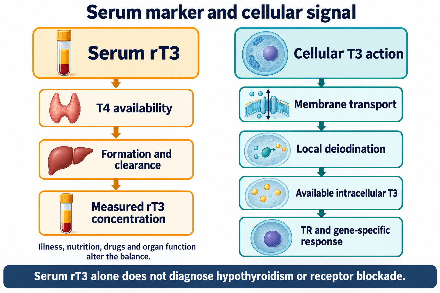
Tissue T3 availability genuinely differs from the hormone concentration in blood. Transporters, local deiodinases, receptors and cellular state determine the response. This complexity does not make one serum rT3 test a measurement of all those processes. Chemistry, local signaling, non-thyroidal illness syndrome (NTIS) and established thyroid failure need separate explanations.
T4, T3 and rT3 share a diphenyl-ether framework with an amino-acid side chain. Standard numbering places positions 3 and 5 on the inner ring and 3′ and 5′ on the outer ring. T4 is 3,5,3′,5′-tetraiodothyronine; T3 is 3,5,3′-triiodothyronine; rT3 is 3,3′,5′-triiodothyronine. T3 and rT3 are positional constitutional isomers, not stereoisomers. Both have formula C15H12I3NO4 and molar mass approximately 650.97 g/mol, while their iodine arrangement and biological properties differ.
Removing one iodine atom does not always activate the molecule. Outer-ring deiodination of T4 produces T3; inner-ring deiodination produces rT3. Conformation also depends on solvent, protonation and the binding protein. A universal ring angle, receptor-cavity volume or affinity cannot be assigned without the relevant experiment. Corinti et al. (2021) studied gas-phase deprotonated ions using spectroscopy and quantum calculations. Their work informs analytical discrimination of isomers; it does not establish aqueous pKa values or clinical receptor blockade.
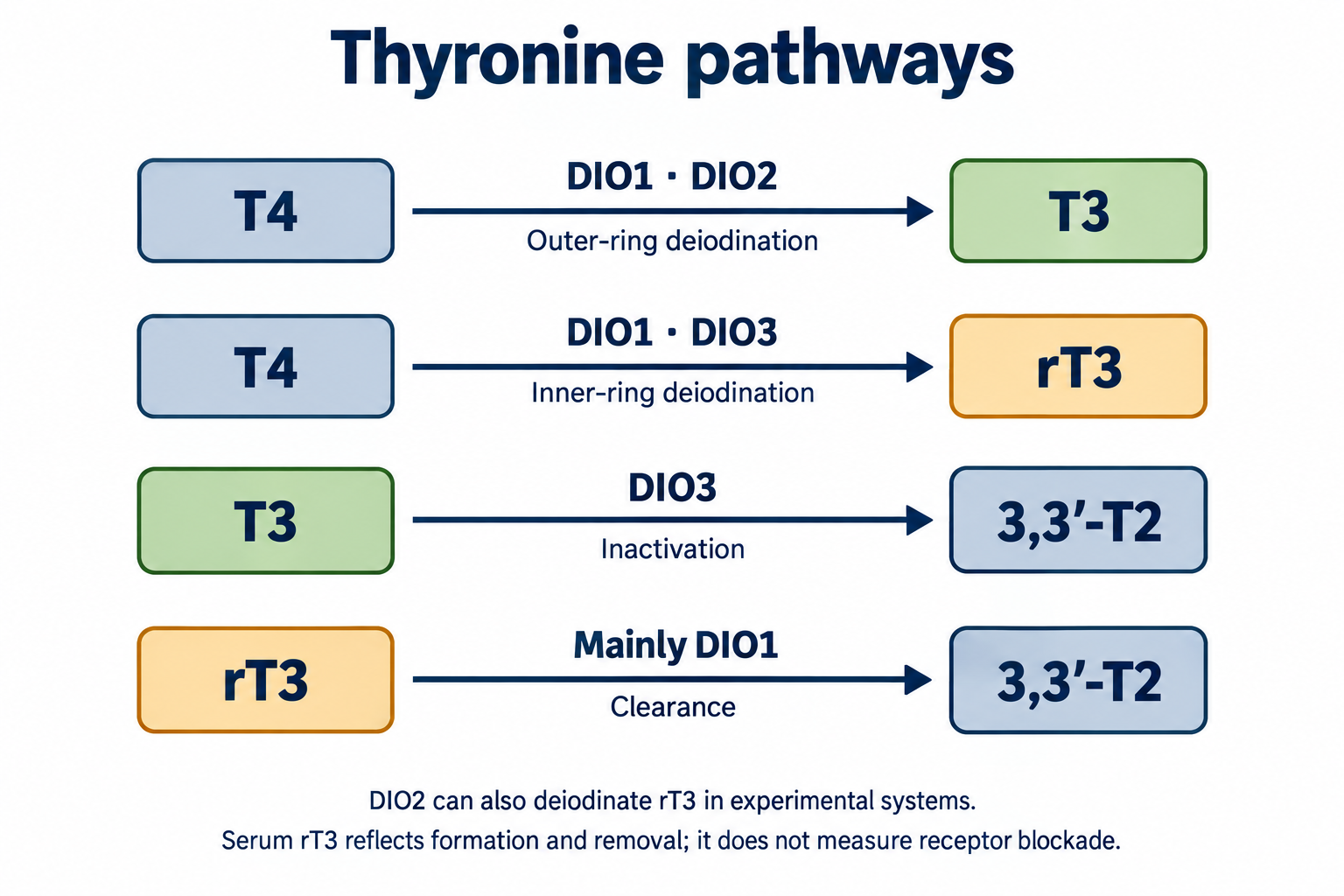
| Enzyme | Principal reactions | Functional context |
|---|---|---|
| DIO1 / D1 | Outer- and inner-ring deiodination; contributes to T4 → T3, T4 → rT3 and especially rT3 removal | Liver, kidney and other pools; circulating T3 and clearance |
| DIO2 / D2 | T4 → T3; also deiodinates rT3 in experimental systems | Local activation, including brain, pituitary and brown fat |
| DIO3 / D3 | T4 → rT3; T3 → 3,3′-T2 | Limits local hormone action during development and in some pathological states |
Expression and activity are not interchangeable. D2 is regulated by ubiquitination and protein turnover, among other mechanisms; substrate availability influences local flux. D3 can directly decrease T3 availability through inactivation, without any need for rT3 receptor antagonism. “D3 only makes rT3, D1 only clears it” is an oversimplification. A small thyroidal secretory contribution to rT3 also exists (Chopra, 1976; Bianco et al., 2002).
Deiodinases are selenoproteins. Catalytic selenocysteine incorporation uses a specialized UGA/SECIS system; dietary selenium is not converted directly into an active enzyme. The active site and thioredoxin-like fold support reductive iodine removal. For a generic molecular residue R, the redox balance is:
Two reducing equivalents cannot be replaced by a single GSH molecule in an unbalanced equation. An illustrative glutathione-coupled balance is:
This describes stoichiometry, not a universal physiological electron donor for all three enzymes. Rodriguez-Ruiz et al. (2022) investigated recombinant D1 redox chemistry, including Cys124/Sec126 and GSH-mediated reduction. Extrapolation to every tissue, D2 or NAC supplementation requires separate evidence. Glutathione reductase uses NADPH to regenerate GSH. The pentose-phosphate pathway is important but is not the only NADPH source. Carbohydrate restriction does not demonstrate its complete shutdown: intracellular substrates and alternative reducing systems remain relevant.
For one substrate, a simplified Michaelis–Menten model is:
S is substrate accessible to the enzyme, v the reaction rate, Vmax the maximum rate under specified conditions, and Km the concentration producing half that rate. Deiodinase parameters depend on enzyme preparation, reductant, cellular environment and assay. An experimental Km measured with an artificial reductant is not a personal blood threshold.
Where competitive inhibition is actually demonstrated, apparent Km may contain the factor 1 + I/Ki, with competitor concentration I and inhibition constant Ki. The formula itself does not establish that physiological rT3 inhibits a particular enzyme or receptor. Appropriate compartment concentrations and measured interactions are necessary.
A simple serum mass-balance model provides another useful distinction:
C is concentration, P production rate, V a hypothetical distribution volume, k the elimination constant and CL clearance. With constant parameters, higher concentration can result from increased production, reduced clearance or both. Chopra’s 1976 study linked cirrhosis to impaired rT3 clearance. A single result cannot yield an individual's half-life or a prescribed “washout” duration. The organism contains multiple connected pools, variable enzymes and protein binding.
TBG, transthyretin and albumin bind thyroid hormones in plasma. Total concentration includes bound and free fractions; the free fraction depends on proteins, patient state and assay. Intracellular availability additionally depends on transport, metabolism and cellular efflux.
MCT8, encoded by SLC16A2, transports T3, T4, rT3 and some T2 species. Friesema et al. (2003) measured transport with rat MCT8 in Xenopus oocytes; their micromolar kinetic parameters are not universal human-brain constants. OATP1C1, SLCO1C1, preferentially transports T4 and rT3; Pizzagalli et al. (2002) reported parameters for a specific experimental system. MCT10, LAT and other transporters contribute according to tissue context.
Sharing a transporter permits interactions in principle, but does not demonstrate a clinically important rT3 “traffic jam.” Comparing total serum rT3 with Km determined under different substrate-access conditions is misleading. Competition depends on free concentrations, localization, transport direction and actual cellular effects.
T3 binds nuclear TRα and TRβ receptors. Interactions with RXR, thyroid-response elements (TREs) and chromatin regulate gene expression. In particular contexts, unliganded receptors associate with NCoR/NCOR1 and SMRT/NCOR2 corepressors and complexes containing HDAC3 and TBL1. Agonist binding changes the ligand-binding domain, including helix H12 and the AF-2 surface; SRC/p160 coactivators with LXXLL motifs and p300/CBP participate downstream. Positive and negative regulation differ with gene, isoform and cell type.
Wagner et al. (1995) and T3-bound TRβ structures support agonist signaling. They do not depict rT3 locking H12 in an inhibitory state. Historical animal nuclear-extract experiments by Wiersinga et al. (1982) do not establish human TR antagonism at ordinary rT3 concentrations.
A competitive-occupancy equation is useful as a hypothetical model only when both ligand interactions are demonstrated:
L and I represent free agonist and verified-competitor concentrations at the receptor; Kd values are their dissociation constants. Substituting total serum rT3 and an invented Kd assumes the disputed mechanism before testing it. Physiologically important TR blockade by rT3, and diagnosis of such blockade using FT3/rT3, are unestablished (Van Uytfanghe et al., 2023).
Severe illness often lowers T3. rT3 may rise, remain normal or fall. TSH frequently does not rise in proportion to low T3, and FT4 can decrease during prolonged severe illness. Recovery may involve transient TSH elevation. Hepatic clearance, nutrition, medications, inflammation, central regulation and tissue deiodinases change together.
| Condition | Typical laboratory relationship | Qualification |
|---|---|---|
| Overt primary hypothyroidism | High TSH, low FT4 | Interpret illness, medications and assay alongside the result |
| Subclinical primary hypothyroidism | Elevated TSH, normal FT4 | Cause, persistence and clinical setting matter more than rT3 |
| Central hypothyroidism | Low FT4 with inappropriately low or normal TSH | Occasionally mildly elevated but biologically inadequate TSH; pituitary context matters |
| NTIS | Often low T3, variable rT3, normal/low TSH | No single profile and no proof of receptor blockade |
Normal TSH does not describe every cell's signaling or exclude central hypothyroidism. This limitation does not make rT3 a universal diagnostic solution (Persani et al., 2018). Thyroid antibodies can help identify an autoimmune cause; their absence does not exclude every thyroid disorder.
Pituitary D2 and TRβ2 contribute to feedback. Hypothalamic tanycytes, TRH, nutritional signals, leptin and inflammation affect different levels of the axis. The pituitary cannot be declared immune to all deiodinase changes, nor every normal TSH explained as rT3 “deception.” During NTIS, tissue hormone concentrations may decrease, remain unchanged or increase according to organ and illness stage (Fliers and Boelen, 2021).
HIF-1-dependent D3 induction and reduced local T3 action are demonstrated in experimental hypoxic systems (Simonides et al., 2008). This is an important local mechanism distinct from receptor competition. Human critical-illness observations also support altered activation and inactivation, without validating diagnosis from one serum marker (Peeters et al., 2003). Rare consumptive hypothyroidism associated with highly D3-expressing hemangiomas is a separate established disorder (Huang et al., 2000).
In the heart, T3 affects calcium handling, contractile proteins, rate and vascular resistance. SERCA2a, ATP2A2, is a calcium-reuptake pump in the sarcoplasmic reticulum, not a receptor. Phospholamban regulates its activity. MYH6/MYH7 responses are species dependent: a human ventricle cannot be described using rodent myosin proportions. HCN channels, adrenergic sensitivity and diastolic relaxation help explain hypothyroid cardiac physiology. Bradycardia alone cannot identify an rT3 syndrome.
In liver, thyroid signaling connects LDLR, cholesterol and bile-acid metabolism, TR–PPAR/LXR interactions and energetics. CYP7A1 regulation differs between models and humans. Muscle responses involve substrate use, GLUT4, fiber composition and ATP demand; nervous-system effects are particularly important in development and myelination. Fatigue, cognitive fog, muscle pain and weight changes are nonspecific. They do not establish rT3 suppression of all these genes or unavoidable weight gain regardless of energy balance (Mullur et al., 2014).
T3 influences mitochondrial biogenesis through PGC-1α, nuclear respiratory factors and TFAM. Nuclear respiratory factor 2, NRF2/GABP, must be distinguished from antioxidant regulator NFE2L2, also commonly called NRF2. TFAM contributes to mtDNA organization and transcription; coordinated nuclear and mitochondrial protein expression supports the respiratory chain.
The proton-motive force includes electrical potential and a pH gradient. Electron transfer, oxygen consumption and ATP synthase are functionally connected; one altered variable does not demonstrate whole-system mitochondrial collapse. ROS generation depends on chain site and conditions, including complexes I and III. The p43 pathway, involving a shortened TRα1 form, was investigated in mitochondria and experimental cells (Casas et al., 1999; Wrutniak-Cabello et al., 2018). It does not prove a fixed rT3-induced PGC-1α decline in patients. The article on R-alpha-lipoic acid and pyruvate dehydrogenase explains related redox energetics; that mechanism does not establish rT3 treatment.
Substrate regulation also involves CPT1A, which participates in mitochondrial entry of long-chain fatty acids through the carnitine system, and tissue-dependent UCP3 changes. This does not mean complete beta-oxidation arrest. The phosphocreatine system buffers muscle ATP demand; its role does not make muscle pain a specific thyroid test. Neurotrophic pathways including BDNF are discussed in experimental thyroid biology, without establishing a universal measured deficit in people with high rT3. Zinc and copper have broad metabolic roles and should be considered according to actual need; their combination is not a proven specific D1-restoration treatment.
LPS/TLR4, Kupffer cells, cytokines, NF-κB and STAT3 can participate in inflammatory changes to hepatic and systemic metabolism. Acute and prolonged responses differ. Hypoxia, organ failure and infection can alter hormone formation and removal. These mechanisms do not yield universal rT3 increases in obesity or sleep apnea, obligatory endothelin-1 effects in every organ, or a validated CPAP benefit through rT3 clearance.
Glucocorticoids affect the thyroid axis and conversion according to dose and condition. A cortisol measurement without context cannot establish continuous DIO1 promoter suppression or obligatory DIO3 activation. Sleep, circadian timing and psychological stress deserve their own assessment. The article on ashwagandha, withanolides and the HPA axis explains that separate system; plant effects on stress outcomes do not prove reversal of thyroid receptor blockade.
Fasting and energy deficit can lower T3 and change rT3. Human feeding studies support roles for energy intake and dietary composition (Danforth et al., 1979). They do not establish that less than 50 g carbohydrate “switches off D1,” or mandate restoring 100–250 g as treatment. Involuntary undernutrition, critical illness, planned energy restriction and personal dietary context differ. Gluconeogenesis, redox metabolism and NADPH availability cannot be reduced to one daily carbohydrate number.
| Exposure | Possible thyroid connection | Unsupported inference |
|---|---|---|
| Amiodarone | Iodine load, thyroid effects and altered deiodination | Any rT3 change requires drug withdrawal |
| Propylthiouracil | Hormone-synthesis inhibition and predominantly D1-related conversion effects | Irreversible blockade of all deiodinases |
| Glucocorticoids, high-dose propranolol | Context-dependent lower T4 → T3; central effects for some drugs | Every beta-blocker has the same effect |
| Dopamine in intensive care | Can suppress TSH | Specific evidence of cellular rT3 blockade |
| Metals and some organic pollutants | Experimental thyroid toxicology | rT3 justifies chelation or a detoxification protocol |
Immunoassays depend on antibodies, cross-reactivity, matrix and possible interference. A fixed fraction of all results cannot be declared erroneous. LC–MS/MS can provide specific measurement with a validated method, but T3 and rT3 have the same molecular mass. Verified discrimination, matrix-effect control, calibration, internal standards and quantification limits remain necessary. Chromatographic separation is common; fragmentation and ion mobility may also provide analytical information. Mass spectrometry does not eliminate every interference.
Laboratory reference intervals depend on method and population; they are not universal “optimal” targets. General treatment thresholds for rT3 or FT3/rT3 are unestablished (Van Uytfanghe et al., 2023). Burmeister (1995) demonstrated limits of rT3 discrimination in hospitalized patients.
A frequent index mixes free T3 in pg/mL with total rT3 in ng/dL:
With illustrative FT3 = 2.4 pg/mL and rT3 = 24 ng/dL, J = 10. The physical free/total molar ratio is nevertheless 2.4/240 = 0.01, because molecular masses coincide:
This is not “10% active hormone” or a ratio of two free intracellular ligands. If both total hormones use identical mass units, their ratio equals the molar ratio, but still does not measure receptor occupancy or deiodination flux. Proposed index cutoffs of 15, 20 or 25 do not become diagnostically valid simply because the division is correct.
Levothyroxine is standard replacement for established hypothyroidism. T3 is not converted to rT3 by removing an iodine atom; that fact does not justify switching everyone from T4 to T3. Stopping LT4 does not instantly remove tissue and protein-bound T4 pools. A universal several-week “washout” with escalating T3 doses is not validated treatment for rT3 blockade.
Jonklaas et al. (2021) discuss selected circumstances for considering LT4/LT3 in patients with persistent symptoms after assessing other causes. This is not selection by an arbitrary rT3 index; trial benefits have been inconsistent. Excess hormone action involves cardiac and skeletal risks, while pregnancy and comorbidities change decisions. Routine thyroid-hormone treatment of critical-illness NTIS has no established advantage; pre-existing hypothyroidism requires separate management (Jonklaas et al., 2014).
Selenium and iron deficiencies can matter for thyroid biology, but their roles differ. Selenoproteins participate in deiodination; iron-dependent thyroid peroxidase contributes to hormone synthesis. These functions establish neither a universal selenium dose nor one ferritin target for reversing rT3. Excess micronutrient exposure can also cause harm. Iron can impair LT4 absorption; drug interactions matter more than assuming every supplement combination activates D1 (Schomburg, 2012; Duntas, 2010).
A human NAC randomized trial exists: Vidart et al. (2014) included 67 acute myocardial-infarction patients and followed hormone and oxidative measures over 48 hours. It supports a redox hypothesis in that acute setting. It does not establish oral NAC treatment of chronic fatigue, improved clinical outcomes or reversal of receptor blockade. Lehnen et al. (2020) separately studied D3 and oxidative stress after infarction in rats. These findings cannot be merged into a validated outpatient protocol.
| Intervention | What was investigated | Translation limit |
|---|---|---|
| Selenium | Thyroid biology and, for example, autoantibodies in Gärtner et al. (2002) | Not treatment of demonstrated rT3 blockade |
| Ashwagandha | A small subclinical-hypothyroidism trial, Sharma et al. (2018) | Thyroid indices rather than established rT3 clearance |
| Guggulu | PTU-induced hypothyroidism in mice, Panda and Kar (2005) | Not human efficacy or dose selection |
| R-ALA, silymarin, artichoke | Separate redox and metabolic mechanisms | No established rT3 treatment or obligatory bile-mediated clearance |
| NAC | Human acute infarction and separate experimental models | Not a universal fatigue course |
Cold can activate sympathetic beta-adrenergic/cAMP/PKA signaling, D2 and UCP1 in brown fat. This is local thermogenic adaptation, not an established way to clear circulating rT3. Light acts through melanopsin retinal cells and the suprachiasmatic nucleus to influence circadian rhythms. TSH usually rises at night; a mandatory morning maximum is an incorrect description. Circadian biology does not establish rT3 treatment with one prescribed illuminance.
MOTS-c is a 16-amino-acid mitochondrial-derived peptide. Lee et al. (2015) linked experimental metabolic effects to folate/purine metabolism and AMPK. Humanin has 24 amino acids; Guo et al. (2003) studied BAX interactions and apoptosis. Intracellular BAX effects should be separated from proposed extracellular receptor complexes. These studies do not validate human injection schedules targeting DIO1 or rT3.
DNA methylation, DNMT enzymes and chromatin regulation can alter expression. Taking SAMe, B12 or 5-MTHF is not targeted DIO1/SLC16A2 “demethylation.” Supplying a methyl donor does not establish release of repression at a particular gene. Such a treatment claim requires direct tissue, target and clinical-outcome evidence.
Hepatic UGT and SULT enzymes conjugate thyroid hormones; intestinal deconjugation can contribute to enterohepatic cycling. Microbiota effects depend on substrate and state, while a fixed “20–30% of rT3 clearance” share is unconfirmed here. rT3 is not a steroid. SIBO treatment, particular probiotic strains, glutamine and zinc-carnosine do not form a verified rT3-removal protocol.
Rare pathogenic MCT8 variants cause severe neurodevelopmental disease with disturbed hormone transport and characteristic peripheral changes (Friesema et al., 2004). This is distinct from common acquired rT3 competition. Rare defects in other transporters cannot be replaced by interpreting common SNPs through unverified percentage losses of function.
DIO2 Thr92Ala, rs225014, is studied as a modifier. Panicker et al. (2009) reported a treatment-response association in a trial analysis but required replication. The variant is not an independently approved LT3 selector, an explanation for every fatigue complaint or an indication for a receptor-unblocking nutraceutical. Genetics does not remove biochemical, clinical and analytical limits.
Persistent symptoms first require context: established thyroid disease, recent severe illness, symptom trajectory, nutrition, medications and measurement method. TSH and FT4 are interpreted together with that context; suspected central dysfunction calls for pituitary-axis assessment rather than an independent conclusion from rT3. Transient illness-related changes may need reassessment after recovery. Depending on symptoms, anemia, sleep disorders, depression and other causes of fatigue are also considered. This sequence is not a newly validated protocol. It explains why diagnosis and the intervention objective precede an attempt to change a surrogate. Improvement in symptoms or one laboratory result does not prove the proposed receptor mechanism.
This narrative scientific review used targeted verification of disputed mechanisms, identifiers and selected findings on 8 October 2026. Primary articles, available author reviews, PubMed records, publisher pages and structural records were consulted. No systematic search, independent GRADE assessment or external peer review was performed. Some full texts were unavailable; accessible metadata and abstracts were used. Exact methods and quantities from inaccessible full text are therefore not presented as verified findings.
rT3 helps explain hormone metabolism and some specialized clinical situations. It is not a universal tissue-energy measurement. Established local T3 inactivation, organ differences and TSH limitations call for precise reasoning rather than replacing thyroid assessment with one ratio or a supplement stack.
No. Production and clearance change with illness, nutrition and drugs. Diagnosis uses clinical context, TSH and FT4, accounting for central disorders.
No validated general treatment threshold exists. Mixing free and total hormones and different units further limits interpretation.
rT3 alone does not justify stopping replacement or switching to T3. Decisions concern the particular thyroid disorder and clinical situation.
Yes. Local D3 and transport can change availability, which differs from an unestablished generalized rT3 receptor blockade.