Ashwagandha: Withanolides, Cortisol, the HPA Axis, and Safety — NutriFit
Ashwagandha: withanolides, the HPA axis, cortisol, and the limits of clinical use
An in-depth examination of withanolide chemistry, GABA signaling, the HPA axis, and the glucocorticoid receptor, distinguishing experimental mechanisms from clinical outcomes and the risks of ashwagandha.
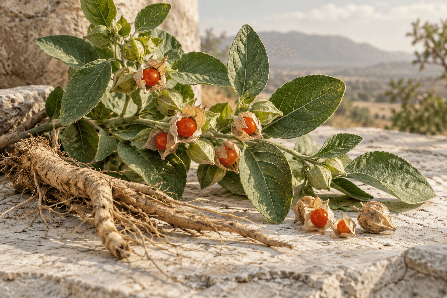
Ashwagandha, Withania somnifera (L.) Dunal, is a shrub of the nightshade family whose roots and leaves provide the raw material for chemically distinct extracts. In clinical studies, some of these extracts reduced perceived stress and improved certain sleep measures. However, there is considerable distance between that finding and a claim of targeted restoration of the hypothalamic–pituitary–adrenal axis: a change in cortisol concentration does not identify the protein targeted or show how communication between the brain and adrenal glands has changed.
The historical concept of adaptogens, associated with the work of N. V. Lazarev and I. I. Brekhman, described an increase in nonspecific resistance to adverse influences. It helps explain the origins of this research program, but does not itself establish a diagnosis, dose, or clinical efficacy. Modern assessment must identify the specific preparation, population, and outcome: a lower questionnaire score, a hormonal change, shorter sleep onset latency, or increased strength during training. These findings cannot all be called “normalization” when neither the normal range nor the significance of the change for the patient has been established.
Three levels of evidence are particularly important for specialists. A chemical reaction between a purified withanolide and a protein characterizes the molecule; an extract's effect in a cell or animal supports a hypothesis; a randomized human study assesses a specific clinical outcome. Moving between these levels requires evidence on exposure and reproducibility. Ashwagandha biochemistry therefore needs to be read alongside the limits of its clinical interpretation.
1. Withanolides: the same formula does not mean the same effect
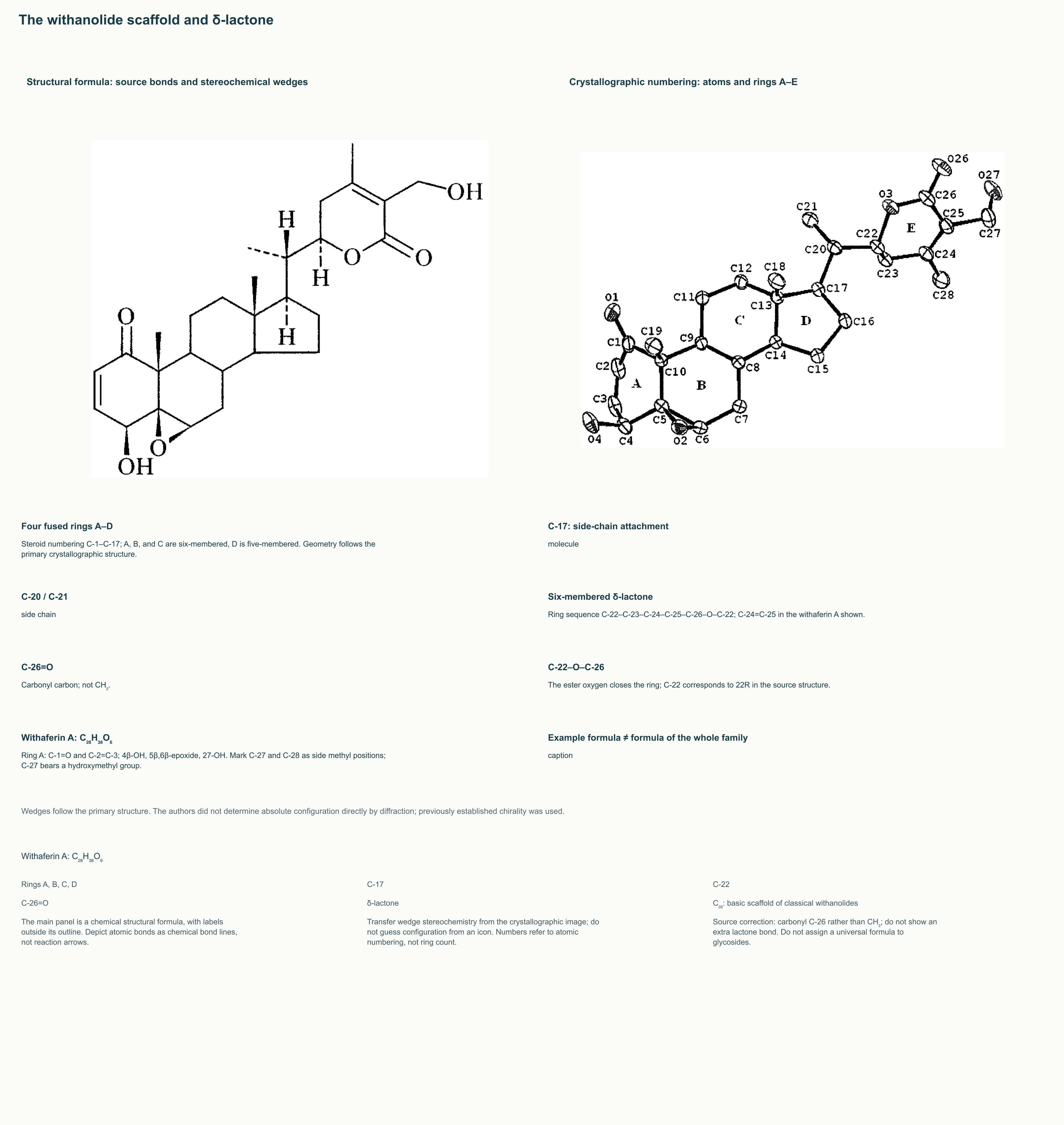
Classical withanolides are oxygen-containing steroidal lactones with a C₂₈ carbon skeleton. It contains the recognizable system of four fused steroid rings A–D, while the side chain at C-17 forms an additional lactone ring. Common representatives feature a six-membered δ-lactone: oxygen connects C-22 to the carbonyl carbon C-26. C-26 is the carbon that carries the lactone carbonyl; depicting it as CH₂, as some simplified diagrams do, is chemically incorrect.
Withaferin A and withanolide A have the formula C₂₈H₃₈O₆, but this describes two specific compounds, not a universal formula for the family. The number of oxygen atoms, degree of saturation, positions of hydroxyl groups and the epoxide bridge, and stereocenter configurations can differ. Glycosylation further changes composition and properties: an aglycone's formula cannot be assigned to a withanoside. A percentage of “total withanolides” likewise does not reveal each molecule's share.
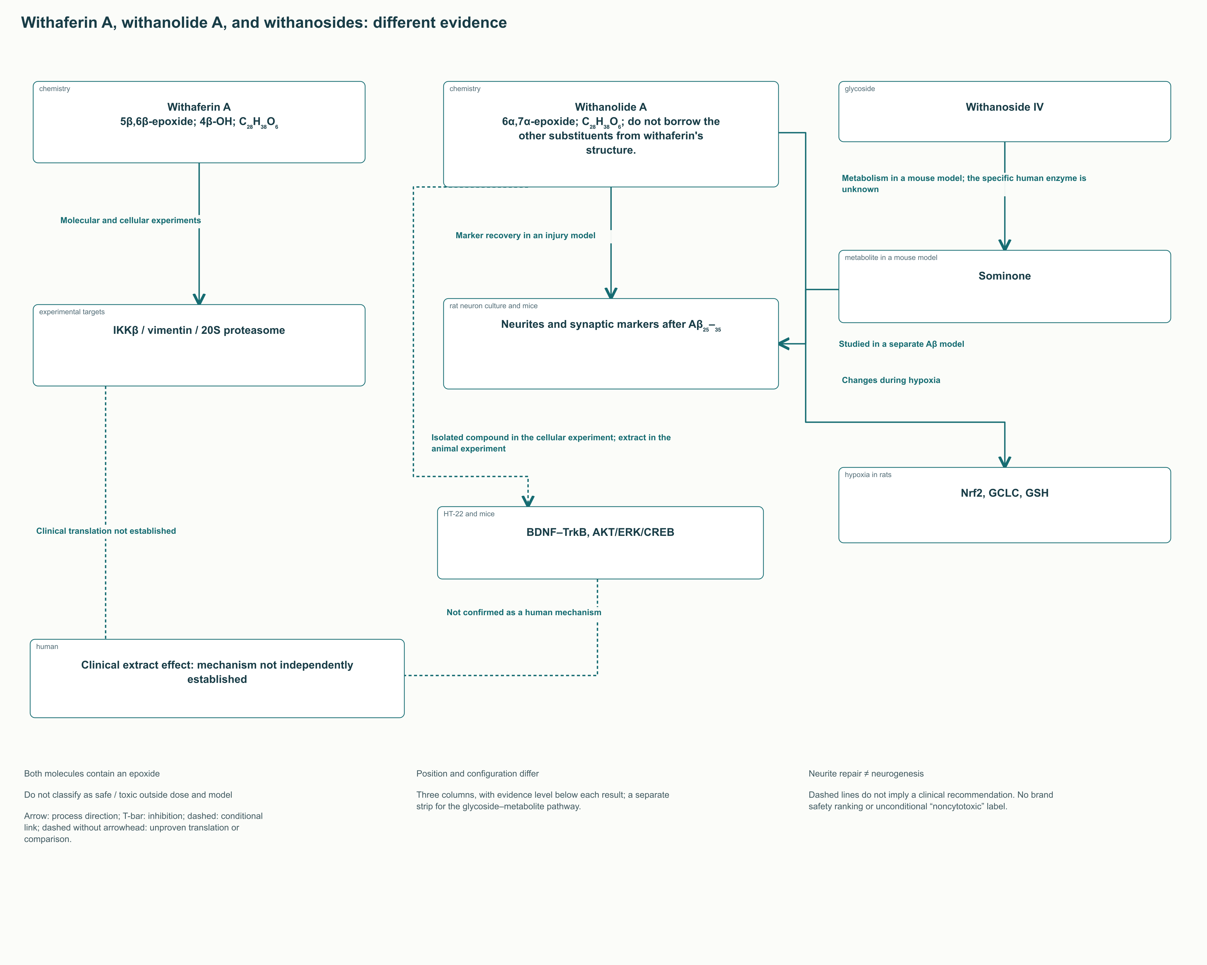
Important features of withaferin A include the 4β-hydroxyl, 5β,6β-epoxide, 27-hydroxyl, and conjugated ketone in ring A. Withanolide A also contains an epoxide, but between C-6 and C-7, with an α orientation. The distinction therefore concerns epoxide position and stereochemistry, rather than its presence in one compound and absence from the other. Experiments with thiol reagents show that these differences can substantially change reactivity.
Dividing the compounds into “cytotoxic withaferin” and “noncytotoxic withanolide” is excessively categorical. Activity depends on concentration, exposure time, the cellular system, and mixture composition. Neither lipophilicity nor a steroid skeleton proves neuroprotection, hormonal action, or the safety of prolonged use. Chemical structure defines possible interactions, but their clinical significance must be established separately.
2. Withaferin A electrophilicity and experimental protein targets
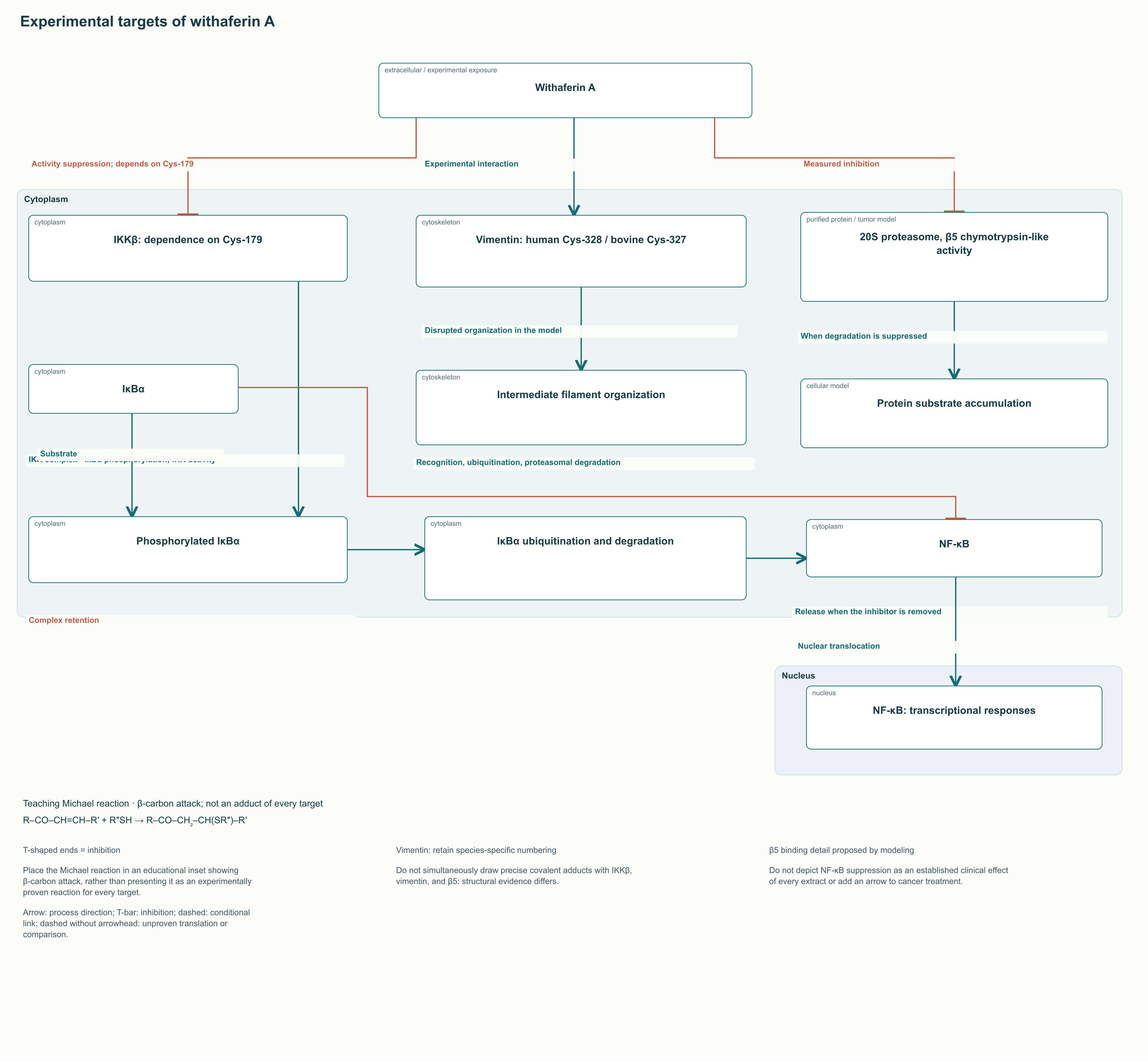
Conjugated unsaturated carbonyl groups can interact with nucleophiles, including protein thiol groups. Michael addition means nucleophilic attack on the β-carbon of a conjugated system, rather than any interaction of a molecule with cysteine. Withaferin A also has a reactive epoxide group; a universal statement that “withaferin alkylates every target” therefore cannot describe all the chemical and cellular findings.
In Heyninck's study, the interaction with IKKβ depended on Cys-179: replacing that residue disrupted the compound's effect. This links NF-κB suppression to a specific element of the signaling complex. In the usual cascade, IKK phosphorylates IκBα, after which the inhibitor undergoes ubiquitination and proteasomal degradation, allowing NF-κB to move into the nucleus. Reduced IKKβ activity may retain IκBα and decrease this translocation. This is, however, an experimental explanation of anti-inflammatory activity; it does not mean that an ordinary oral dose of any extract produces the same concentration in human tissues.
NF-κB transcriptional responses are associated with regulation of inflammatory mediators and enzymes, but the set involved depends on the cell and stimulus. TNF-α, IL-1β, and IL-6 are cytokines; COX-2 and iNOS are enzymes, not additional cytokines. Changing one signaling step does not establish equal suppression of all these products in every tissue.
Another line of research concerns vimentin, an intermediate filament protein. Cys-328 is discussed for human vimentin; the corresponding residue in the bovine protein used is designated Cys-327. This numbering difference must be retained when comparing experiments. Disruption of filament organization and effects on angiogenesis or tumor cells should not be converted into recommendations to treat tumors with a botanical supplement.
In Yang's work, withaferin A reduced the chymotrypsin-like activity of purified 20S proteasomes and affected tumor models. A possible interaction with the β5 catalytic site was discussed using molecular modeling. Measured inhibition and a proposed atomic binding geometry have different evidential strength. Protein accumulation during proteasome inhibition also explains why activity useful for experimentally damaging a tumor cell is not automatically desirable in a healthy neuron or hepatocyte.
This biochemistry justifies analytical control of composition, but does not allow brands to be ranked by clinical hepatotoxicity. That assessment requires data on actual compound content, exposure, other components, and adverse outcomes. The cause of liver injury reported in humans has not yet been reduced to a single molecule or protein target.
3. Neuronal models: neurite repair, BDNF, and withanoside metabolites
Kuboyama's study examined withanolide A after neuronal injury caused by the amyloid fragment Aβ₂₅–₃₅. Axons, dendrites, and synaptic markers were assessed in cultured rat cortical neurons, while memory and neuronal network changes were assessed in a mouse model. This was not a corticosterone injury model. Neurite growth and an increased area of synaptic protein immunoreactivity should be described as findings from that specific model; they do not prove the formation of new neurons in the adult human brain.
Later studies do link the actions of certain ashwagandha preparations to BDNF, TrkB, ERK, AKT, and CREB. A study using corticosterone in HT-22 cells and unpredictable chronic mild stress in mice investigated a root extract containing withanolide A, with the isolated compound also studied in the cellular experiment. Changes in signaling protein phosphorylation accompanied neuroprotective effects. This design maintains interest in the BDNF–TrkB pathway, but does not establish that it explains reduced anxiety in a person taking the supplement.
Opposite ERK changes in different tissues must not be conflated. In an injured neuron, stronger survival-related signaling may be beneficial; in another experiment, reducing excessive stress activation may be considered protective. Saying “ashwagandha activates ERK” or “ashwagandha blocks ERK” without specifying the cell, model, and observation period loses its meaning.
Withanosides IV and VI are glycosylated compounds. Sugar residues alter solubility and metabolism, but do not justify treating all glycosides as one pharmacological fraction. An active metabolite of withanoside IV, sominone, was described in mouse experiments. It should not be renamed “somonoside” or confused with somniferine. A role for intestinal metabolism is plausible, but the specific enzyme, contribution of microbiota, and conversion rate in humans require their own investigation.
In a separate rat model of hypobaric hypoxia, withanolide A affected Nrf2, the catalytic subunit of glutamate–cysteine ligase GCLC, and reduced glutathione. This broadens the mechanistic picture beyond BDNF. Nevertheless, hypoxia, amyloid injury, and psychological stress are different exposures; combining them into a single clinical “neuroregeneration” is unjustified. None of these findings demonstrates unrestricted penetration of all withanolides across the human blood–brain barrier or prolonged sedation attributable to withanosides.
4. GABA receptors and chloride conductance: the extract's effect and the molecule's effect
The GABA_A receptor is a ligand-gated anion channel. The common α₁β₂γ₂ variant is typically a pentamer with two α, two β, and one γ subunit; other variants have different compositions and pharmacology. GABA and classical benzodiazepines have distinct binding sites. This does not mean that an additional unique withanolide site has already been identified: their interaction geometry and the active components of different extracts remain research questions.
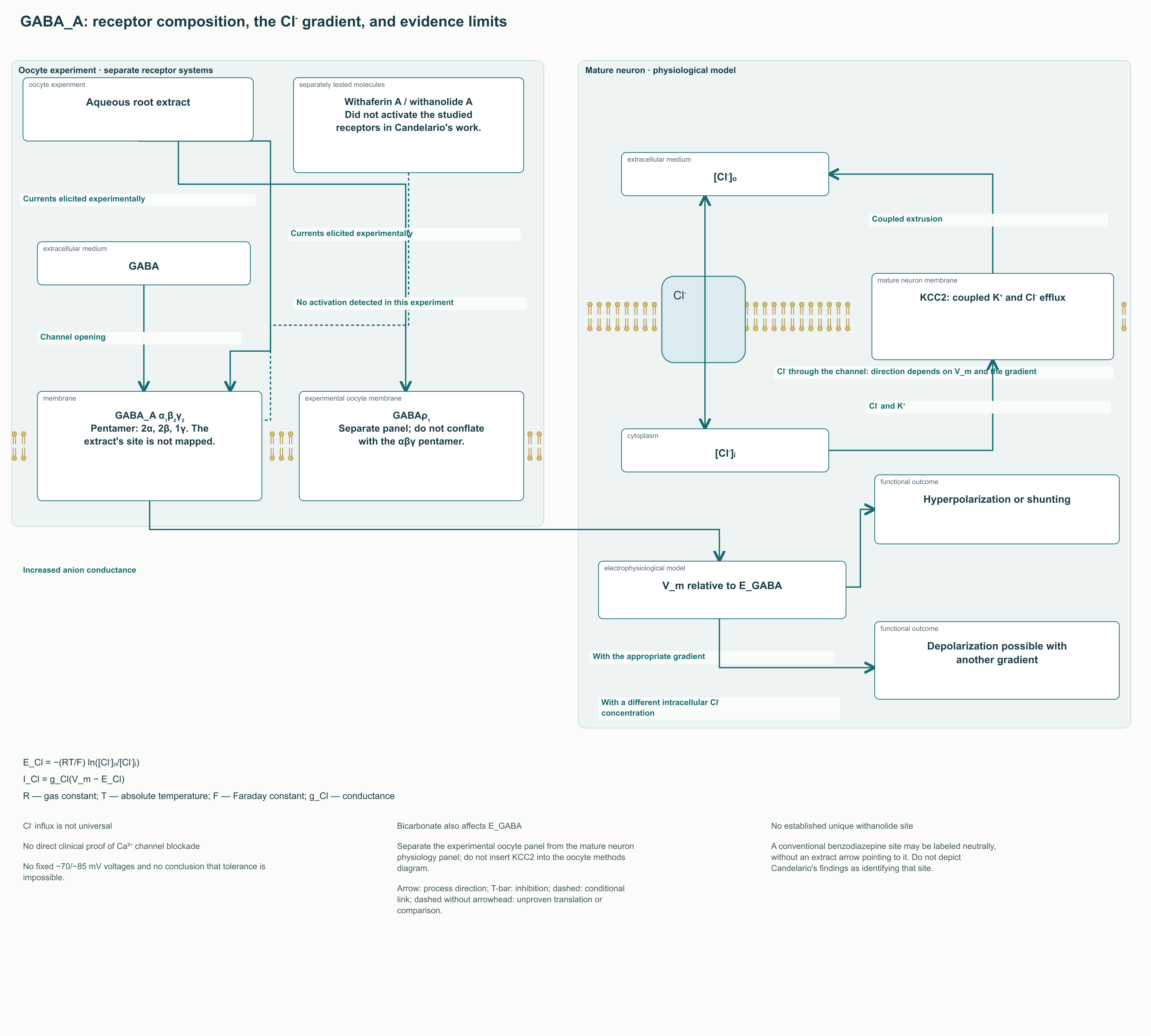
Candelario used two-electrode voltage clamp in Xenopus oocytes with transplanted rat brain receptors or an expressed ρ₁ receptor. An aqueous root extract elicited currents, but separately tested withaferin A and withanolide A activated neither GABA_A nor GABAρ₁ receptors. The mixture's effect in this study therefore cannot be attributed to these two substances. The ρ receptors are ionotropic GABA receptors, but differ from the usual αβγ complexes, including in their sensitivity to antagonists.
Cl⁻ flux direction depends on the electrochemical gradient, not merely on channel opening. In the chloride approximation, the equilibrium potential is given by:
E_Cl = −(RT/F) ln([Cl⁻]ₒ/[Cl⁻]ᵢ).
Here R is the gas constant, T is absolute temperature, F is the Faraday constant, and the o and i subscripts denote external and internal concentration. The minus sign reflects the chloride ion's negative charge. The formula describes equilibrium for one ion, not the exact membrane voltage after taking an extract. The actual reversal potential of the GABA_A current also depends on bicarbonate permeability.
In a simple electrical model, I_Cl = g_Cl(V_m − E_Cl), where g_Cl is chloride conductance and V_m is membrane potential. A positive or negative electrical current cannot be equated with “inward chloride flux” without specifying the sign convention: the ion carries a negative charge. In mature neurons, KCC2 contributes to maintaining a low intracellular chloride concentration. Channel opening can hyperpolarize the membrane or shunt excitatory currents; with an altered gradient, depolarization is possible. Universal statements that “−65 becomes −80 mV” with ashwagandha are therefore incorrect.
Reduced excitability may lower the probability of voltage-gated calcium channel opening, but does not prove that extract components directly block VGCCs. Similarly, failure of one antagonist to block an effect does not establish the absence of habituation, dependence, or cross-tolerance in humans. Nor are the precise changes in GABA K_d or single-channel open time attributed in the original descriptions to withanolide A and withanoside IV confirmed.
Another experimental component is triethylene glycol, TEG. Kaushik identified sleep-inducing activity in an aqueous leaf fraction and studied increased NREM sleep in mice. This observation does not confirm the clinical efficacy of purified TEG or establish an action through somatostatin receptors. Independent use of an industrial chemical does not follow from a plant study; findings for a leaf fraction cannot be applied to every root extract.
5. From neuronal activation to cortisol: what the HPA axis actually shows
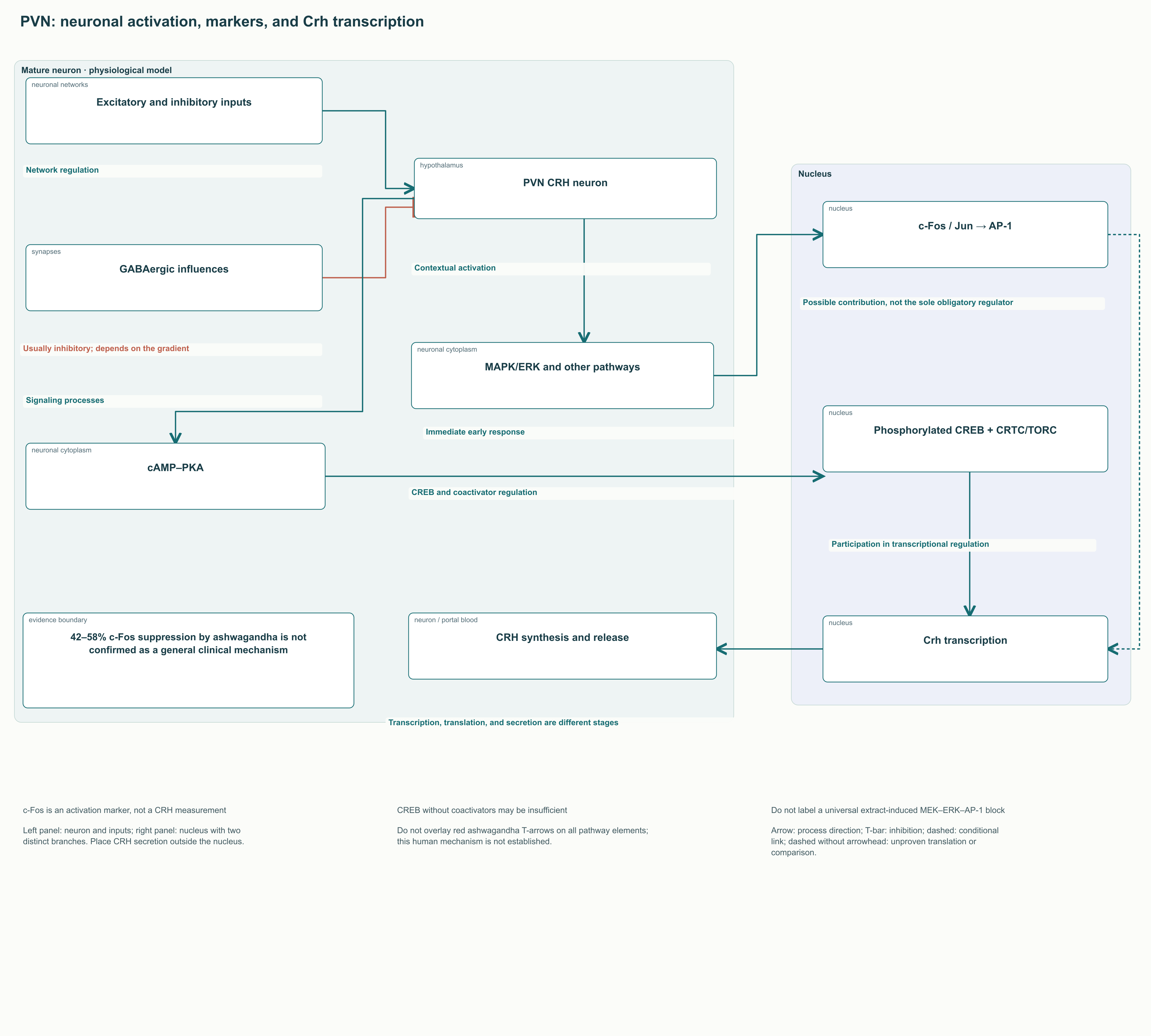
The stress response is coordinated by neuronal networks and the hypothalamic–pituitary–adrenal axis, HPA. Inputs to the hypothalamic paraventricular nucleus, PVN, include excitatory and inhibitory influences; the amygdala and hippocampus participate through complex, often indirect connections. Not every stress state can be depicted as the same GABA deficiency or obligatory persistent hypercortisolemia.
c-Fos is an immediate early response protein often used as a marker of recent cell activation. Together with Jun family proteins, it contributes to AP-1 formation. However, c-Fos staining is not equivalent to measuring CRH secretion and does not demonstrate that AP-1 alone initiates Crh transcription. Primary studies of hypothalamic cells identified important roles for phosphorylated CREB and its CRTC coactivators, historically called TORC; CREB activation alone could also be insufficient.
Claims of 42–58% c-Fos suppression in the PVN, sometimes attributed to “standardized withanolides,” lack sufficient primary support for such a general statement. Nor has a sequence of obligatory MEK–ERK–AP-1 blockade been established in people taking ashwagandha. The cell activation diagram remains useful when the measured marker, transcriptional regulator, and hormonal output are distinguished.
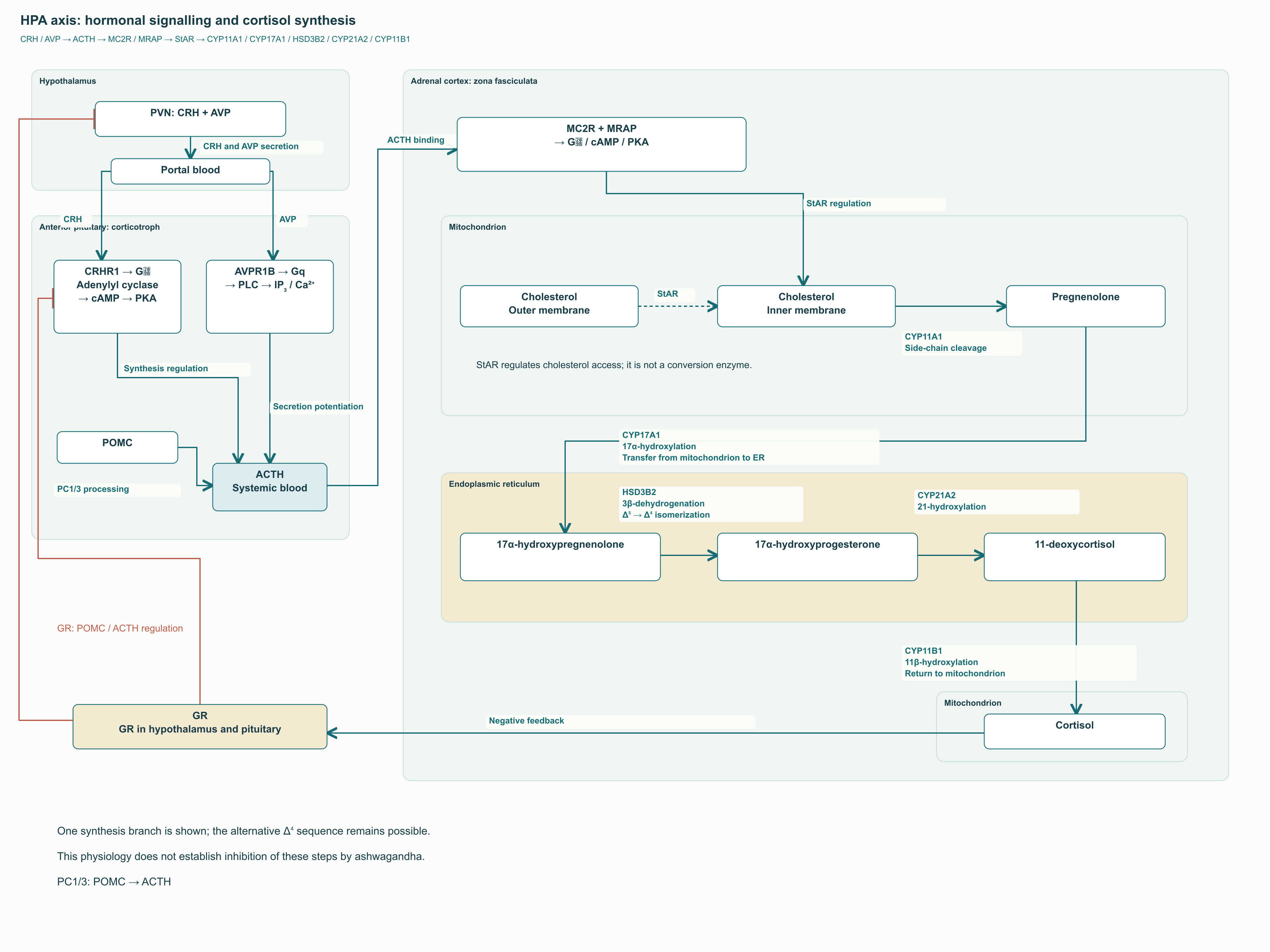
The endocrine sequence is as follows: CRH and arginine vasopressin enter the pituitary portal circulation from the hypothalamus; anterior pituitary corticotrophs release ACTH; the adrenal cortex synthesizes cortisol. CRHR1 couples to G_s, adenylyl cyclase, cAMP, and PKA. AVP augments the response through its own receptor and a different signaling pathway. POMC is the ACTH precursor; its processing, involving PC1/3, and release of the already formed hormone are distinct events. They cannot be reduced to a single instantaneous cAMP-induced proteolysis.
In the adrenal zona fasciculata, ACTH acts through MC2R, whose normal function requires the accessory protein MRAP. cAMP–PKA signaling regulates cholesterol access to steroidogenesis. StAR facilitates cholesterol transfer from the outer to the inner mitochondrial membrane; it is not an enzyme that converts cholesterol into a hormone. CYP11A1 produces pregnenolone, after which the cortisol pathway involves CYP17A1, HSD3B2, CYP21A2, and CYP11B1, distributed between the endoplasmic reticulum and mitochondria.
Cortisol participates in negative feedback at several levels. Evaluating this feedback requires attention to circadian and pulsatile changes, free and bound hormone fractions, receptor sensitivity, and the organism's state. Lower morning cortisol in an intervention group is compatible with an altered stress response, but does not prove direct inhibition of all three axis components, CYP11B1 inhibition, or restoration of a daily rhythm. Maximizing cortisol suppression is not a goal either: normal secretion is necessary for adaptation to exertion and maintenance of metabolism.
6. The glucocorticoid receptor: chaperones, transport, and the limits of the restoration hypothesis
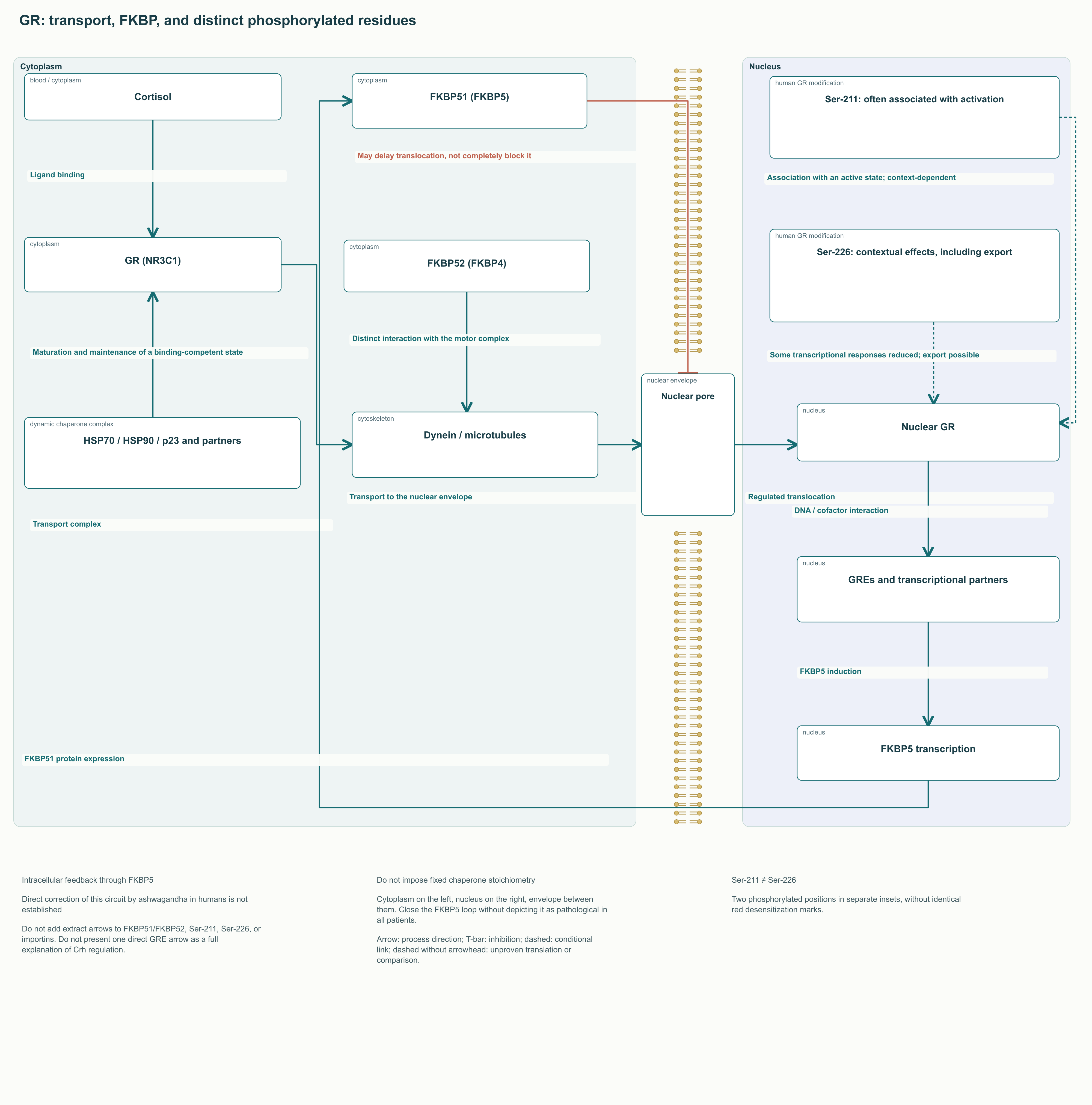
The glucocorticoid receptor, GR, is encoded by NR3C1. Its functional maturation depends on dynamic interactions with HSP70, HSP90, p23, and other cofactors. There is no single immutable stoichiometric assembly that can be drawn identically at every stage. Chaperones help maintain a ligand-binding state, while rearrangement of the complex accompanies changes in the receptor's intracellular location.
The immunophilins FKBP51 and FKBP52, products of FKBP5 and FKBP4, affect this process differently. In cell experiments, increasing FKBP51 reduced GR activity and delayed nuclear translocation; differences in dynein interactions helped explain the effect. Microtubules, motor proteins, and nuclear pore transit mechanisms participate in transport, but elevated FKBP51 does not permanently switch off every receptor. The outcome depends on the cell, complex composition, and hormone exposure.
GR can induce FKBP5, creating intracellular negative feedback on its own sensitivity. Changes in this circuit under stress have genetic and epigenetic components. However, the known role of FKBP5 does not mean that every patient with fatigue has pathological overexpression, or that lower cortisol confirms its correction.
Phosphorylation likewise cannot be treated as one uniform desensitization process. In human GR studies, Ser-211 phosphorylation correlated with activation and nuclear localization, whereas Ser-226 was linked to a reduction in some transcriptional responses and nuclear export. The effect depended on the target gene and cofactors. Labeling Ser-211 and Ser-226 as two equally pathological marks and promising to “normalize” both with a chemically defined extract is therefore misleading.
In the nucleus, GR interacts with glucocorticoid response elements, GREs, and transcriptional partners. HPA feedback is more complex than GR simply binding directly to a single Crh promoter: experiments in hypothalamic neurons also discussed indirect regulatory mechanisms. Primary clinical evidence is insufficient to show that ashwagandha corrects the FKBP51/FKBP52 ratio, preserves a particular chaperone complex, activates importins, or changes Ser-211/Ser-226 in a way that explains symptom relief. These questions retain research value, but are not indications for prescribing the supplement.
7. KSM-66, Sensoril, and Shoden: compare the preparation and analytical method
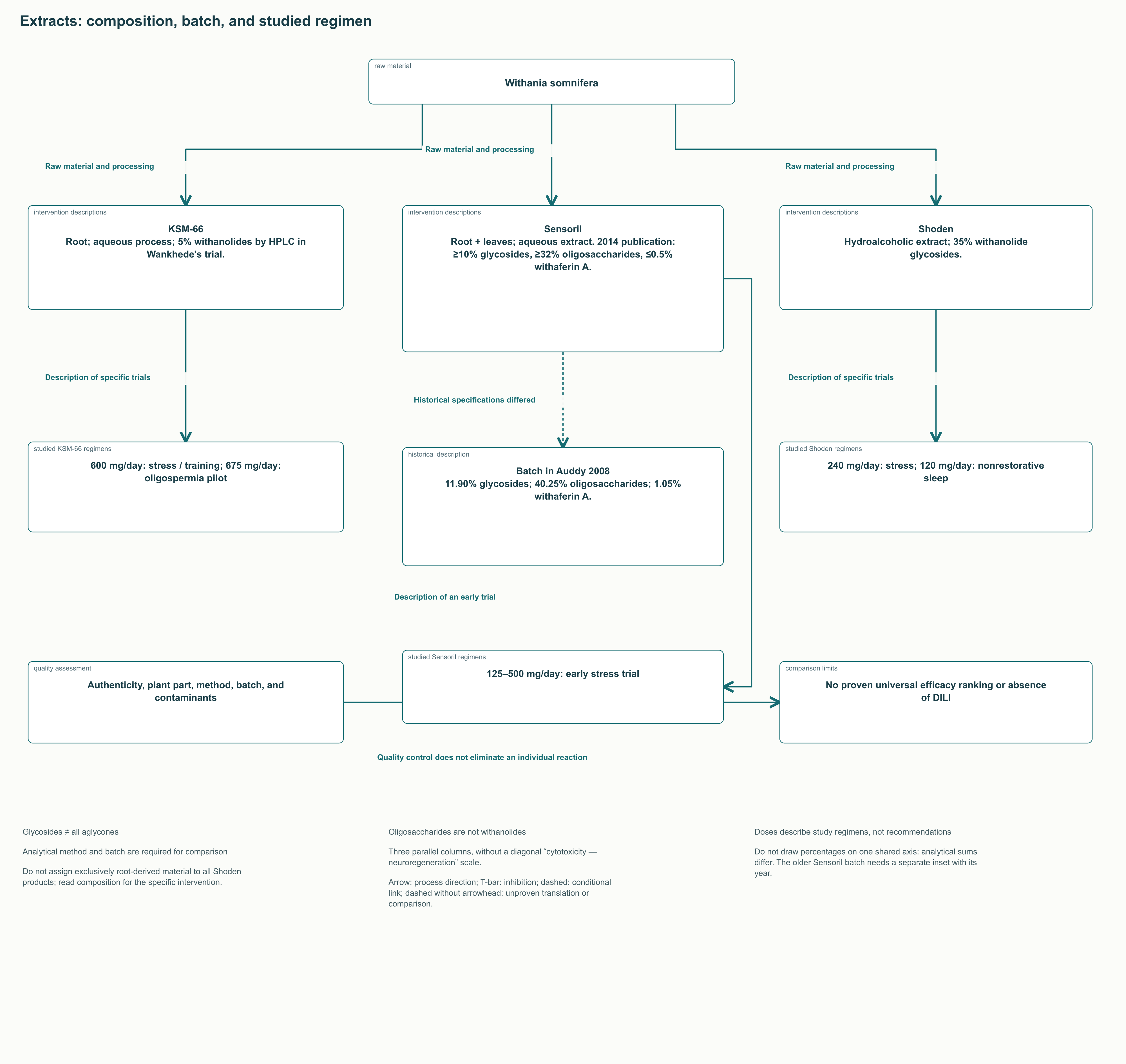
A botanical species name does not ensure pharmaceutical equivalence. Roots and leaves differ in composition; chemotype, cultivation, extraction, subsequent processing, and standardization method affect the final product. Even the same percentage on a label can refer to different sums of analytical components. Oligosaccharides are not withanolides, and the glycoside fraction cannot be equated with total aglycones without specifying the method.
Characteristic
KSM-66 in the publications examined
Sensoril in the publications examined
Shoden in the publications examined
Raw material
Root
Root and leaves
Root and leaf preparations; read the description of each intervention separately
Extraction
Aqueous process in the strength training study
Aqueous process, rather than universally hydroalcoholic
Hydroalcoholic extraction specified in the clinical description
Analytical characteristics
5% withanolides by HPLC in Wankhede's study
In the cognitive function study: ≥10% glycosides, ≥32% oligosaccharides, ≤0.5% withaferin A; older publications report different parameters
35% withanolide glycosides in stress and sleep studies
Examples of studied regimens
600 mg/day for stress or training; 675 mg/day in the oligospermia pilot
125–500 mg/day in the early stress study; a description of trials, not a general prescribing standard
240 mg/day for stress; 120 mg/day for nonrestorative sleep
What the name cannot establish
Absence of liver injury risk or universal hormonal efficacy
Obligatory “deep sedation” or high clinical cytotoxic risk
Superiority for every purpose and safety of continuous long-term use
In Auddy's early work, the tested root and leaf extract batch contained 11.90% withanolide glycosides, 1.05% withaferin A, and 40.25% oligosaccharides. Later publications described Sensoril with different limits for withaferin content. This illustrates why a historical batch specification cannot become an immutable brand characteristic. Identifying the supplier and analytical protocol is more useful than a diagram claiming “one brand is cytotoxic, the others neuroregenerative.”
Practical comparison requires the plant part, extract mass, raw-material-to-extract ratio, list of assayed markers, analytical method, and batch-specific data. Authentication and testing for residual solvents, heavy metals, and microbial contamination concern product quality. They reduce uncertainty about composition, but do not eliminate a possible individual reaction to an authentic extract. Neither withanolide percentage nor a quality certificate replaces clinical risk assessment.
8. Pharmacokinetics: what has been measured in humans and what remains hypothetical
Glycosylation may change solubility and metabolism, but a glucose residue does not demonstrate transport of the whole withanoside through SGLT1. Universal logP values of 2.5–3.8, plasma protein binding of 82–88%, and appearance of withanolide A in the human brain within 60 minutes are likewise unconfirmed. Lipophilicity, interaction with albumin in vitro, and detection in animal tissue address different questions.
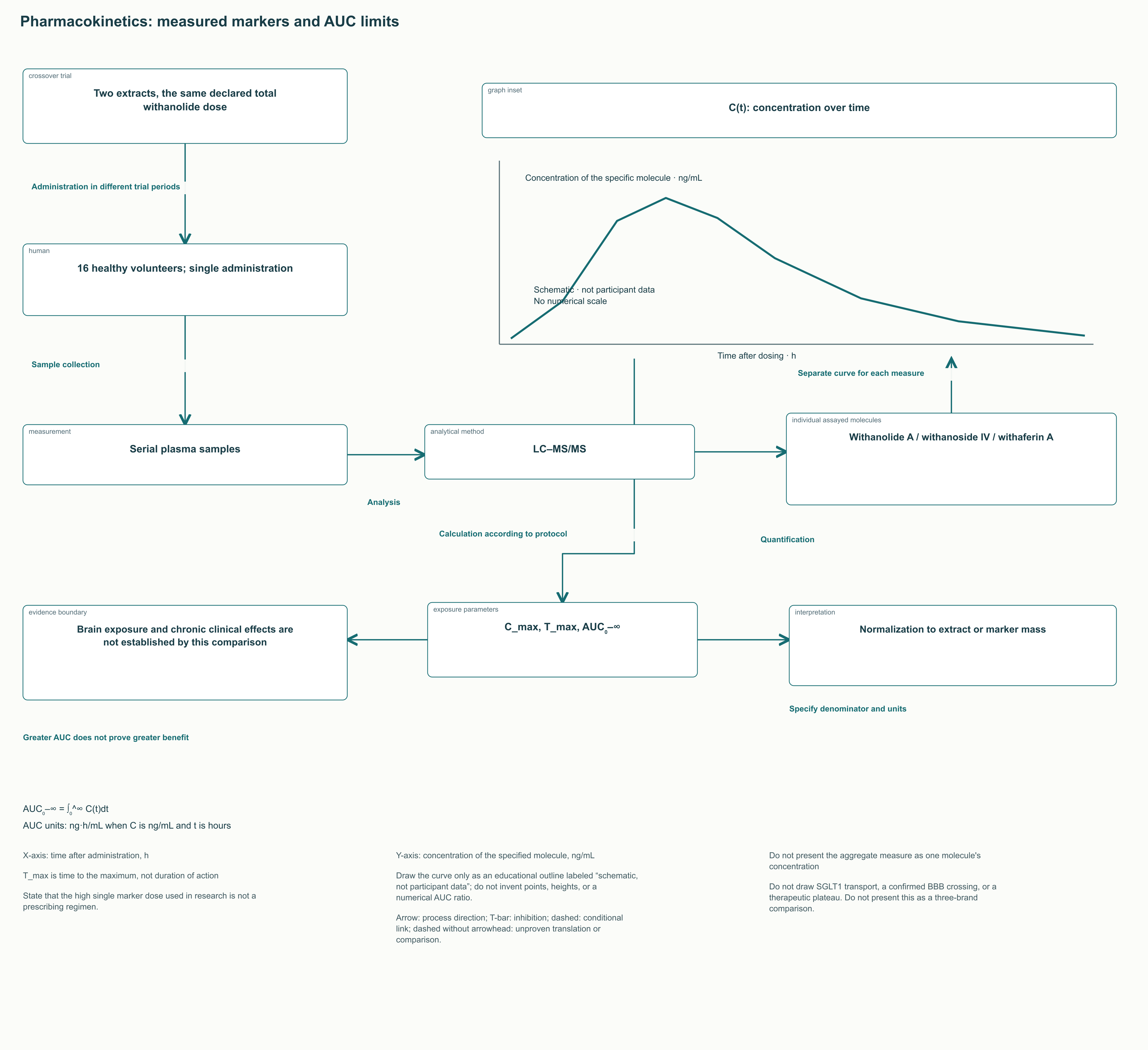
Kim's primary crossover study enrolled 16 healthy volunteers and compared two extracts after a single dose with the same declared total withanolide dose. LC–MS/MS measured plasma withanolide A, withanoside IV, withaferin A, and an aggregate measure. Exposure differed, confirming the importance of composition and formulation. However, this was not a direct three-way comparison of KSM-66, Sensoril, and Shoden for chronic clinical effects; the high marker dose used cannot be transferred to routine prescribing.
C_max is the highest measured concentration, T_max is the time at which it is reached, and AUC₀–∞ is the area under the concentration curve from administration to extrapolated infinite time. AUC₀–∞ = ∫₀^∞ C(t)dt emphasizes integrated exposure rather than duration of action. The measure requires concentration and time units, the method used to calculate the curve's tail, and precise identification of the molecule analyzed.
Comparing AUC normalized per milligram of extract includes differences in marker concentrations within the products themselves. It cannot be presented as a pure absorption ratio for one identical molecule. Moreover, greater AUC does not prove better efficacy or safety: greater exposure can intensify both desired and unwanted effects. A “uniform therapeutic plateau” and three- to fourfold AUC superiority of one brand require specific comparative data; they do not follow from general phytochemistry.
9. Stress and anxiety: a clinical signal and the limits of its explanation
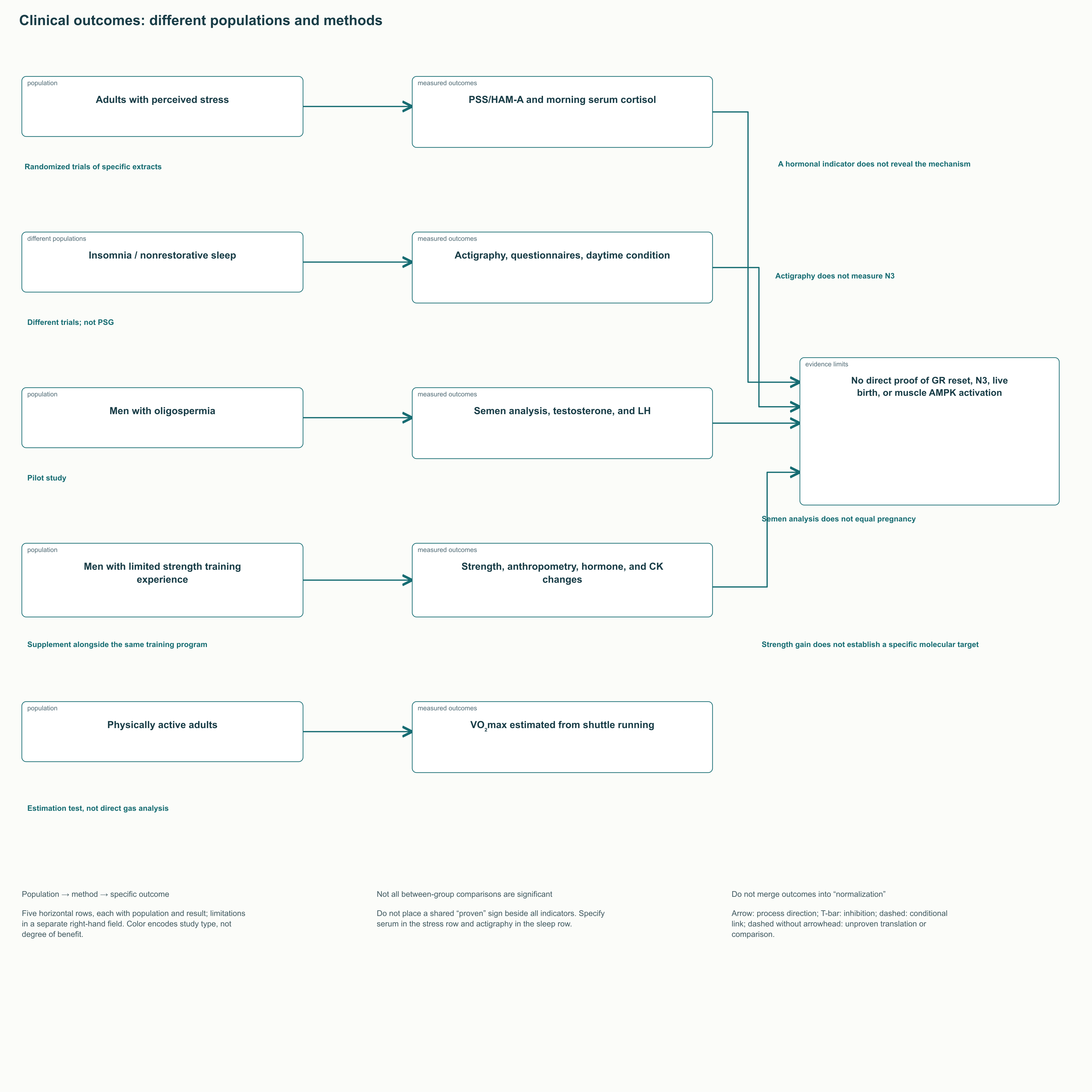
Chandrasekhar's study randomized 64 adults with chronic stress to 300 mg of root extract twice daily or placebo for 60 days; 61 completed participation. Perceived stress measured by PSS decreased by 44% in the extract group and 5.5% with placebo. Morning serum cortisol decreased by 27.9% versus 7.9%, with a between-group p = 0.002. GHQ-28 and DASS also changed favorably. These percentages describe changes in scores and a hormone in a specific sample, not the proportion of patients cured or the strength of a universal anxiolytic effect.
In Lopresti's study, 60 adults received 240 mg of standardized Shoden extract or placebo for 60 days. The advantage on the HAM-A anxiety scale was statistically significant, whereas the between-group DASS-21 difference did not reach the chosen significance threshold. Morning cortisol decreased by approximately 23%, versus a 0.5% increase with placebo. This was fasting serum collected around eight in the morning, not salivary cortisol or daily CRH secretion. A single measurement cannot establish restoration of a circadian profile.
DHEA-S decreased in that same study, whereas Auddy's earlier study reported an increase. Opposite directions are especially important for interpretation: “cortisol must go down, DHEA-S must go up” does not describe all clinical findings. Of Auddy's 130 enrolled participants, 98 completed the study, and analysis relied on completers. Such attrition increases uncertainty. The studies also differed in preparation, dose, baseline condition, and recording procedures.
Salve compared 250 and 600 mg/day of root extract with placebo in 60 adults over eight weeks. The work supports interest in perceived stress and morning cortisol, but does not establish that increasing the dose of any supplement automatically increases efficacy. Direct between-group analysis, adverse events, and the intervention's analytical characteristics matter for comparison. A small study does not establish a universal dose for patients with an anxiety disorder.
The clinical signal is consistent with experimental behavioral studies of glycoside fractions in animals, but this consistency does not reveal the human mechanism. Comparing a botanical intervention with lorazepam or imipramine in an animal test does not prove equivalence in treating anxiety or depression. The initial population is crucial for clinicians: a person with subjective stress and a patient with a diagnosed mental disorder require different clinical decisions.
A small Choudhary study in adults with chronic stress also assessed food cravings and body weight. These findings support investigation of connections between stress and eating behavior, but do not make ashwagandha a treatment for obesity, diabetes, or “metabolic decompensation.” Questionnaire improvement and weight changes should be considered separately from established approaches to nutrition and metabolic disease treatment.
Several trials involved extract suppliers or investigators affiliated with manufacturers. This does not invalidate the findings, but heightens the importance of independent replication, preregistered outcomes, and complete accounting for withdrawals. Small samples, multiple compared scales, and short follow-up limit confidence in effect size. Statistical significance does not replace assessment of how noticeably a person's functioning and quality of life improved.
10. Sleep: onset, efficiency, and architecture are different outcomes
Langade's study published in 2019 enrolled 60 patients with insomnia, allocated 2:1 to extract and placebo. A root preparation at 600 mg/day was studied for ten weeks, with sleep measures recorded by actigraphy. Mean sleep onset latency changed from 41.62 to 29.00 minutes with the extract and from 41.95 to 33.95 minutes with placebo; the final between-group p was 0.019. This represents a shorter time to fall asleep, not a promise that everyone will fall asleep within a particular number of minutes.
Sleep efficiency increased from 75.63% to 83.49%, compared with 75.14% to 79.68% with placebo. However, final between-group comparisons for total sleep time and wake after sleep onset had p = 0.076 in the published table. Claiming that every objective measure improved significantly against placebo is therefore too strong. The Pittsburgh Sleep Quality Index, PSQI, also changed favorably relative to placebo, but a subjective index and actigraphic parameters assess different aspects of sleep. Within-group change must be distinguished from advantage over control, and discrepancies between narrative conclusions and tabulated analyses considered.
Sleep efficiency is described by SE = 100 × TST/TIB, where TST is total sleep time and TIB is time in bed; both are expressed in the same units. SE can increase because of less wakefulness in bed and does not equal the proportion of deep sleep. An actigraph estimates sleep from movement and a device algorithm, but does not identify N1, N2, N3, and REM stages with polysomnographic accuracy. This methodological boundary rules out a conclusion that the study proved increased N3.
A separate Langade study published in 2021 enrolled 80 people: healthy volunteers and patients with insomnia. It assessed sleep during an eight-week intervention, not pharmacokinetics. Deshpande's study examined 120 mg of Shoden in 150 adults with nonrestorative sleep for six weeks, using subjective assessments and actigraphy. Its findings cannot be relabeled as confirmation of “deep sedation” or proven preservation of sleep architecture compared with benzodiazepines: neither that direct comparison nor sleep staging was performed.
For specialists, useful outcomes are those assessable in routine practice: sleep onset latency, awakening frequency, morning well-being, and daytime functioning. Cognitive behavioral therapy for insomnia remains the principal evidence-based approach to chronic insomnia. Before discussing a supplement, it is important to identify sleep apnea, circadian rhythm disorders, restless legs syndrome, and effects of alcohol and medications. Drowsiness from a product does not itself establish restorative sleep and may compromise driving safety.
11. Male hormones and spermatogenesis: do not substitute a biomarker for a clinical outcome
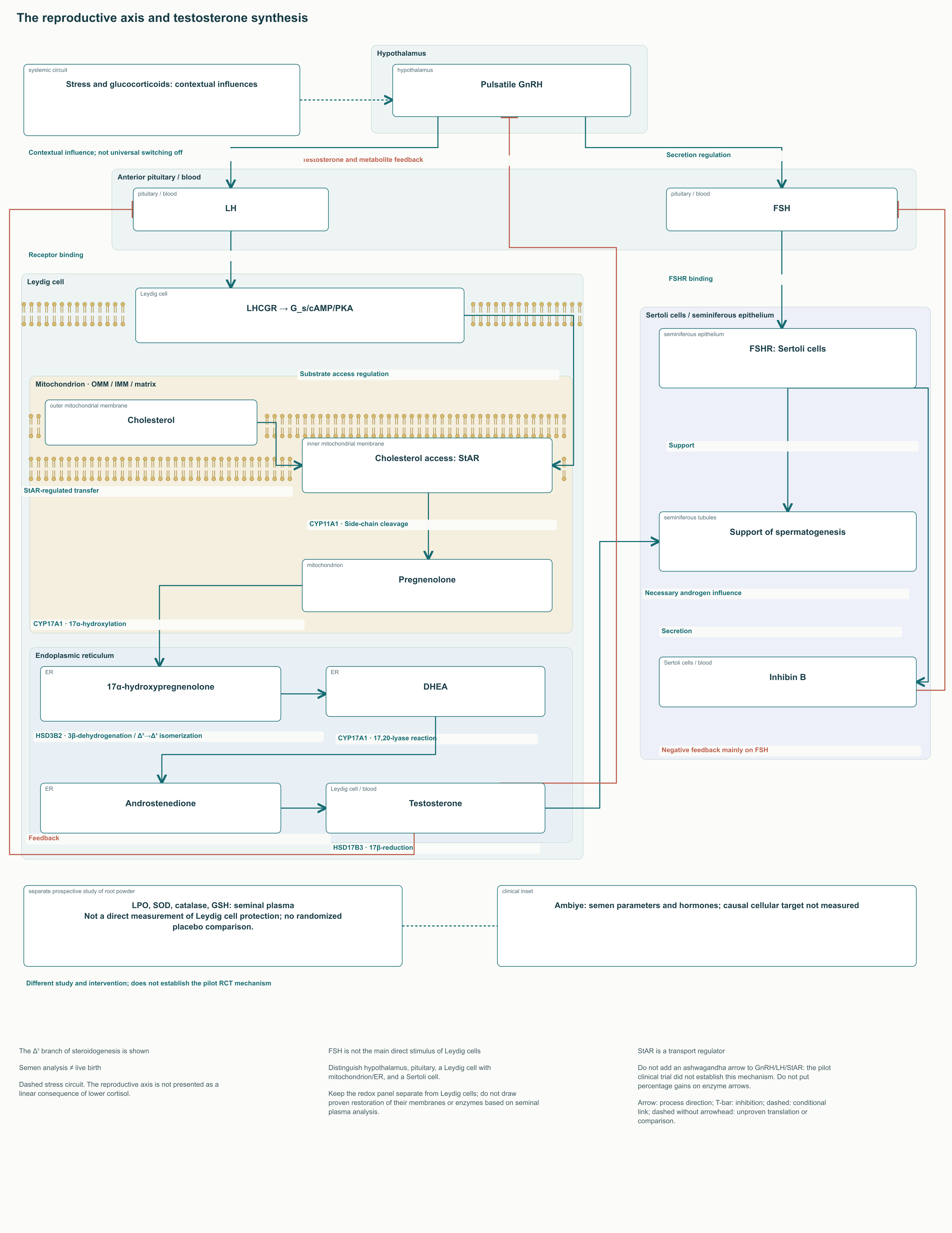
The reproductive axis is regulated by pulsatile GnRH secretion, which sustains pituitary LH and FSH release. LH acts on Leydig cells by binding LHCGR and activating cAMP–PKA; StAR and steroidogenic enzymes participate in testosterone formation from cholesterol. FSH acts mainly on Sertoli cells, which support spermatogenesis. Testosterone and its metabolites participate in feedback, while inhibin B mainly regulates FSH. Cortisol and stress may affect this circuit, but it is not a simple switch between “catabolic” and “anabolic” hormones.
Ambiye's pilot study enrolled 46 men with oligospermia and examined 675 mg/day of root extract for 90 days. Sperm concentration in the intervention group increased from 9.59 to 25.61 million/mL; increases in ejaculate volume, motility, testosterone, and LH were also reported. The often-cited increases of 167% in concentration, 53% in volume, 57% in motility, 17% in testosterone, and 34% in LH are changes from baseline within that group. They are neither the magnitude of between-group superiority nor the probability of pregnancy.
An oligospermic sample does not represent all men, and semen parameters are not live birth rates. The study did not establish direct stimulation of StAR, particular Leydig cell enzymes, or release of the human hypothalamus from cortisol inhibition. Hormonal and semen changes allow several explanations, including measurement variability and changes in overall health.
A separate line of research concerns seminal plasma oxidative status. In Mahdi's prospective study, infertile men received root powder; lipid peroxidation products, SOD and catalase activity, GSH, and antioxidant vitamins were assessed before and after intervention. Reported changes support interest in redox mechanisms, but the lack of a randomized placebo comparison limits causal inference. Powder composition cannot be equated with a standardized extract, nor seminal plasma measures with proven Leydig cell protection. The work does not establish direct StAR stimulation or prove that an antioxidant pathway explains the hormonal result of every clinical trial.
Participants with baseline stress in Lopresti's study produced a different result: the within-group testosterone increase in men was not accompanied by statistically significant between-group superiority. This is a useful counterweight to the generalization that “ashwagandha raises testosterone.” Suspected hypogonadism requires symptoms and persistently low morning testosterone confirmed by repeat measurement, followed by investigation of the cause with LH and FSH considered. A supplement does not replace infertility assessment or treatment of an established hormonal disorder.
12. Strength, recovery, and endurance: what was actually measured
Wankhede's study randomized 57 men with limited strength training experience; 50 completed the program. With the same eight-week training program, 600 mg/day of root extract was compared with placebo. Bench press performance increased by approximately 46.1 kg versus 26.4 kg, and leg extension by approximately 14.5 kg versus 9.8 kg. These are gains in kilograms within a specific program, not percentages or the expected response of an experienced athlete.
Total testosterone increased by approximately 96 ng/dL versus 18 ng/dL with placebo. Muscle size measures were assessed mainly using circumferences and skinfolds; they do not establish changes in individual muscle fiber area. Creatine kinase analysis concerned changes between measurements at 24 and 48 hours after exercise at the start and end of the program. This cannot be turned into a universal claim of a particular percentage reduction in muscle damage: CK depends on workload, sampling time, and individual response.
In Choudhary's study of physically active adults, VO₂max was estimated from a twenty-meter shuttle run. Reported gains with 600 mg/day exceeded those with placebo, but this was an estimate of aerobic performance, not direct oxygen consumption measurement by gas analysis. Translating the findings into changes in mitochondrial number, hemoglobin synthesis, or muscle AMPK and PGC-1α expression in humans requires measurement of those mechanisms.
Meta-analyses are useful for pooling trials, but do not increase the size of each original study or remove differences between preparations. Bonilla's review included 13 studies, of which 12 entered quantitative analysis; the VO₂max review published a year earlier covered five studies. The original description of these reviews as combining “dozens” of randomized trials is therefore incorrect. Their conclusions should be read alongside primary protocols, population heterogeneity, and measurement methods.
Sports specialists can use this knowledge to assess product claims: ask whether participants were novices, whether training and diet were matched, and how body composition and aerobic capacity were measured. Adequate energy and protein, load management, and sleep retain priority. Even a reproducible supplementary effect does not permit diagnosis of testosterone deficiency from post-training fatigue or explanation of every strength gain by hormonal change.
13. The thyroid: a clinical signal does not identify a cellular target
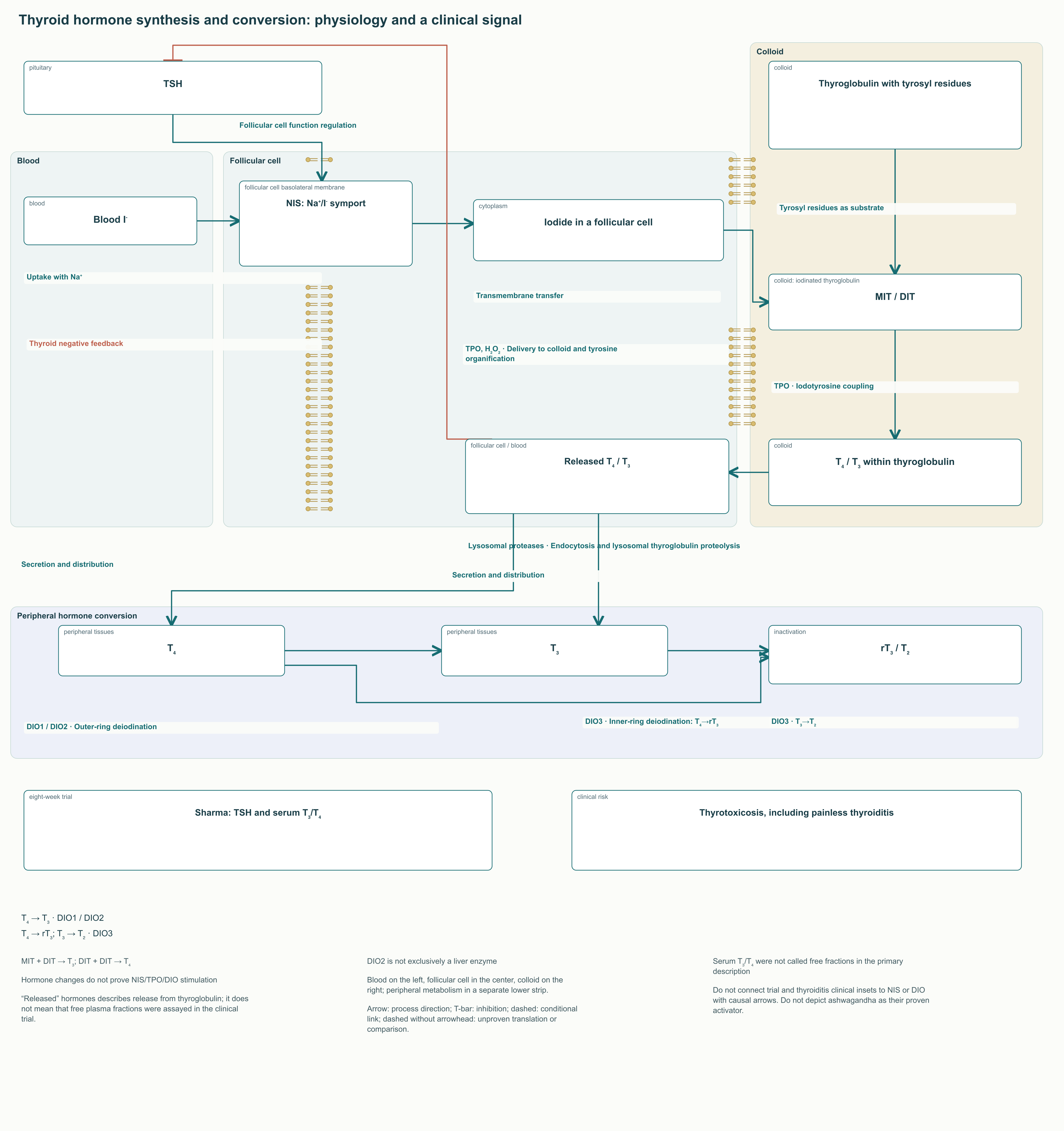
A thyroid follicular cell takes up iodide from blood through the sodium–iodide symporter, NIS, on its basolateral membrane. At the apical side, thyroid peroxidase, TPO, uses hydrogen peroxide to support iodine organification and iodotyrosine coupling within thyroglobulin. Thyroglobulin proteolysis releases T₄ and T₃. Deiodinases DIO1 and DIO2 participate in T₄-to-T₃ conversion, while DIO3 inactivates hormones. Expression depends on tissue; DIO2 cannot be described as exclusively a liver enzyme.
Sharma studied 600 mg/day of root extract in 50 adults with subclinical hypothyroidism and baseline TSH of 4.5–10 μIU/mL for eight weeks. The work reported between-group changes in TSH and serum T₃ and T₄. The available primary description does not identify these hormones as free fractions, so labeling them free T₃ and free T₄ is unjustified. Significance values also differed: TSH p < 0.001, T₃ p = 0.0031, and T₄ p = 0.0096. Unsupported exact percentage changes should not be reconstructed from secondary accounts.
This result does not prove human NIS, TPO, or DIO1/DIO2 stimulation by withanolides. Those targets were not measured. A small short-term study also does not establish superiority over observation or standard therapy, effects on complications, or safety in autoimmune thyroid disease.
An opposite clinical outcome has also been described: thyrotoxicosis during ashwagandha use, including a case of painless thyroiditis with low radiotracer uptake. A possible hormonal increase therefore cannot be explained solely by increased synthesis or necessarily attributed to Graves' disease. Follicular destruction and release of stored hormone require a different diagnostic interpretation.
For people with thyroid disease or taking levothyroxine, supplement discussion requires the treating clinician's involvement. Palpitations, tremor, heat intolerance, unexplained weight loss, and markedly increased anxiety warrant stopping use and assessing thyroid status. Reducing levothyroxine independently to compensate for a supplement's action is inappropriate. Repeat testing intervals are selected according to diagnosis, treatment changes, and symptoms, rather than a single schedule for an ashwagandha course.
14. Interactions, adverse reactions, and liver injury
Reported adverse effects include drowsiness, abdominal discomfort, diarrhea, and vomiting. Potential combinations with sedatives, alcohol, anticonvulsants, and some medications affecting blood pressure or glycemia require assessment of the overall pharmacological context. It cannot be claimed that respiratory depression from a particular combination has been proven through an established withanolide binding site. Nevertheless, marked sedation and the known risks of combining CNS depressants are sufficient grounds for a cautious clinical decision.
Experimental findings on AMPK, GLUT4, and immune cells do not establish the magnitude of a drug interaction in a patient. With insulin or sulfonylureas, the possibility of an additional glycemic change should be considered; after transplantation and during immunosuppressive therapy, discussion of a botanical product should involve the specialist team. Ashwagandha is unsuitable for unsupervised use in autoimmune disease, pregnancy, or breastfeeding. Lack of data in these situations cannot be replaced by assurances of a “gentle natural effect.” Particular caution is also necessary with hormone-dependent prostate tumors because of possible hormonal effects.
Why rare liver injury needs separate attention
Björnsson's series described five cases from Iceland and the US DILIN network. Symptoms appeared after 2–12 weeks, predominantly with cholestatic or mixed injury, jaundice, and itching. Recovery over several months was documented in four patients; one did not complete follow-up. The series cannot provide an incidence among all users because the relevant denominator is absent.
Philips' Indian series published in 2023 separately examined eight cases associated with single-ingredient products. Five patients had chronic liver disease; three with acute decompensation on that background died. A blanket statement that outcomes are invariably favorable is therefore incorrect. Severity and prognosis also depend on baseline liver health, and a case collection does not characterize the average risk in healthy populations.
The mechanism remains unresolved. HLA-B*35:01, known in the context of certain other herbal liver injuries, has not been confirmed as a specific clinical genetic predictor for ashwagandha. Nor has the universal sequence “leaf withaferin A — GSH depletion — BSEP blockade — cholestasis” been proven to explain the published cases. Analysis of some suspected products did not identify relevant toxic contaminants or adulteration. An authentic preparation can also cause an adverse reaction; absence of contamination does not justify continued use when liver injury is suspected.
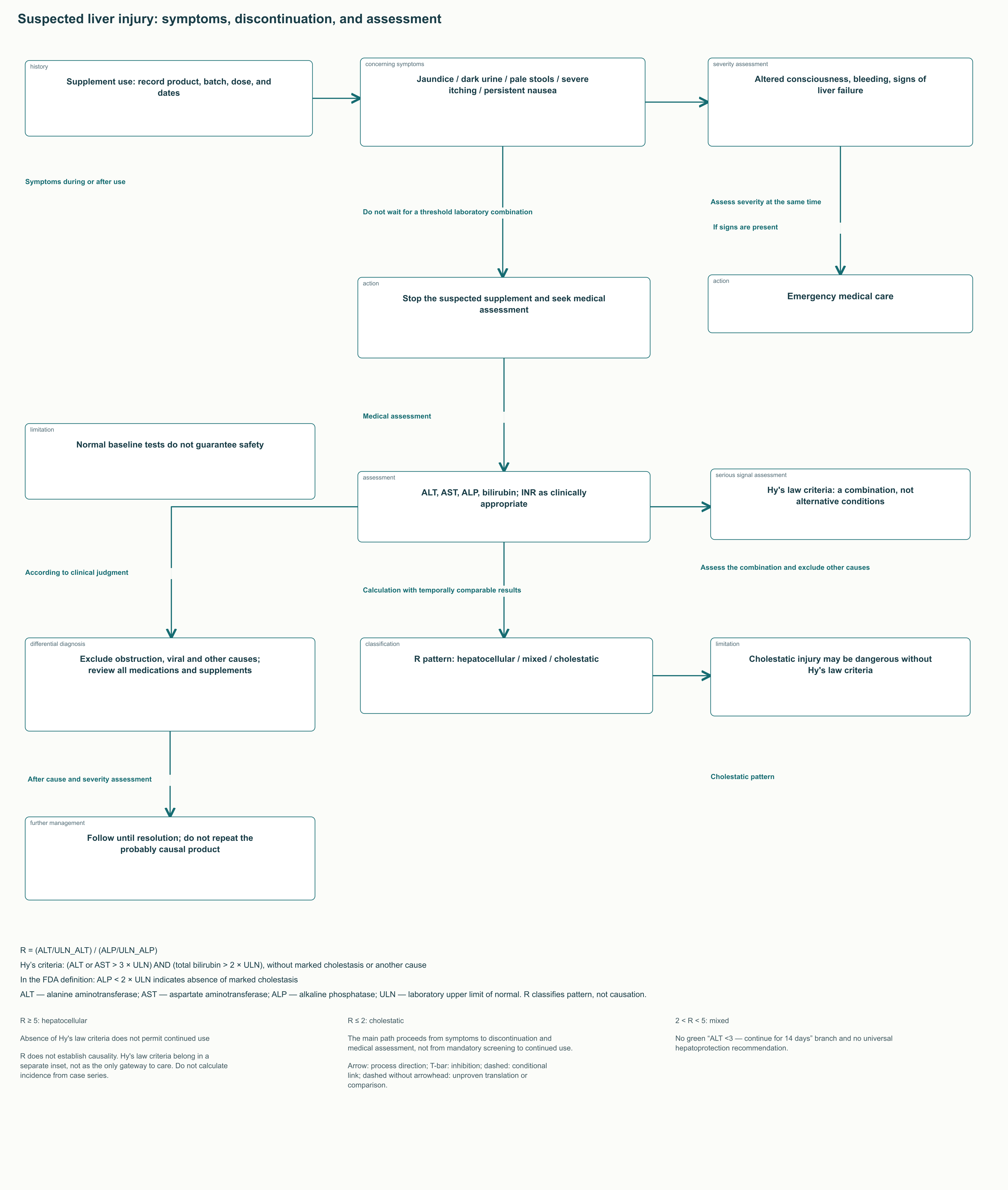
The injury pattern is described using R = (ALT/ULN_ALT)/(ALP/ULN_ALP), where ULN is the particular laboratory's upper limit of normal. R ≥ 5 corresponds to a hepatocellular pattern, R ≤ 2 to cholestatic injury, and intermediate values to a mixed pattern. Calculation requires temporally comparable results and assessment of whether the elevated ALP is of hepatic origin. It classifies the pattern, but does not establish supplement causality.
Hy's law criteria describe a dangerous combination of substantial hepatocellular injury and raised bilirubin: ALT or AST above three times ULN together with total bilirubin above twice ULN, without marked cholestasis or a convincing alternative cause. FDA guidance associates absence of marked cholestasis with ALP below twice ULN. The logical conjunction is “and,” not “or.” These criteria assess a serious signal, but their absence neither excludes dangerous cholestatic injury nor permits continued use until the thresholds are met.
Jaundice, dark urine, pale stools, severe itching, or persistent nausea require discontinuation of the suspected supplement and timely medical assessment. The clinician measures ALT, AST, ALP, bilirubin, and liver function indicators, including INR as clinically appropriate, and excludes obstruction, viral causes, and other explanations. Altered consciousness, bleeding, or signs of liver failure require emergency care. Universal “hepatoprotection” is not a proven way to neutralize continued exposure, and rechallenge after a probable reaction creates an unjustified risk.
Follow-up duration and long-term tolerability
Most randomized trials discussed lasted weeks or months. A prospective observation of 191 adults taking 600 mg/day of a standardized root extract for one year has also been published. It adds information on long-term tolerability in a selected population, but lacked a randomized control comparison and excluded several serious diseases. Such a sample cannot rule out rare idiosyncratic liver injury or establish equal safety across all extracts.
Recommendations to pause after 8–12 weeks, follow a 5/2 or 3/1 schedule, or begin with a week of titration are not confirmed methods of preventing GR desensitization or hepatotoxicity. Seven initial symptom-free days do not exclude a reaction several weeks later. A limited trial period may help assess benefit and avoid purposeless prolonged use, but should not be presented as a proven biological protective mechanism.
15. How specialists can apply this knowledge
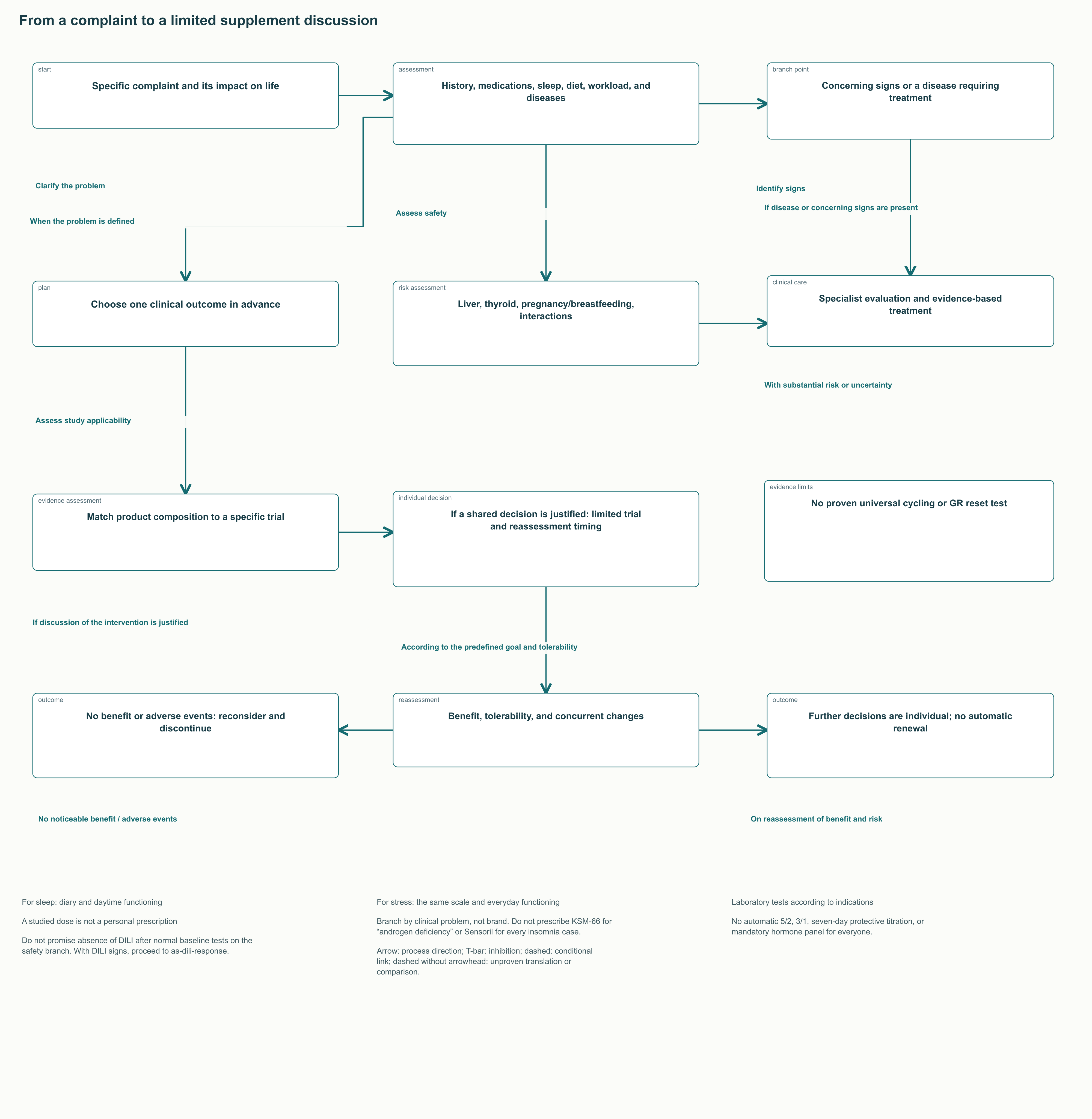
A mechanistic article's practical value is to sharpen the selection of questions and outcomes. Start with the complaint: difficulty falling asleep, daytime anxiety, declining training performance, or impaired fertility. Then establish its duration, functional consequences, medications, alcohol, sleep schedule, diet, and existing diseases. Sometimes this reveals a problem for which a supplement is not an appropriate answer at all: sleep apnea, thyrotoxicosis, severe depression, a medication adverse effect, or inadequate energy availability in an athlete.
If a limited supplement trial is discussed after this assessment, define its aim in advance. For sleep, this could be a change in diary-recorded latency and morning functioning; for subjective stress, the same validated scale alongside everyday functioning. Compare the result with baseline and concurrent changes. Simultaneous changes in training schedules, psychotherapy, and several supplements make causal interpretation particularly weak.
Match the product to a specific studied intervention rather than choosing it on an abstract “brand sedation” scale. Extract mass and marker percentage are needed together; transferring a dose from a 35% glycoside extract to a 5% preparation is incorrect. Regimens in the comparison table describe trials. Prescribing, if justified at all, depends on the patient, medications, and available product quality; evidence does not provide a universal “complaint — brand — dose — time of day” algorithm.
Clinical situation
Information that may be needed
What it allows and what it does not prove
Subjective stress without endocrine signs
History, one selected scale, sleep, and daytime functioning
Assessment of symptoms and change; random cortisol does not diagnose an HPA axis disorder
Signs of thyroid disease or levothyroxine treatment
TSH and free T₄, additional measures at the clinician's discretion
Clarification of thyroid function; does not measure supplement-induced NIS or deiodinase stimulation
Symptoms of possible liver injury
ALT, AST, ALP, bilirubin, and, where needed, INR and investigation of causes
Assessment of pattern and severity; normal baseline values do not exclude a future reaction
Suspected hypogonadism
Repeat morning testosterone under appropriate conditions, LH/FSH, and further evaluation
Confirmation and investigation of the cause; a hormonal increase does not equal improved fertility
Identification of inadequate recovery causes; a single CK or cortisol value does not define “anabolic status”
Laboratory tests are ordered according to indications, not on an identical calendar for every user. For suspected hypercortisolism, clinicians use validated diagnostic methods, such as late-night salivary cortisol, 24-hour urinary free cortisol, or a dexamethasone suppression test as appropriate. Endocrine Society guidance does not recommend random serum cortisol as initial screening for Cushing's syndrome. Four saliva samples to assess supplement efficacy and the cortisol/DHEA-S ratio are not established tests of GR restoration.
For nutrition professionals, the boundary follows their competence: assess diet, sleep, and product composition, and refer people with concerning symptoms to a physician. Endocrinologists need to avoid mistaking a biomarker change for proven disease treatment. Sports physicians should distinguish a possible additional effect from ordinary training adaptation and recovery quality. Sleep specialists should not substitute sedation for insomnia therapy or promise more deep sleep based on actigraphy.
Reassessment should address the predefined goal and tolerability. No noticeable benefit, drowsiness, palpitations, gastrointestinal symptoms, or signs of liver injury require reconsideration, rather than escalating a dose to achieve a desired laboratory number. Re-prescribing after probable herbal liver injury is excluded from routine practice. In people with chronic liver disease, severe outcome data provide a strong reason to avoid unsupervised experimentation.
Ashwagandha remains a subject of substantial chemical, cellular, and clinical research. Some standardized extracts show short-term benefit for individual stress and sleep symptoms and warrant further study in sports and reproductive medicine. Confident application of this knowledge requires simultaneous consideration of composition, measurement method, population, and risk. Neither the term “adaptogen” nor a molecular pathway diagram turns these findings into a proven universal protocol for restoring the neuroendocrine system.
FAQ
Does lower cortisol mean the HPA axis has been restored?
No. It shows a change in a specific measured indicator. Daily rhythm, CRH secretion, GR sensitivity, and receptor transport were not established as the mechanism in these clinical trials.
Do withanolides directly activate GABA receptors?
This is not proven for all withanolides. Candelario found activity in the extract, while separately tested withaferin A and withanolide A did not activate the receptors. An active mixture cannot be equated with an individual molecule.
How should KSM-66, Sensoril, and Shoden be compared?
Compare plant part, extraction, analytical markers, the specific batch, and a trial in an appropriate population. Glycoside percentage does not equal total aglycone percentage; no universal brand hierarchy of efficacy and safety is proven.
Is increased deep sleep proven?
The trials discussed mainly used actigraphy and questionnaires. They cannot establish increased N3 sleep or preserved sleep architecture compared with benzodiazepines.
Does ashwagandha treat low testosterone and infertility?
Small studies measured hormones and semen parameters in specific populations. They do not replace hypogonadism or infertility diagnosis and do not establish improved pregnancy or live birth outcomes.
Can a one-week trial or cycling prevent liver injury?
There is no evidence of such protection. A reaction can develop after several weeks of good tolerability. 5/2 or 3/1 breaks and mandatory cycling are not confirmed prevention methods for DILI or GR desensitization.
When should use be stopped?
Jaundice, dark urine, pale stools, severe itching, persistent nausea, or thyrotoxicosis symptoms require supplement discontinuation and medical assessment. Signs of liver failure require emergency care; do not wait for Hy’s law criteria.
Does everyone need cortisol, DHEA-S, and hormone monitoring?
No. Evaluation is selected according to symptoms and clinical risk. These measures are not universal tests of HPA restoration or supplement efficacy, and normal baseline tests do not guarantee future safety.