Lion’s Mane: NGF, BDNF, Erinacines, and Clinical Evidence — NutriFit
Lion’s mane: erinacines, NGF, BDNF, and the limits of the neurotrophic hypothesis
A detailed examination of Hericium erinaceus chemistry, neurotrophic pathways, myelination, and clinical studies. What has been demonstrated in cells and animals, what findings come from humans, and how professionals can assess raw materials, expected effects, and safety.
The neurotrophic hypothesis: why lion’s mane deserves attention and why claims of neurogenesis are premature
Lion’s mane, Hericium erinaceus, interests neurobiology for reasons beyond its use as an edible mushroom. Different groups of compounds isolated from its mycelium and fruiting bodies affect neurotrophin production, neurite growth, and cellular resistance to injury in experimental systems. However, three separate evidentiary boundaries must be crossed: a chemical can act in cell culture, reach an animal’s brain, and produce a measurable clinical effect in a human. Confirmation of one stage does not establish the other two.
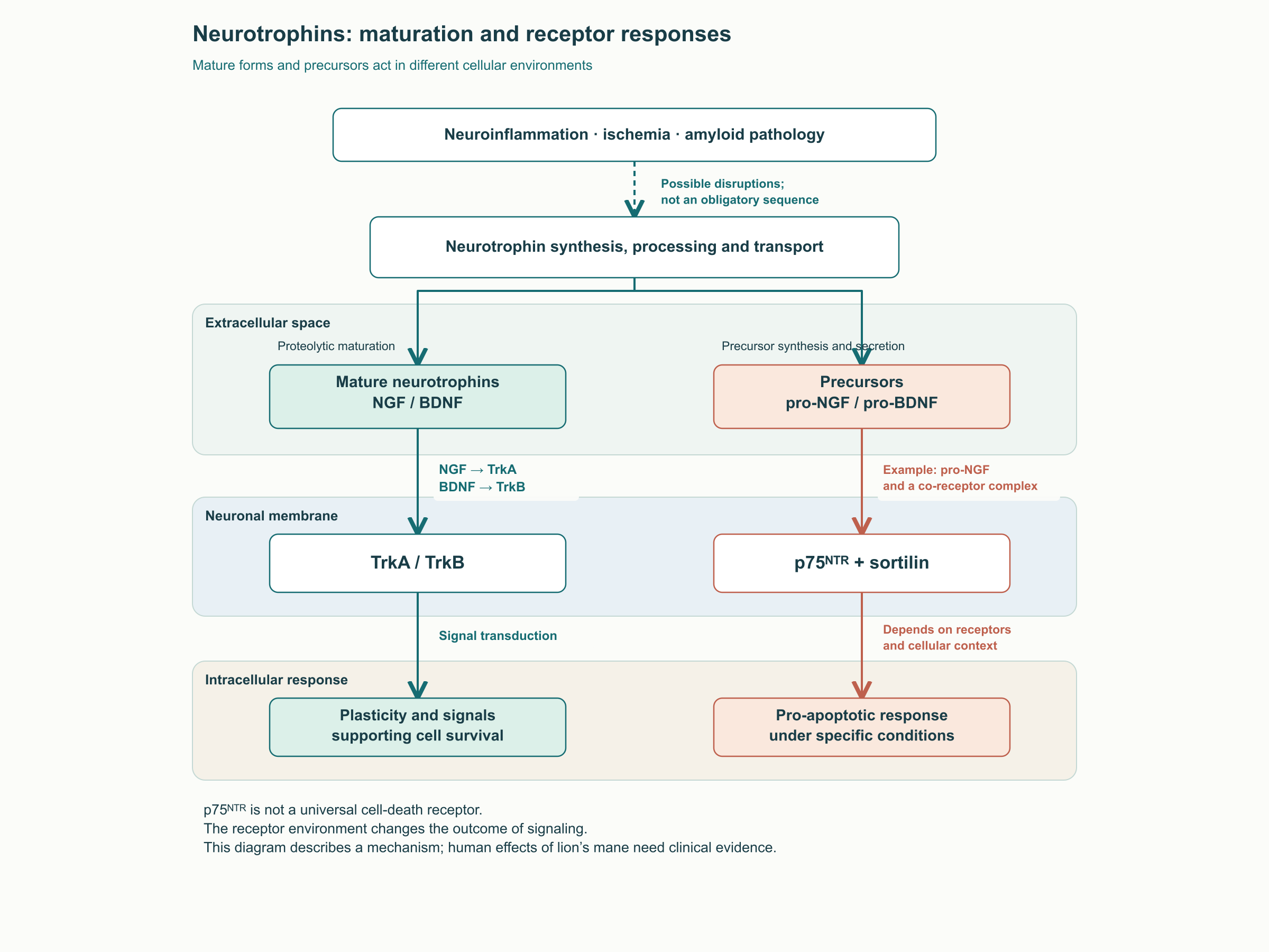
NGF, nerve growth factor, supports particular populations of peripheral sensory and sympathetic neurons and is associated with the viability of the cholinergic system in the forebrain. BDNF, brain-derived neurotrophic factor, participates in synaptic activity, learning, and plasticity. Their signals depend on receptors, local concentrations, transport, and conversion of precursors into mature proteins. Neurotrophic support therefore cannot be reduced to the principle that more NGF or BDNF is always better.
Neurotrophin maturation changes its interactions with the surrounding receptors. Under certain cellular conditions, pro-NGF forms a complex with p75ᴺᵀᴿ and sortilin and initiates a proapoptotic signal; this mechanism has been established experimentally. But p75ᴺᵀᴿ has several functions, including interactions with Trk receptors, and is not a universal switch that turns off survival. Neuroinflammation, ischemia, and amyloid pathology can disrupt the neurotrophic environment through different mechanisms. The sequence “any injury → reduced transcription → accumulation of pro-forms → death” does not describe every condition in the aging brain.
Replacement administration of NGF does not automatically solve the problem either. Mature β-NGF is a dimeric protein with a mass of approximately 26 kDa, so systemic administration does not provide unrestricted passive delivery across the blood–brain barrier. Separate methods of local delivery and control of distribution are needed. Subcutaneous rhNGF administration in humans produced pain and hyperalgesia; a large diabetic polyneuropathy trial did not demonstrate an advantage on its primary outcome, although early findings had been encouraging. This illustrates the difficulty of translating a trophic mechanism into treatment.
Stimulating endogenous neurotrophin production with small molecules is a reasonable research direction. It is not the only possible approach and does not guarantee safe, selective stimulation in the right cell. Lion’s mane provides preclinical grounds for such research, but convincing evidence of restoring lost neurons, preventing dementia, or treating neuropathy in humans remains insufficient.
Different parts of the mushroom yield chemically different preparations
Mycelial erinacines and fruiting-body hericenones
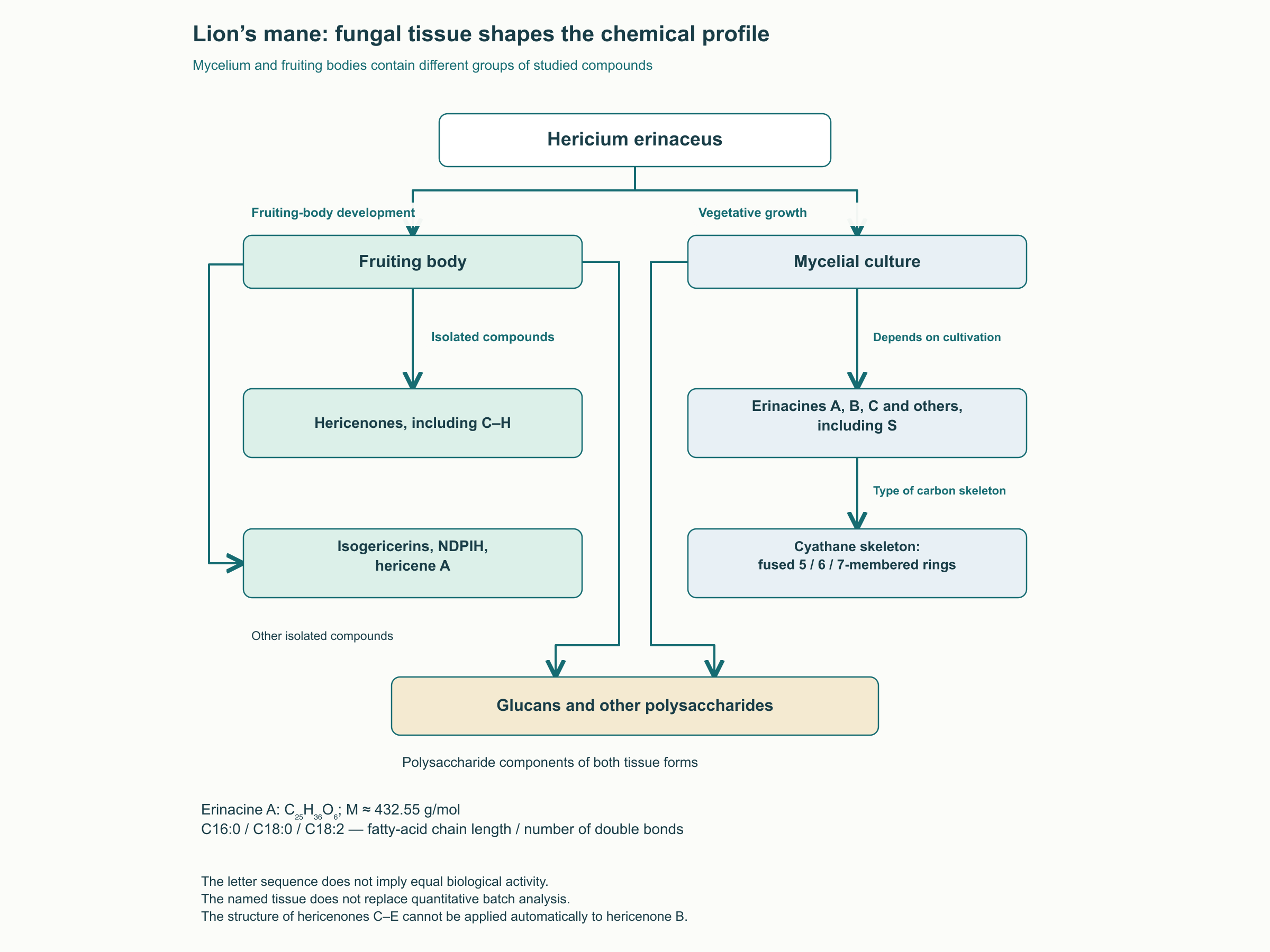
The mycelium and fruiting body belong to the same organism but differ in developmental conditions and secondary-metabolite profiles. Erinacines are studied primarily as compounds of mycelial cultures, and hericenones as constituents of fruiting bodies. Their content nevertheless depends on the strain, growth medium, cultivation time, and processing. A label stating “fruiting body” or “mycelium” identifies the material’s origin but does not replace quantitative chemical analysis.
Erinacines are diterpenoids with a cyathane scaffold, distinguished by fused five-, six-, and seven-membered rings. “Cyathane” describes the structural class rather than a physiological target. Investigated members include erinacines A, B, C, and others, including S; the alphabetical list does not imply equal activity or equal ability to reach the brain. In work with mycelial cultures, A, B, C, and several other compounds altered neurotrophic responses in cellular systems. The effect depended on the substance and model.
Erinacine A has the molecular formula C₂₅H₃₆O₆, corresponding to a molar mass of approximately 432.55 g/mol. The formula specifies elemental composition, not stereochemistry, substituent positions, or pharmacological activity. The sugar moiety and oxygen-containing groups influence polarity; calculated logP and polar surface area depend on the calculation method. Without specifying that method, precise values cannot serve as a bioavailability profile. Numbering of aldehyde, hydroxyl, and glycosidic groups should be taken from the relevant structural publication rather than reconstructed from the formula.
Hericenones C, D, and E are derivatives of an aromatic scaffold with an isoprenoid portion and fatty-acid residues; differences in those residues alter molecular properties. C16:0, C18:0, and C18:2 describe saturation: the first number indicates carbon-chain length and the second the number of double bonds. These designations describe the fatty-acid residue, not the entire hericenone molecule. The structural scheme for C–E cannot be extended to every family member, particularly hericenone B, which has a different structure.
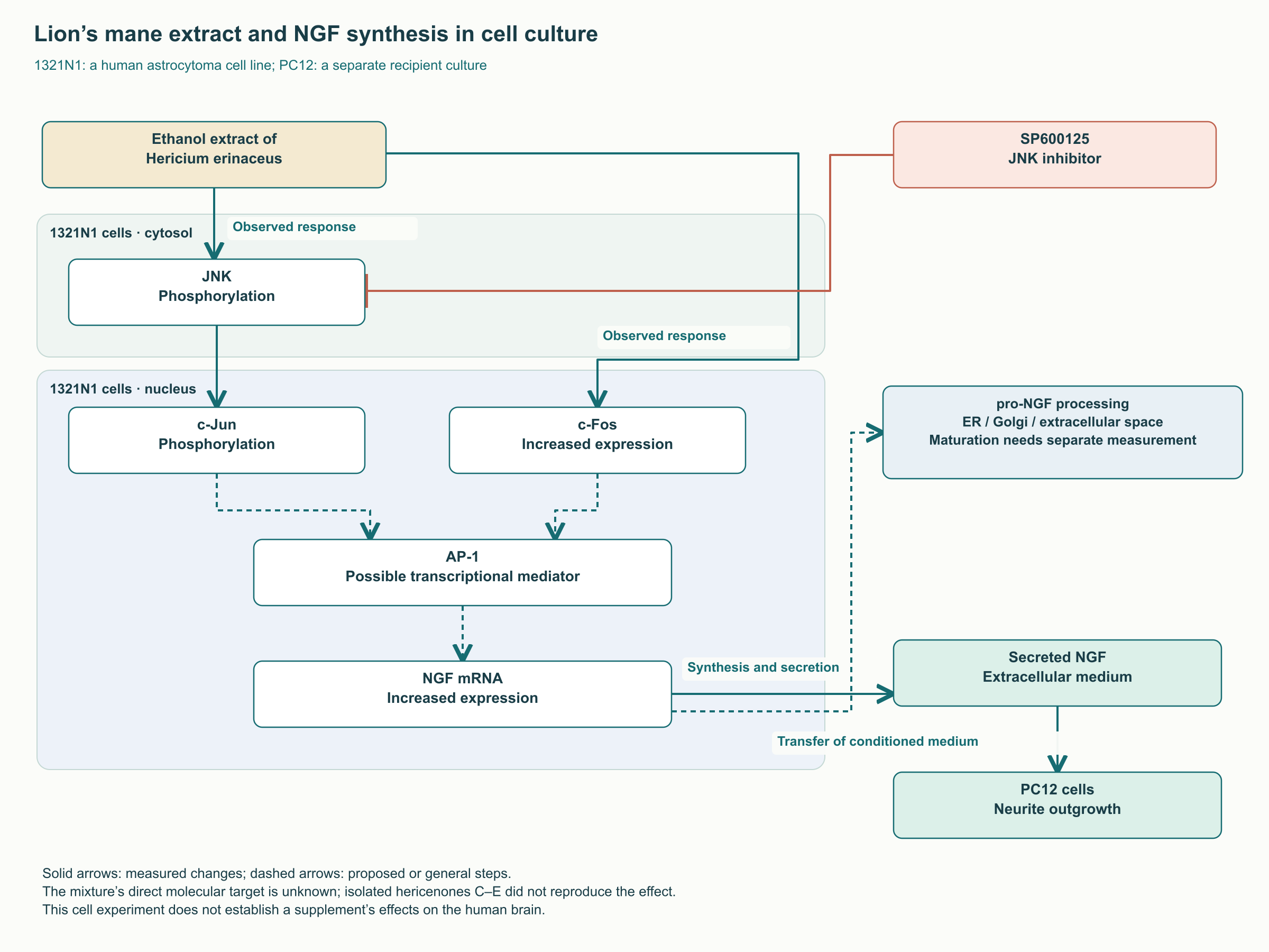
Early experiments considered hericenones as NGF stimulators, but results proved dependent on the cellular system. In Mori’s study, hericenones C, D, and E did not increase NGF expression in the human astrocytoma line 1321N1, although the mushroom’s ethanolic extract did. Thus, lack of activity of individual molecules does not disprove activity of the mixture, and activity of the mixture does not identify hericenones as its sole active constituents.
Fruiting bodies also contain other small molecules. Martínez-Mármol and colleagues examined N-de-phenylethyl isohericerin, NDPIH, and hericene A in detail. In hippocampal neuron cultures, they enhanced neurite growth and branching and changed growth-cone morphology—the mobile tips of growing processes. The work included mouse experiments and signaling-pathway analyses; it did not establish an effective human dose. The name “hericene A” must be distinguished from “hericenone” and “erinacine.”
What is actually known about the blood–brain barrier
Molecular mass and lipophilicity help estimate distribution but do not form a rigid filter with a 450–500 Da cutoff. Hydrogen bonding, ionization, protein binding, efflux transporters, and metabolism also matter. High lipophilicity can facilitate membrane interactions while reducing solubility and the free concentration. Evidence of brain penetration therefore requires measurements, not calculated logP alone.
Parameter
Erinacine A
Hericenone C
Recombinant NGF
Chemical nature
Cyathane diterpenoid
Small-molecule aromatic derivative with a fatty-acid residue
Protein neurotrophin
Size
C₂₅H₃₆O₆; approximately 432.55 Da
Larger than erinacine A; size alone does not establish delivery limitations
Mature dimer of approximately 26 kDa
Brain-distribution evidence
Measured in rat brain after an oral dose
Comparable convincing human data are unavailable
Unrestricted passive delivery from blood is limited
Oral pharmacokinetics
Absolute bioavailability of 24.39% was estimated for one preparation in rats
A reliable quantitative human estimate has not been established
Not used as an ordinary oral protein supplement
Can a human dose be derived?
No; animal pharmacokinetics alone are insufficient
No
No
In Tsai’s study, rats received a mycelial extract equivalent to 50 mg/kg of erinacine A, and an intravenous dose of the compound was used separately to compare exposure. Erinacine A was detected in the brain after one hour, with the highest measured concentration after eight hours. Free-fraction analysis supported predominantly passive distribution. These findings belong to a particular model using a high experimental dose; they do not demonstrate equivalent exposure in the human hippocampus or establish detection of erinacine B in human cerebrospinal fluid.
The quantitative meaning of bioavailability can be expressed as:
F = (AUCₚₒ × Dᵢᵥ) / (AUCᵢᵥ × Dₚₒ) × 100%.
Here AUC is the area under the plasma concentration–time curve, D is the administered dose, po denotes oral administration, and iv intravenous administration. Dose adjustment is necessary because the routes are compared using different quantities of the compound. The formula estimates systemic availability, not the proportion of the dose reaching the brain. Its applicability assumes an appropriate analytical method and comparable dose dependence. Rat pharmacokinetic data with brain detection have also been published for erinacine S; they do not apply to all erinacines.
Hypotheses involving hericenone hydrolysis by esterases, albumin binding, and effects through the gut or vagus nerve warrant investigation. Without comparable measurements, however, these processes cannot be claimed to explain the fruiting body’s effects in humans. This is particularly important when comparing supplements: absence of a measured marker and absence of biological activity are different things.
From a cellular response to neuronal signaling
JNK, c-Jun, and NGF production: an experimental sequence
Astrocytes contribute to neurotrophic support but are not the only source of NGF. In the 1321N1 line, an ethanolic lion’s mane extract increased NGF expression and protein secretion. Medium from treated cells enhanced neurite growth in PC12 cells. A JNK inhibitor attenuated the transcriptional response, while investigators observed JNK and c-Jun phosphorylation and increased c-Fos expression. Together, these findings support involvement of JNK signaling but do not establish direct binding of erinacine A to the kinase.
JNK belongs to the stress-activated MAP kinases. Kinases transmit signals through phosphorylation, while c-Jun and c-Fos can form part of AP-1, a complex regulating transcription. Subsequent NGF production involves transcription, translation in the secretory pathway, and proteolytic maturation. The extent of processing cannot be inferred from a single measurement of total NGF. Mature and precursor forms, and intracellular and secreted protein, require separate assays.
The original mechanistic model additionally included p38, specific amino-acid residues, and a precisely defined promoter motif. Such details create an impression of complete proof, although the cited mixture study did not determine every link. For professionals, the more important distinction is between an inhibitor experiment and evidence of a direct target. An inhibitor can help identify a necessary response branch, but its selectivity and effects on cell viability must also be considered.
TrkA and p75ᴺᵀᴿ: the receptor environment determines the effect
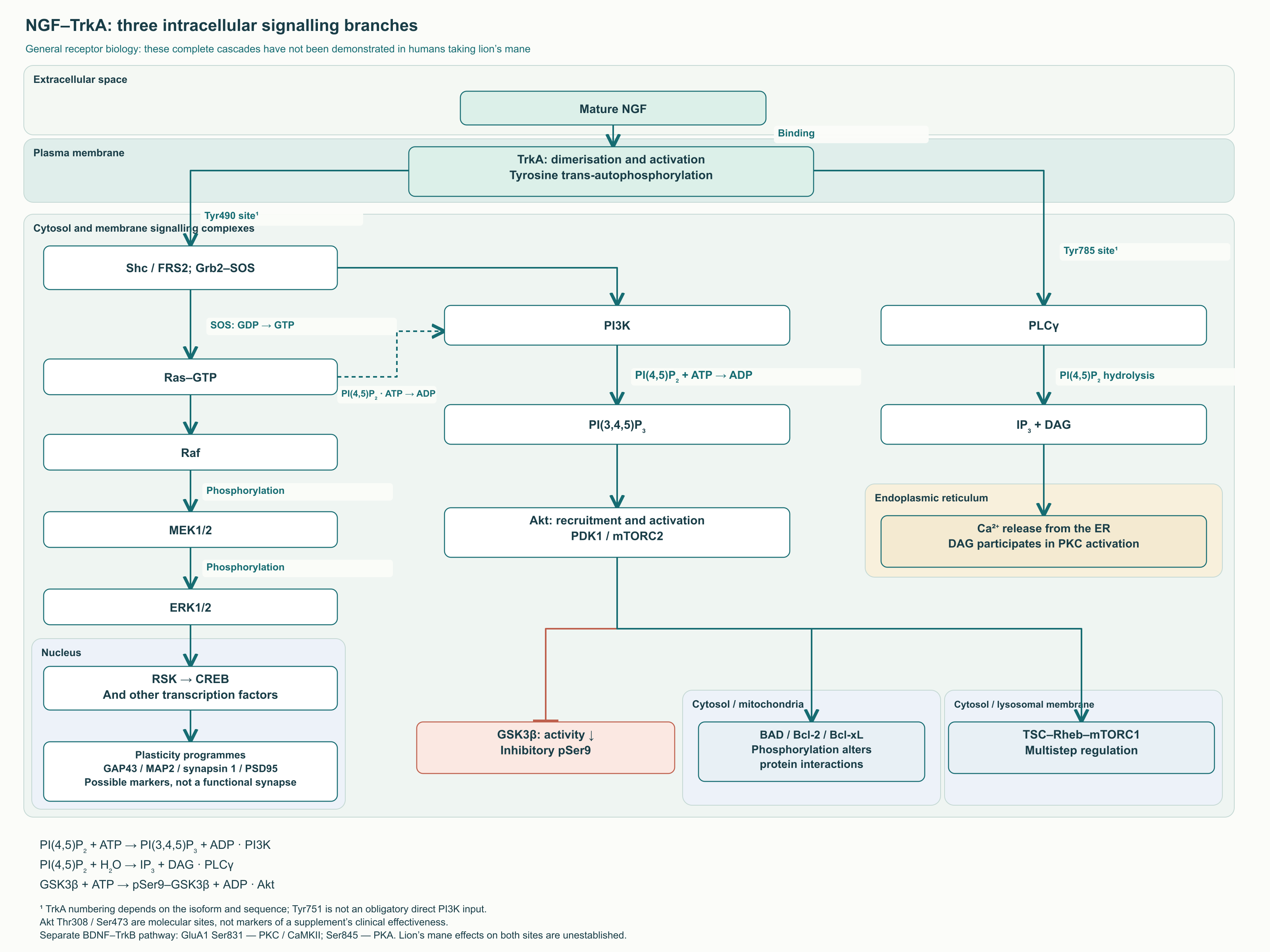
Mature NGF binds TrkA, a receptor tyrosine kinase. Receptor activation creates phosphotyrosine sites for interactions with adaptors and enzymes, directing signals toward process growth, metabolism, and survival. p75ᴺᵀᴿ lacks intrinsic tyrosine-kinase activity and uses partner proteins. A proapoptotic response is possible when pro-NGF, p75ᴺᵀᴿ, and sortilin are present, but it is not inevitable in all neurons.
Functionally investigated TrkA sites are traditionally designated Tyr490 and Tyr785: the former is associated with Shc/FRS2 adaptors and the latter with PLCγ. The isoform and reference sequence must be specified when numbering residues. Tyr751 cannot automatically be drawn as an independently established PI3K input: primary studies treated it as a candidate, and its phosphorylation was not detected in every system. PI3K activation can occur through adaptor complexes and Ras.
Three effector branches: differentiation, survival, and calcium
In the Ras–Raf–MEK–ERK branch, the Shc adaptor interacts with Grb2 and the exchange factor SOS. SOS promotes replacement of GDP by GTP in Ras; active Ras initiates the Raf, MEK1/2, and ERK1/2 kinase cascade. Through RSK and other intermediates, ERK can influence CREB and, through CREB and other factors, differentiation and plasticity programs. GAP-43, MAP-2, synapsin-1, and PSD-95 are useful molecular indicators of growth or synaptic organization, but altered expression is not equivalent to formation of a functional synapse, much less improved memory in a patient.
The PI3K branch includes the chemical reaction PI(4,5)P₂ + ATP → PI(3,4,5)P₃ + ADP. PI3K adds a phosphate to the third position of the lipid’s inositol ring, creating a membrane signal for recruitment of Akt and associated proteins. PDK1 and mTORC2 contribute to Akt activation, commonly described through Thr308 and Ser473. These sites concern Akt biochemistry, not a fully established response of the human brain to lion’s mane.
One Akt substrate is GSK-3β. Phosphorylation of its Ser9 decreases enzyme activity: GSK-3β + ATP → pSer9–GSK-3β + ADP, with Akt serving as the kinase. This is a well-studied regulatory principle, but GSK-3β has many substrates and is regulated in other ways as well. Detection of pSer9 therefore does not establish prevention of tau pathology or neurofibrillary tangles.
Akt can also phosphorylate BAD and change its interactions with the antiapoptotic proteins Bcl-2/Bcl-xL. Preservation of mitochondrial function involves control of outer-membrane permeability and cytochrome c release, rather than Bcl-2 literally “closing pores.” Through TSC–Rheb regulation, Akt is linked to mTORC1 and protein synthesis. Neither increased synthesis nor suppression of an apoptotic marker alone guarantees restoration of a damaged network.
The third branch begins with PLCγ, which hydrolyzes a membrane phosphoinositide: PI(4,5)P₂ + H₂O → IP₃ + DAG. IP₃ promotes Ca²⁺ release from the endoplasmic reticulum, while DAG contributes to PKC activation. Unlike PI3K, PLCγ cleaves the lipid rather than adding a phosphate; the reactions must be shown separately. Their effects on the cytoskeleton and excitability depend on signal timing and location.
BDNF/TrkB, AMPA receptors, and the limits of “neurogenesis”
BDNF/TrkB participates in long-term potentiation, LTP, and dendritic-spine organization. In neuron cultures, BDNF altered AMPA-receptor expression and delivery, particularly GluA1. Caldeira’s work linked the effect to Ser831 phosphorylation through PKC and CaMKII. Ser845 is a recognized GluA1 regulatory site associated with PKA, but it cannot automatically be added as an established consequence of BDNF or lion’s mane intake.
In chronic-stress experiments in mice, erinacine A-enriched mycelium altered behavioral measures and BDNF/PI3K/Akt/GSK-3β signaling. Correlation between biochemistry and behavior supports a mechanistic hypothesis, but the forced-swim test does not diagnose human depression or demonstrate antidepressant efficacy of the supplement.
In the work on hericene A and NDPIH, neurotrophic responses converged on ERK1/2 but were not explained by simple direct TrkB agonism. This distinction is fundamental: a shared downstream kinase does not imply an identical primary target. Changes in growth cones, branching, and neurite area describe morphological plasticity, not the birth of new neurons.
Neurogenesis requires differentiation between precursor proliferation, neuronal differentiation, survival, and integration of a cell into the network. In aging mice, Ratto’s work used PCNA and DCX, among other measures, and recognition-memory testing. These findings belong to a preclinical model. The extent and measurement of neurogenesis in the adult human hippocampus remain methodologically challenging; studies of human tissue have produced different results. None of the clinical lion’s mane studies discussed measured the appearance and functional integration of new neurons in participants.
Myelin, peripheral nerves, and pain: three different research questions
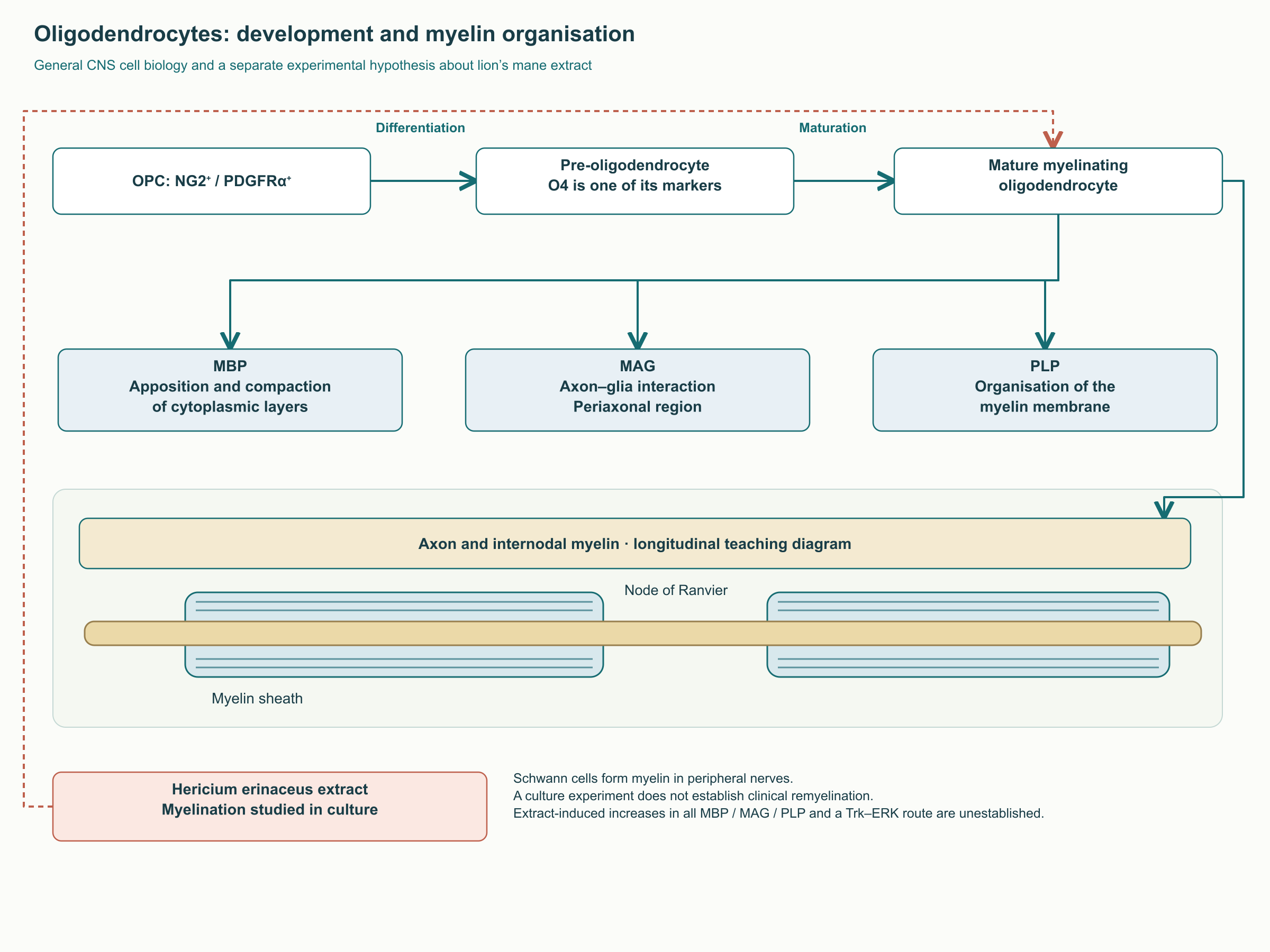
Myelin electrically insulates axons and supports saltatory conduction between nodes of Ranvier. Oligodendrocytes produce it in the CNS, and Schwann cells in the peripheral nervous system. Oligodendrocyte precursor cells, OPCs, are often characterized by NG2 and PDGFRα; later stages have different marker sets, including O4. MBP contributes to compaction of cytoplasmic layers, MAG to axon–glia interactions, and PLP to organization of the myelin membrane. These functions explain the choice of measurements but do not establish that lion’s mane increases every one of them.
Kolotushkina’s study examined myelination in cerebellar cell culture after addition of mushroom extract. It supports discussion of effects on myelin development in vitro. The available primary record does not allow simultaneous reconstruction of the original claims of exactly 21–22 versus 25 days and quantitative increases in MBP, MAG, and PLP. Those numbers and molecular effects therefore cannot be presented as established findings of that study without the corresponding experimental tables. Even fully confirmed acceleration of culture myelination would not demonstrate remyelination in a person with multiple sclerosis.
After peripheral nerve crush, the distal segment undergoes Wallerian degeneration, while Schwann cells and macrophages establish conditions for regeneration. Preservation of guiding structures in the crush model makes it substantially different from complete nerve transection or longstanding diabetic polyneuropathy. The speed of motor recovery in a rat cannot be translated into a time to recovery of sensation or strength in a patient.
In Wong’s primary study, an aqueous fruiting-body extract accelerated recovery of the injured peroneal nerve’s function according to the footprint index. Baseline measures returned 4–7 days earlier than in controls; recovery times were 10–14 days in extract groups and 14–17 days in controls. This differs from the original claim of “24 versus 13–14 days.” The authors also examined neurofilaments and neuromuscular junctions. A 75% CMAP increase cannot be attributed to this study: it did not establish that result.
Neuropathic pain is a separate problem. Changes in channels, ectopic activity, and peripheral and central sensitization contribute to its development; disrupted trophic transport does not explain every case. CGRP and substance P are important in nociception, and NF-κB, iNOS, and cytokines can participate in inflammatory responses. A regeneration study, however, cannot establish normalization of these mediators by erinacines or effective pain relief in humans. NGF itself can, moreover, enhance nociceptive sensitivity. An erinacine A experiment in ischemic injury examining iNOS/p38 and CHOP characterizes a particular cellular-stress model, not treatment of radiculopathy.
Amyloid pathology and erinacine A-enriched mycelium have also been studied in transgenic APPswe/PS1dE9 mice. This is a useful model of particular pathological processes but does not reproduce the full sporadic human Alzheimer’s disease. Changes in amyloid deposits, glia, or clearance enzymes in such an experiment do not mean that an ordinary supplement removes plaques in a patient.
Clinical studies: preparation, outcome, and the correct comparison matter
Mild cognitive impairment and follow-up after discontinuation
Mori and colleagues randomized 30 participants aged 50–80 with mild cognitive impairment. For 16 weeks, the experimental group took four 250 mg tablets three times daily; the tablets contained 96% dried lion’s mane powder. This amounted to 3000 mg of tablets, or 2880 mg of mushroom powder per day, not 3000 mg of a pure compound. Four weeks of follow-up without intake followed.
Period
What the available primary publication confirms
Interpretation
Initial allocation
Two groups of 15 participants
Small study with limited precision
Weeks 8, 12, and 16
Advantage on an HDS-R-based cognitive scale
Efficacy signal for a particular powder
Four weeks after discontinuation
Lower scores in the experimental group
Duration of effect requires separate investigation
Exact means ± SD in the original table
Cannot be reconstructed from the primary abstract that was read
Not published as reliable numerical data
Follow-up after discontinuation establishes neither the lifespan of new synapses nor a need for lifelong intake. The authors did not measure neurogenesis, brain NGF, or loss of structural changes. A declining score can have several explanations; it should be retained as a clinical observation rather than converted into a universal biological law.
A pilot study in mild Alzheimer’s disease
Li’s study randomized 49 people, of whom 41 completed the trial. Participants took three 350 mg mycelium capsules daily for 49 weeks; erinacine A content was 5 mg/g. The daily marker quantity was 1.05 g × 5 mg/g = 5.25 mg. This calculation describes preparation composition, not the amount reaching the brain. Some authors were linked to the manufacturer, which matters alongside the small sample and multiple outcomes.
Measure
Observation at week 49
Important limitation
MMSE
Within-group improvement in the mycelium group
The between-group comparison at that point was not significant
CASI
More favorable direction of change in the mycelium group
No convincing between-group difference
IADL
A between-group difference was detected
One of several investigated outcomes
Contrast sensitivity
A separate difference for the left eye
Does not establish general visual recovery
Neuroimaging and blood
Exploratory DTI measures and biomarkers
Not direct evidence of neurogenesis or clinical slowing of disease
The study title includes “prevention,” but participants already had mild Alzheimer’s disease. It cannot therefore be cited as a prevention study in healthy people. The work provides no basis for claiming that OCT demonstrated preservation of the peripapillary RNFL: the ophthalmic measures studied were different. These distinctions are especially important when a clinical text informs treatment choices.
Mood, sleep, and healthy young participants
Vigna and colleagues included 77 people with overweight or obesity and complaints assessed by questionnaires. A low-calorie diet was compared with diet plus supplementation; no placebo was used. After eight weeks, improvements in selected mood and nighttime-rest measures were reported. Serum pro-BDNF increased, while mature BDNF did not change significantly over that period. This establishes neither increased brain BDNF nor “harmonization” of the ratio between the two forms. The scales used were Zung and SCL-90; the original references to STAI and PSQI do not match the study description.
Nagano also examined mood in a small four-week study in women. It supports further investigation of subjective symptoms but does not replace trials in diagnosed depression or anxiety disorders. Questionnaire results, a peripheral biomarker, and animal experiments cannot be combined into a single proof of an antidepressant mechanism in humans.
Docherty’s pilot involved 41 healthy adults aged 18–45. With 1.8 g/day, faster Stroop-test performance was observed after a single dose; after 28 days, the main overall effect on subjective stress was borderline, p = 0.051. The authors also reported null findings and some unfavorable results. The appropriate conclusion is therefore a preliminary signal for selected measures rather than a proven general “nootropic boost.”
Saitsu’s study in older adults broadens the clinical investigation of cognition but does not overcome the problem of small samples and differing preparation compositions. Studies should be compared by participants, product, duration, and prespecified outcome. Grams of fruiting-body powder, freeze-dried mycelium, and extract are not interchangeable doses.
Extraction and standardization: what professionals should see in product documentation
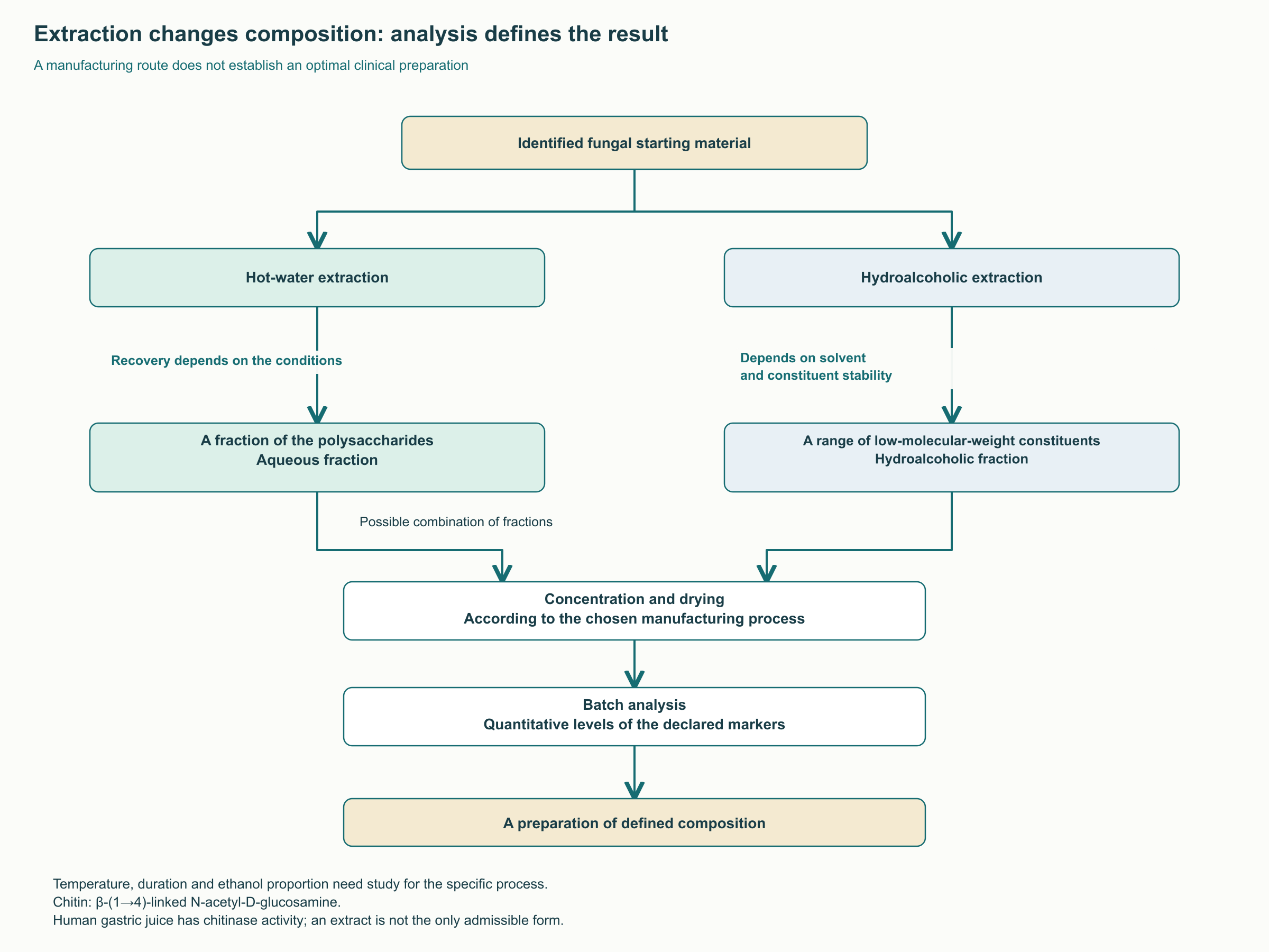
The fungal cell wall contains chitin—a polymer of N-acetyl-D-glucosamine with β-(1→4) linkages—and glucans of different structures. Milling, heating, and extraction alter component accessibility, but the claim that powder passes through completely unchanged because humans lack chitinase is incorrect. Chitinase activity has been demonstrated experimentally in human gastric juice, with marked individual variability. This does not imply complete digestion of the fungal wall, but it rules out that absolute explanation.
Hot water can extract some polysaccharides, while hydroalcoholic systems extract a different spectrum of small molecules. Ethanol concentration, temperature, time, and the ratio of material to solvent should be selected according to the analytical goal and compound stability. “Dual extraction” describes a technology but does not establish clinical superiority. The fruiting-body powder in Mori’s work and the mycelial biomass in Li’s work themselves show why hydroalcoholic extract cannot be declared the only studied form.
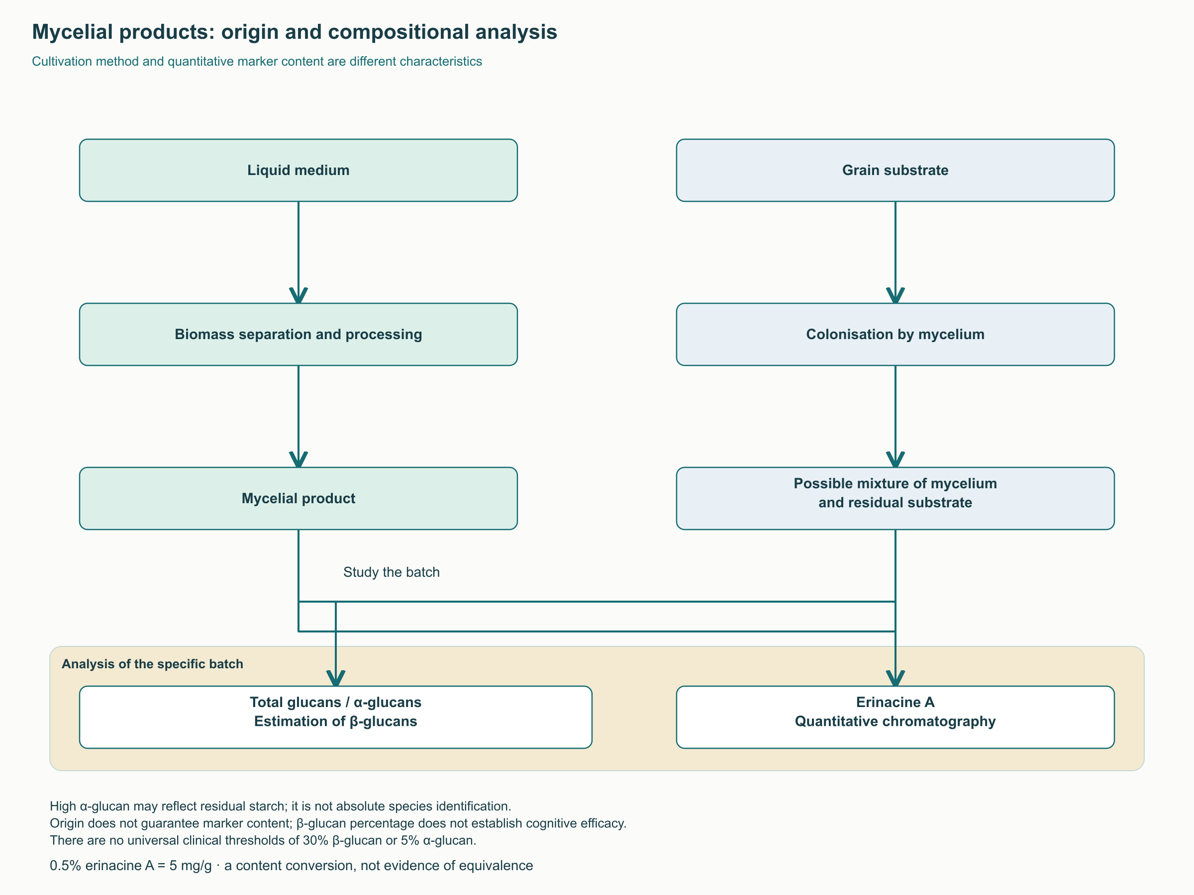
Mycelium can be grown in a liquid medium followed by biomass separation, or on a solid grain substrate. In the latter case, the final product can contain substantial residual starch. Liquid fermentation, however, guarantees neither “100% purity” nor a uniform erinacine A concentration; information on medium separation, additives, and the batch is required. Technological origin helps formulate questions rather than deliver a final quality verdict.
Analytical studies of mushroom products distinguish total glucan and α-glucan; β-glucan content is estimated using the selected method. α-Glucan is not exclusively a grain marker, and β-glucan establishes neither mushroom species nor erinacine A quantity. A threshold of “at least 30% β-glucans” has not been established as a clinical criterion for neurotrophic efficacy.
Professionals benefit more from a batch-specific dossier: species and mushroom-part identification, a list of carriers, the method for measuring β- and α-glucans, quantitative chromatography for erinacine A or other claimed markers, units on a dry-weight basis, and controls for microbial contamination, heavy metals, and residual solvents. A “10:1” ratio alone states only the claimed relationship between starting material and resulting product; without extraction yield and markers, it does not allow comparison of potency.
Clinical topic
What was actually used
What cannot be inferred
Mild cognitive impairment
3000 mg of tablets/day, 96% powder; 16 weeks, Mori
An optimal dose of dual extract
Mild Alzheimer’s disease
1050 mg of mycelium/day, 5 mg/g erinacine A; 49 weeks, Li
Equivalence of any mycelium or treatment of dementia
Mood and sleep in overweight participants
Supplementation alongside a diet; eight weeks, Vigna
An established depression-treatment protocol
Healthy young adults
1.8 g/day; 28 days, Docherty
Sustained improvement in every cognitive function
Neuropathy and remyelination
Cellular and animal models were considered
A patient dose of 2–3 g or a 12–24-week course
The table’s compositions and doses describe studies rather than prescriptions. For example, 0.5% = 5 mg/g, but equal analytical concentrations do not establish equivalent impurities, metabolites, solubility, or exposure. Professionals should separately consider tolerability, whether the studied composition is achievable, and the clinical importance of the expected outcome.
The possibility that lipophilic compounds depend on the food matrix is biochemically plausible. However, comparative human evidence requiring MCT oil, lecithin, omega-3, or avocado specifically for lion’s mane is insufficient. Likewise, there is no reliable basis for prescribing it exactly 20 minutes before food or converting circadian BDNF changes into a morning-intake rule. Vivid dreams and excitation should not be described as established consequences of cortical neurotrophic stimulation.
Combinations: a component’s mechanism does not establish synergy
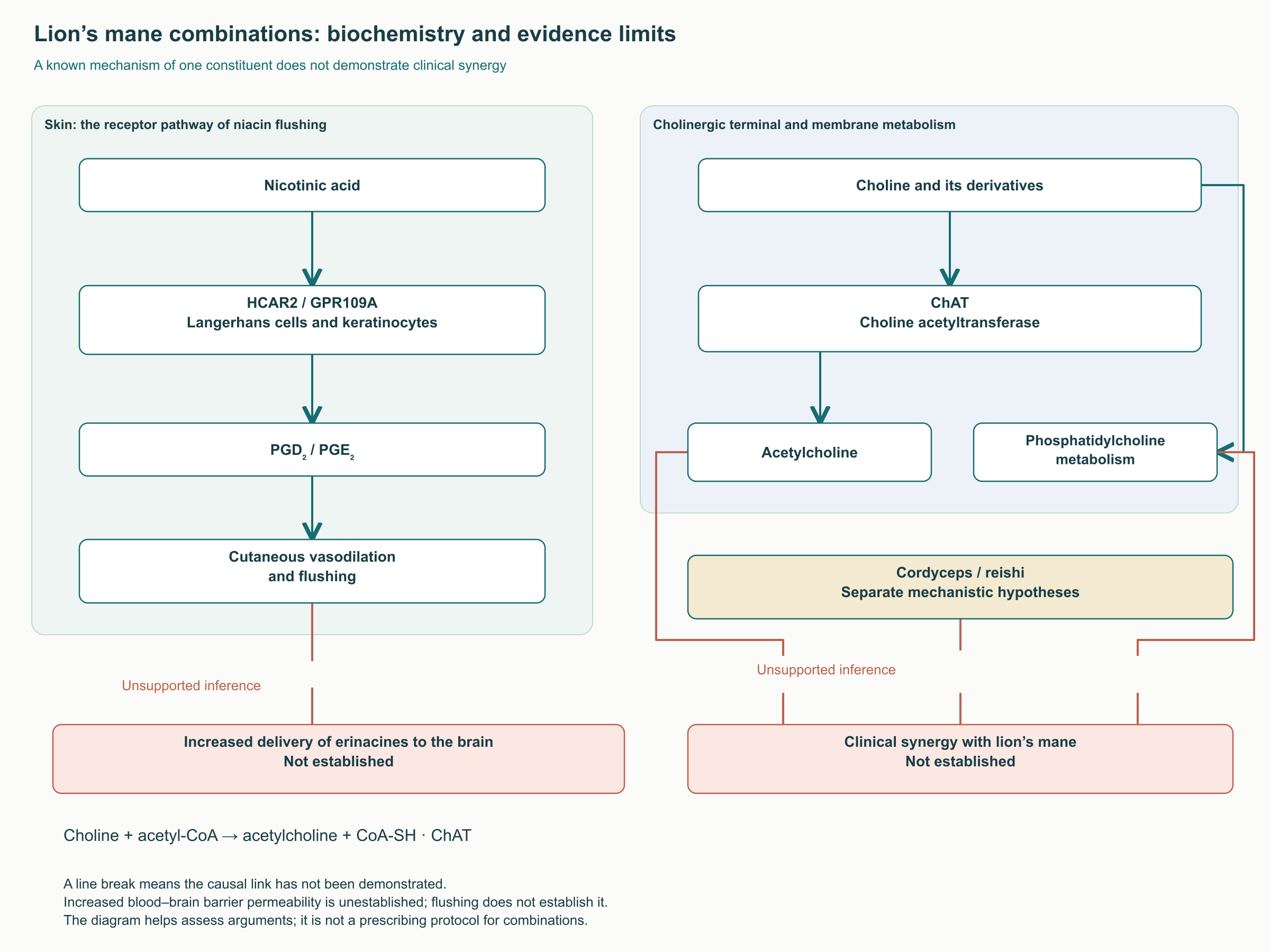
Discussions of the “Stamets stack” often mix different combinations; in public usage, the term is also associated with psilocybin microdosing. Combining lion’s mane with nicotinic acid cannot be considered a clinically established way to increase erinacine delivery. Niacin flushing involves HCAR2/GPR109A, skin cells, and prostanoids including PGD₂ and PGE₂. Research on this response establishes neither increased brain perfusion nor increased blood–brain barrier permeability or erinacine A concentration. Skin redness measures none of these processes.
Choline and its derivatives participate in membrane and acetylcholine metabolism. The biochemical reaction choline + acetyl-CoA → acetylcholine + CoA-SH is catalyzed by choline acetyltransferase, ChAT. NGF’s role in cholinergic biology does not, however, mean that a person taking lion’s mane develops a substrate deficiency that must be corrected with α-GPC or citicoline. Likewise, cordycepin, AMPK signaling, or reishi triterpenes do not establish synergy between mushroom combinations in preserving synapses. Such conclusions require combination studies with clinical outcomes and safety monitoring, not separate publications on individual components.
Practical patient care and safety boundaries
For neurologists, the value of these findings lies in discussing the request of a patient already interested in the supplement accurately. The first task is to define the problem: subjective forgetfulness, objective cognitive impairment, consequences of nerve injury, and depressive disorders require different diagnostic approaches. Deteriorating daily function, rapidly progressing symptoms, or focal neurological deficits cannot be explained by “low NGF,” with assessment postponed for a mushroom course.
Discussion of intake should specify the product, the expected measurable outcome, and the timing of reassessment. For cognitive complaints, changes in daily activity and standardized assessment in the relevant clinical context are more useful than repeated serum BDNF. For pain and neuropathy, injury cause, neurological status, and functional change matter. An unchanged or increased blood neurotrophin does not establish nerve regeneration.
Researchers should measure molecules and processes separately: NGF and pro-NGF, BDNF and pro-BDNF, neurite growth and cell division, myelin markers and functional conduction. Preparation comparisons require batch and composition data. In clinical studies, a prespecified primary outcome and between-group comparison protect against a few chance-positive results emerging from numerous tests. This is particularly important in small pilot studies.
Safety data are limited by the preparation and observation period. Li’s study reported gastrointestinal complaints and skin rash. Long-term use of different extracts, pregnancy, breastfeeding, and childhood have not been studied comparably. Allergic reactions require discontinuation of exposure and clinical assessment; tolerating the edible mushroom does not guarantee tolerating a concentrated supplement.
NOAEL is the level at which no adverse effect was observed in a particular toxicological experiment. Doses up to 3000 mg/kg/day were studied for particular preparations in rats over 28 days; a 90-day experiment with a β-glucan preparation reported a NOAEL of 2000 mg/kg/day. These are not safe human doses and do not establish that all extracts are nontoxic.
Hericenone B inhibited collagen-induced platelet aggregation in laboratory experiments, including experiments with human platelets. This provides grounds to discuss potential relevance during anticoagulant or antiplatelet use, but not a demonstrated supplement interaction with warfarin or direct anticoagulants. A universal “10–14-day” discontinuation period does not follow from these experiments; before surgery, supplements should be assessed with the treating team. Prescribed antithrombotic therapy should not be stopped independently.
Immune effects of mushroom polysaccharides likewise do not justify automatically predicting multiple-sclerosis or systemic-lupus flares through Dectin-1 and Th1. In autoimmune disease or immunosuppression, the practical question concerns the shortage of interaction data and the particular product. It requires individual assessment, not a universal promise of immune enhancement or a universal prohibition based on a cellular diagram.
Lion’s mane remains a substantial subject in pharmacognosy and neurobiology: it contains chemically distinct compounds, produces experimental neurotrophic responses, and has several clinical signals. The most useful professional application is to identify precisely where measured findings end and hypotheses begin, without replacing standard patient care with a promise of neurogenesis.
FAQ
Can lion’s mane be considered an Alzheimer’s disease prevention aid?
No. The enriched-mycelium pilot included patients who already had mild Alzheimer’s disease. It does not establish prevention in healthy people or replacement of treatment.
Which is preferable: mycelium or fruiting body?
They differ in composition and have no universal efficacy hierarchy. Comparison requires identification of the material, quantitative markers, and correspondence to a particular studied product.
Does increased NGF mean new neurons are growing?
No. Neurotrophin production, neurite growth, precursor proliferation, and functional integration of new neurons are different processes.
Should niacin or choline be added?
Evidence of clinical synergy and increased erinacine delivery across the blood–brain barrier is insufficient for these combinations. They do not follow automatically from the neurotrophic hypothesis.
Should BDNF be measured to choose a supplement?
The studies discussed do not establish serum BDNF or pro-BDNF as tools for individual prescribing or monitoring neurogenesis.