
Glycoxidation and AGEs: methylglyoxal, RAGE and mitochondria
The chemistry of distinct AGEs, mitochondrial targets, the ROS–PARP–GAPDH model, the glyoxalase cycle and clinical limits of interventions.
The chemistry of distinct AGEs, mitochondrial targets, the ROS–PARP–GAPDH model, the glyoxalase cycle and clinical limits of interventions.
Mitochondrial proteins continually encounter reactive products of metabolism. Some arise from sugars, others from lipid oxidation or the breakdown of other substrates. When formation exceeds detoxification, chemical protein modification can alter respiration, signaling and the quality of the cellular proteome. Yet glycation, glycoxidation, dicarbonyl stress and advanced glycation end products, or AGEs, describe distinct, overlapping processes. These distinctions matter particularly when a biochemical model is turned into a clinical protocol. [1–4,11]
In water, glucose predominantly exists in cyclic forms. This describes the combined anomers; it does not mean that more than 99% of molecules are specifically β-D-glucopyranose. In the β-anomer, the large substituents of the pyranose ring occupy favorable equatorial positions. Mutarotation nevertheless maintains a small open-chain aldehyde population whose carbonyl carbon can react with nucleophilic protein groups.
Even a small reactive fraction can matter during prolonged exposure. Sugar concentration, amino-group accessibility, pH, time and protein turnover all contribute. Intracellular glycation is not automatically fatal: its consequences depend on the balance among adduct formation, carbonyl detoxification, proteolysis, repair and new protein synthesis. Long-lived collagen and crystallins accumulate modifications differently from rapidly renewed enzymes. [3,4,12]
Glycosylation is enzymatic attachment of carbohydrate groups and supports many protein functions. In addition to the secretory pathway, regulated intracellular O-GlcNAc modification exists. Glycation encompasses nonenzymatic reactions of reducing sugars or derived carbonyls with biomolecules. Glycoxidation is the oxidation-linked portion of glycation chemistry. Consequently, not every AGE is exclusively a glycoxidation product, and AGE formation does not always require the same combination of oxygen and transition metals. [1,2,11]
Glycation can change a side chain's charge, active-site structure, protein interactions or susceptibility to proteolysis. Finding an adduct does not by itself establish functional loss: its location, site occupancy and functional effect must be determined. Cross-linking two chains is a separate outcome of particular reactions, rather than a universal property of all AGEs.
The open-chain aldehyde form of a sugar reacts with an accessible primary amino group, such as a lysine ε-amino group or an N-terminal amino group. A carbinolamine forms first, followed by a reversible Schiff base. This generalized scheme illustrates the chemistry rather than every glucose state in solution:
For aldoses, subsequent rearrangement yields an Amadori product, a ketoamine. The corresponding ketose route is called the Heyns rearrangement. Rates depend on the microenvironment and the fraction of unprotonated amino groups. One reduced pKa cannot be assigned to every lysine, nor one glycation rate constant to every protein. Arginine's guanidino group is particularly important in dicarbonyl reactions, but its chemistry is not simply the primary-amino-group scheme above. [3,11,12]
HbA1c exemplifies early glycation: the N-terminal valine of hemoglobin's β-chain is modified. It is not a terminal AGE. Its time profile reflects the erythrocyte population, whereas glycated serum proteins reflect a shorter exposure window. Separating early products from AGEs is necessary both for laboratory interpretation and for defining an intervention's chemical target.
Sugar autoxidation, traditionally discussed in connection with the Wolff pathway, and oxidative breakdown of early glycation products can generate reactive carbonyls. Fragmentation routes of early intermediates are also discussed in Maillard chemistry under the Namiki mechanism. These names do not mean that every stage is the same oxidative cleavage of an Amadori product. Several competing routes contribute, with their relative importance changing with conditions. [3,11]
Redox-active iron can amplify oxidative chemistry. One component is the Fenton reaction:
However, CML does not arise only from glucose: glyoxal and lipid-oxidation products can also lead to lysine carboxymethylation. Tissue CML therefore does not specifically prove one sugar source or a single reaction sequence.
| Compound | Principal chemical feature | Interpretation |
|---|---|---|
| CML, Nε-carboxymethyllysine | Lysine monoadduct | Not a cross-link; several formation routes exist |
| CEL, Nε-carboxyethyllysine | MGO-associated lysine adduct | Different from MG-H1 and not representative of every MGO modification |
| MG-H1 | Methylglyoxal-derived arginine hydroimidazolone | Changes guanidino-group properties; adduct location determines its effect |
| Pentosidine | Fluorescent lysine–arginine cross-link | Fluorescence does not quantify all AGEs |
| Glucosepane | Nonfluorescent lysine–arginine cross-link | Important in long-lived human collagen; its route differs from pentosidine's |
| Pyrraline | Lysine product associated with 3-deoxyglucosone | A distinct monoadduct class |
| Argpyrimidine | Fluorescent MGO-derived arginine product | Not interchangeable with hydroimidazolones |
| MOLD / MODIC | MGO-derived Lys–Lys / Lys–Arg cross-links | Distinct from glyoxal-derived GOLD / GODIC |
Chemical studies established pentosidine's structure and glucosepane's presence in human dermis; MGO-derived adducts have also been studied in the lens. These findings demonstrate a heterogeneous class rather than identical toxicity among its members. Cross-links can increase matrix stiffness and impede renewal, but a monoadduct should not be described as a universal polymer blocking every channel and enzyme. [13–15]
Methylglyoxal, MGO, has the structure , while glyoxal is . Along with 3-deoxyglucosone, they are reactive dicarbonyls. Aqueous MGO also exists in hydrated states: drawing two carbonyls does not mean that the entire dissolved population always has only that structure. [3,4]
An important MGO source is the side conversion of glyceraldehyde-3-phosphate and dihydroxyacetone phosphate. These triose phosphates arise after aldolase cleavage of fructose-1,6-bisphosphate and interconvert through triosephosphate isomerase. Their chemical decomposition and side reactions produce MGO; its entire production need not be assigned to one obligatory enzyme intermediate. A larger available triose pool can increase the burden, but the fraction of glycolytic flux diverted into MGO depends on the cell and conditions.
Dicarbonyl stress means disruption of the balance between dicarbonyl formation and removal. It is not synonymous with hyperglycemia: glyoxalase activity, the thiol pool, alternative carbonyl reduction and oxidation, renal elimination of products and protein turnover also change. [3,4,32]
Aminoacetone can be oxidized to MGO, including through semicarbazide-sensitive amine oxidase, SSAO/VAP-1, encoded by AOC3. A route from acetone through acetol involving CYP2E1 is also discussed. These pathways do not make every form of ketosis equivalent to clinically meaningful carbonyl injury; flux, concentration and tissue-exposure data are needed.
Transferring the threonine-dehydrogenase route from animals to humans requires particular caution: human TDH is an expressed pseudogene. It therefore cannot automatically be described as a major human source of aminoacetone and MGO. [33]
MGO can modify arginine to form hydroimidazolones, including MG-H1. MG-H2 and MG-H3 are other structural products, rather than three names for one tautomer. Other arginine products include argpyrimidine. Lysine reactions yield CEL and particular cross-links, while cysteine can form reversible thiohemiacetals. Rates, yields and stability differ among products. [3,4,15]
For an individual bimolecular reaction, the initial rate can be represented approximately as:
Here denotes an accessible reacting group, and applies to specified conditions and a particular product. Claiming that MGO always reacts tens of thousands of times faster than glucose obscures dependence on nucleophile, pH and method. Likewise, free plasma MGO, total MGO in an extract and local matrix exposure are different quantities.
Arginine modification can disrupt a salt bridge or substrate binding. This explains potential active-site sensitivity but does not prove that every detected MG-H1 occupies a catalytically essential arginine.
The matrix of a functioning mitochondrion is often more alkaline than the cytosol, influencing nucleophile protonation. For a simple amino group, the Henderson–Hasselbalch approximation gives the unprotonated fraction:
Local protein pKa, site accessibility and organelle state nevertheless vary. High protein density alone does not imply exponential acceleration of glycation: crowding can alter accessibility and diffusion as well. New-protein import, proteases, redox defenses, network dynamics and mitophagy oppose damage accumulation.
Experiments with isolated renal mitochondria demonstrated respiratory sensitivity to added MGO. In another diabetic-rat study, MGO modifications accompanied reduced oxidative phosphorylation, while hydroimidazolone abundance correlated with reduced complex III activity. [16,17]
These are important experimental foundations, but they do not establish a universal clinical injury concentration. Proteomic protein identification, adduct immunoreactivity and causal demonstration of a particular modified residue are distinct levels of evidence. The cited 2005 study should not be used as proof exclusively concerning complex I or a precise shift in its Fe–S-center redox potentials.
In complex I, NADH transfers reducing equivalents to FMN in the NADH-binding module associated with NDUFV1; iron–sulfur centers and ubiquinone participate downstream. NDUFS1 is an important part of the hydrophilic module. N2 transfers single electrons; N1a should not be presented as an obligatory member of one linear chain containing every cluster.
Carbonyl modification can affect NADH binding, assembly, stability and subunit interactions. However, claiming that MG-H1 on NDUFV1/NDUFS1 always increases by a specified factor or shifts a particular center's potential by a fixed amount requires separate mapping and functional validation. Such details cannot be inferred from general inhibition of NADH-linked respiration. [16,17]
One-electron oxygen reduction is represented by:
Its probability depends on redox-center states and conditions; an injured mitochondrion does not necessarily increase ROS production with every substrate combination.
Complex III's Q cycle includes Qo and Qi sites, cytochrome b hemes bL/bH, the Rieske iron–sulfur protein and cytochrome c1. Core proteins UQCRC1 and UQCRC2 help maintain complex structure. Altered interfaces can influence electron transfer and side-reaction probability. However, the cited studies do not establish that an AGE on Core1 necessarily locks Rieske-domain motion for a precisely measured interval. [17]
Cytochrome c interacts with respiratory partners through a surface containing positively charged residues. Carbonyl modification can alter these contacts. Interaction with cardiolipin and altered heme coordination can also influence cytochrome c's peroxidase activity. Yet any AGE cannot be assumed sufficient to disrupt Fe–Met80 coordination and inevitably trigger apoptosis. Lipid oxidation and repair are discussed separately in GPX4 and lipid peroxides.
The ATP-synthase F1 catalytic head contains α- and β-subunits. Rotation and coupling to Fo link ATP synthesis to the proton gradient. An arginine finger contributes to catalytic organization, while OSCP connects stator elements. Their functional importance makes interface disruption a plausible injury target, but does not prove selective glycation of a particular Arg373 or a universal 70–80% activity reduction.
MICOS, including MIC60, and OPA1 help organize cristae. TOM/TIM complexes, including TOM40 and TIM23, import many nuclear-encoded proteins. Injury to these systems could disturb proteostasis and membrane architecture. Establishing that MGO modification of a particular site triggers those changes in a given tissue requires direct evidence. A protein's structural role and its status as a demonstrated carbonyl target are different matters. [3,4,16,17]
| Node | Functional basis for susceptibility | Evidence needed beyond general dysfunction |
|---|---|---|
| Complex I | NADH binding, FMN/Fe–S transfer and assembly | Adduct mapping and site-function testing |
| Complex III | Q cycle and subunit interfaces | Distinguish modification correlation from a blocking mechanism |
| Cytochrome c | Electrostatic interfaces and heme coordination | Test binding, electron transfer and peroxidase activity separately |
| ATP synthase / OSCP | Catalysis, rotation and mechanical coupling | Identify the altered site and its occupancy |
| MICOS / OPA1 / TOM–TIM | Cristae and protein import | Separate direct glycation from a secondary stress response |
In the hyperglycemic-injury model, excess substrate increases carrier reduction and changes mitochondrial energetics. High potential can hinder further electron transfer, but there is no single millivolt threshold beyond which every tissue catastrophically generates ROS. The negative sign of must also be distinguished from the magnitude of polarization. [1,2,8,24]
In an experimental endothelial system, increased mitochondrial superoxide was linked to DNA injury, PARP activation and GAPDH inhibition. MnSOD or UCP1 overexpression prevented several changes, while PARP manipulation was tested separately. This supports a causal sequence in that model, rather than proving an identical origin for every diabetic complication. [7,8]
PARP1 uses NAD+ for ADP-ribosylation. Strong activation changes NAD+ consumption, the DNA-damage response and protein function. Du and colleagues observed GAPDH poly(ADP-ribosyl)ation; assigning the modification to a particular catalytic cysteine without mapping is unjustified. Reduced GAPDH activity increases availability of upstream intermediates and can redistribute metabolic flux. [7]
| Branch | Substrate and mechanism | Interpretation limit |
|---|---|---|
| Polyol pathway | Aldose reductase uses NADPH to reduce glucose to sorbitol; sorbitol dehydrogenase uses NAD+ | NADPH is consumed directly, whereas GSH is affected through redox balance, not as aldose-reductase substrate |
| DAG–PKC | Metabolic changes can increase DAG formation and activate PKC | Isoforms and effects depend on tissue |
| Hexosamine pathway | Fructose-6-phosphate supports UDP-GlcNAc production and regulated O-GlcNAc modification | This is enzymatic modification, not an AGE reaction |
| Dicarbonyl burden | Accumulated triose phosphates increase opportunities for MGO formation | The simultaneous detoxification response matters |
Endothelial cells, podocytes, retinal cells and neural tissues differ in glucose transport and metabolic constraints. GLUT1 is important at the blood–brain barrier, for example, whereas GLUT3 is significant in neurons. Saying that every insulin-independent cell uses one transporter erases that distinction.
Respiratory injury can subsequently amplify metabolic and redox stress, creating feedback. Metabolic memory includes long-lived matrix modifications, epigenetic changes and inflammation. It does not mean that improved glycemic control is useless or that complications inevitably continue despite it. [1,2,24]
RAGE, encoded by AGER, belongs to the immunoglobulin superfamily. It contains extracellular V, C1 and C2 domains, one transmembrane segment and a short cytoplasmic tail. It has no intrinsic catalytic kinase domain. Binding depends on domain surfaces, charge and receptor organization. [9,10,25,31]
Ligands include particular AGE-modified proteins, HMGB1, members of the S100 family and amyloid structures. Not every free AGE adduct automatically becomes an equivalent agonist. Modification structure and density, protein aggregation, experimental conditions and impurity controls matter. Total CML measurement therefore does not describe the entire RAGE-signaling burden.
RAGE's intracellular tail interacts with signaling partners. DIAPH1/mDia1 is linked to small-GTPase signaling; a separate experimental system demonstrated involvement of phosphorylated RAGE, TIRAP and MyD88. [34,35]
Downstream branches include Rac1, NADPH oxidases, MAPK and NF-κB, but they are not necessarily the same obligatory chain in every cell. NOX2 assembly depends on regulatory subunits; NOX4 does not simply repeat the Rac1–p47phox mechanism. p38, ERK and JNK can change inflammatory and stress responses. IKK, altered IκB and NF-κB regulate cytokines and adhesion molecules, including IL-6, TNF, MCP-1, ICAM-1 and VCAM-1. [9,25,27,31]
Increased AGER expression and inflammatory mediators can sustain feedback. However, the receptor is not entirely unregulated, and its actions are not confined to disease. Crosstalk among cellular ROS, mitochondrial function, calcium and inflammation depends on tissue, exposure and ligands.
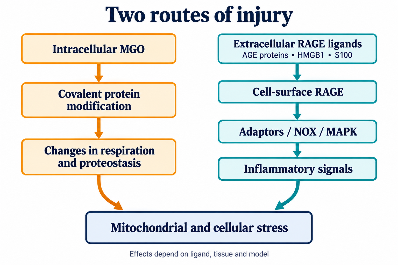
Direct covalent protein modification and RAGE-mediated signaling are interacting but distinct mechanisms. Arrows show a possible sequence under studied conditions, rather than an identical outcome for every person.
Kang and colleagues detected mitochondrial RAGE in a tumor model and linked HMGB1/RAGE to enhanced complex I activity and ATP. The study does not establish a universal pore-forming RAGE–VDAC–ANT–CypD complex. [36]
Even receptor detection in a mitochondrial fraction does not establish its precise topology in every tissue. Protease protection in that study distinguished the investigated population from simple outer-membrane localization; it cannot be converted into an established TOM/TIM-import scheme with integration into both membranes. Tumor bioenergetics also differs from inevitable apoptosis or pyroptosis in diabetes.
RAGE-associated inflammation can affect mitochondria through signaling without requiring a universal mitochondrial localization. Sustained inner-membrane permeability is discussed separately in mPTP and cyclophilin D, where the pore's molecular identity likewise requires careful interpretation.
Glutathione reacts reversibly and nonenzymatically with MGO to form a hemithioacetal. GLO1 converts it to S-D-lactoylglutathione, and GLO2 hydrolyzes the thioester, producing D-lactate and regenerating GSH. [3,32]
Overall, MGO is converted to D-lactate with water participation. GSH is a regenerated participant, rather than a stoichiometrically consumed substrate in the completed cycle. This does not mean that the whole cellular thiol pool always remains intact: oxidation, conjugation, synthesis and export occur in parallel.
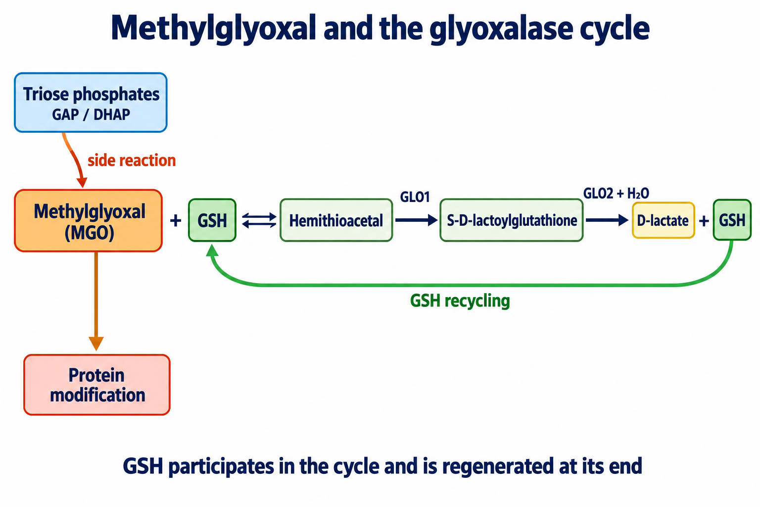
GLO1 acts on the hemithioacetal, and GLO2 regenerates GSH. MGO formation and removal are shown as separate pathways.
Human GLO1 is a metal-dependent enzyme, commonly discussed as a zinc-containing dimer; metal coordination and acid–base catalysis enable substrate rearrangement. GLO2/HAGH belongs to the metal-dependent hydrolases. One invariant metal-center composition cannot be assigned to every enzyme preparation, and constants measured with hemithioacetal cannot simply be applied to free MGO. [3,32]
For a simplified hemithioacetal-formation equilibrium:
This description requires explicit definitions of the measured MGO forms and conditions. Actual cycle flux depends on available GSH, both enzymes and other removal routes. and depend on the preparation and assay; one pair of values cannot establish universally diffusion-limited organismal protection.
Lower GSH can reduce hemithioacetal formation and weaken redox defenses. But no fixed GSH concentration at which every mitochondrion experiences catastrophe has been established. Glutathione reductase uses NADPH to reduce GSSG; new GSH is synthesized sequentially by glutamate–cysteine ligase and glutathione synthetase. NAC can supply cysteine rather than directly activate the final step. D-lactate is subsequently metabolized, including through LDHD, and should not be called harmless at every concentration. [3,32]
Nrf2 regulates antioxidant and detoxification programs. Xue and colleagues demonstrated Nrf2-dependent GLO1 control through a relevant regulatory element. [5]
KEAP1 controls Nrf2 availability, and electrophilic compounds can alter this response. Inducing one gene nevertheless does not guarantee identical cycle activity in every tissue. Age, inflammation, epigenetic changes and microRNAs can influence expression, but a uniform 40–60% GLO1 decrease in all older adults has no general foundation. Compensatory carbonyl reductases and other enzymes also matter when assessing GLO1 deficiency.
Diabetic renal disease involves podocyte, endothelial and tubular injury, altered matrix and inflammation. Modification of collagen IV and laminin can change basement-membrane properties. Signaling changes affect the actin apparatus and podocyte proteins, including synaptopodin. Yet albuminuria cannot be explained solely by loss of negative charge or one AGE modification. Hemodynamics, the filtration barrier, mesangial responses and fibrosis interact. [17,23,28]
Renal impairment also changes elimination of glycation products. Increased concentrations can therefore reflect both formation and reduced clearance; cross-sectional association does not determine causal direction. [26,28]
Retinal pericytes, endothelial cells and neurons have different functions and sensitivities. Matrix glycation and altered integrin contacts, inflammation, barrier dysfunction and VEGF activity are considered in retinopathy mechanisms. Pericyte loss, edema and neovascularization are not reducible to one MGO–RAGE event. Experimental retinopathy prevention in the benfotiamine study does not establish prevention of every human microvascular complication. [6,24,27]
Polyol flux, redox balance, vascular supply and axonal metabolism can jointly impair nerve function. Glycation of neurofilaments, tubulin and motor interfaces is discussed as a possible contribution to axonal-transport disruption. Kinesin, dynein and neurotrophic-signal transport matter, but not every disturbance is proven to result directly from a mapped AGE residue. Sensory loss and pain also have different mechanisms. [18,20,24,30]
Cross-links in long-lived collagen I/III can influence vascular-wall mechanics. Pulse-wave velocity describes stiffness, rather than specifically measuring AGE burden. Matrix, titin, its isoforms and phosphorylation influence passive myocardial stiffness; every case of diastolic dysfunction cannot be treated as proof of titin glycation. [1,12–14]
Modification of ApoB and other LDL components can alter lipoprotein interactions with receptors and macrophages. These processes overlap with oxidation and inflammation. AGE-LDL should not be depicted as one chemically uniform particle behaving identically in all models. [2,25,27]
Carbonyl stress can affect neuronal energy metabolism, as tested in cellular MGO-exposure models. Glycation of amyloid- and tau-related systems is discussed alongside aggregation and inflammation. A fixed hundredfold acceleration of Aβ aggregation or identical blockade of every tau-dephosphorylation mechanism cannot be generalized across tissues. Microglial RAGE responses do not prove that AGEs are the sole cause of Alzheimer's disease. [25,30,31]
Aminoguanidine, or pimagedine, can react with dicarbonyls but also has other chemical and pharmacological actions. ACTION I did not demonstrate statistically significant benefit on its primary renal endpoint. [23]
This differs from saying that every trial was stopped early. The ACTION II paper in the original bibliography describes design and baseline characteristics, rather than substituting for an outcome report. Potential adverse actions, immune reactions and interactions with other pathways constrain translation of laboratory aminoguanidine efficacy into practice. [22,23]
Pyridoxamine, a vitamin B6 form, has been studied as an inhibitor of carbonyl and oxidative conversions. The subsequent randomized Pyridorin trial did not confirm primary-endpoint improvement across the studied population. [37]
That is fundamentally different from attributing lack of use solely to regulatory barriers. A favorable subgroup or early-phase finding requires independent confirmation.
Carnosine, β-alanyl-L-histidine, can react with carbonyls. Oral use nevertheless depends on absorption, tissue exposure and hydrolysis by serum carnosinase CNDP1. Associations between CNDP1 variants and complication risk do not prove that a particular oral amount saturates the enzyme and prevents nephropathy. Test-tube trapping does not establish a dose that competes effectively with protein nucleophiles in tissue. [19]
Benfotiamine is a thiamine precursor. Metabolic conversion can increase availability of thiamine diphosphate, a transketolase cofactor. This is a cofactor mechanism, rather than direct, universal allosteric opening of a metabolic shunt. In the nonoxidative pentose-phosphate pathway, transketolase works with other enzymes, including transaldolase. An appropriate overall carbon-redistribution scheme is:
Both sides retain 15 carbon atoms. A direct F6P + GAP → ribose-5-phosphate + sedoheptulose-7-phosphate equation cannot be used as a balanced two-substrate scheme. Flux direction depends on cellular state, rather than supplement presence alone.
Hammes and colleagues demonstrated protection in experimental retinopathy. Human BEDIP and BENDIP studies mainly investigated short-term neuropathy measures, requiring distinctions among analyses and endpoints. BEDIP was a three-week pilot, rather than a 12-week, 130-participant study. [6,18,38]
The longer BOND trial did not confirm benfotiamine benefit on the primary corneal-nerve-fiber-length measure. [39]
A cellular transketolase effect and a short symptom signal therefore cannot establish nerve regeneration or nephropathy and retinopathy prevention in every population.
Sulforaphane and other electrophilic compounds can influence KEAP1–Nrf2 and detoxification programs. This explains research interest but does not prescribe an identical GLO1-induction percentage for every formulation or dose. [5]
In the tRES–HESP crossover trial, 29 participants completed assessment; 90 mg trans-resveratrol and 120 mg hesperetin were studied daily for eight weeks. A post hoc BMI >27.5 kg/m² subgroup showed a reported 27% GLO1 increase and 37% MGO decrease, with improved OGIS and vascular measures. These are group- and endpoint-specific findings, rather than universal dose effects. [21]
The study does not prove prevention of vascular complications. A pterostilbene combination is a different combination: substituting it for hesperetin while retaining the tRES–HESP citation is invalid. Discussion of SIRT1, Nrf2, bioavailability and polyphenol effects must account for the compound, exposure and model, rather than presenting guaranteed general neuroprotection.
α-Lipoic acid and its reduced form participate in redox chemistry; lipoyl groups within enzyme complexes have a separate cofactor role. SYDNEY 2 studied the short-term effect of oral α-lipoic acid on diabetic-polyneuropathy symptoms. [20]
A symptom endpoint does not prove reduction of every AGE or axonal regeneration. The trial cannot automatically be renamed as testing only the R-enantiomer or a particular salt. Supporting GSH depends on synthesis, cysteine availability and NADPH-dependent reduction; lipoic acid is not a guaranteed replacement for glutathione reductase.
NAC is one approach to increasing cysteine availability. This is a biochemically plausible way to support GSH synthesis, but the effectiveness of combining NAC, carnosine, benfotiamine, polyphenols and lipoic acid specifically against AGE complications must be demonstrated in a combination study. Adding individual mechanisms does not establish clinical synergy, dosing times or an optimal dose.
Soluble RAGE includes products of extracellular-domain shedding, involving proteases, and secreted alternative-splice variants. esRAGE is part of this diversity rather than a synonym for every measured sRAGE population. Ligand binding attracts interest as a decoy mechanism, but circulating concentrations also depend on production, inflammation and clearance. [25,31]
Azeliragon/TTP488 was studied as a RAGE antagonist. STEADFAST parts A and B failed their co-primary endpoints. This does not justify its use for mitochondrial protection. [40]
Cleavage of pre-existing cross-links was investigated, for example, by Vasan and colleagues. Those chemical findings do not prove that every proposed AGE breaker, including historically discussed alagebrium, cleaves all major human-collagen cross-links. Experimental model cross-links must particularly be distinguished from glucosepane in long-lived tissue. [14,29]
| Intervention level | Examples | What needs confirmation |
|---|---|---|
| Reduce substrate burden | Metabolic-state management, transketolase-flux research | Complication changes, beyond laboratory flux |
| Trap carbonyls | Aminoguanidine, pyridoxamine, carnosine | Achievable tissue exposure, selectivity and clinical benefit |
| Increase MGO removal | GLO1 induction, adequate thiol-cycle function | Coordinated GLO1/GLO2 action and changes in burden and outcomes |
| Support redox balance | Approaches to GSH synthesis and regeneration | Functional effects in the relevant tissue |
| Modify RAGE signaling | Soluble forms, experimental antagonists | Ligand specificity, benefit and adverse effects |
| Address existing cross-links | Experimental AGE-cross-link breakers | Reaction with relevant human cross-links and altered tissue function |
This matrix describes research levels. It does not establish a multilayer therapeutic stack or mandatory premeal intervals. Medication selection and interaction assessment in diabetes depend on the clinical situation; research mechanisms do not replace established complication treatment.
| Marker | Principal meaning | Limitations |
|---|---|---|
| HbA1c | Hemoglobin glycation and integrated glycemic exposure | Influenced by erythrocyte turnover, hemoglobinopathies and assay system |
| Fructosamine / glycated albumin | Shorter serum-protein exposure | Influenced by protein synthesis, loss and turnover |
| MGO | Dicarbonyl burden under specified sample preparation | Postcollection formation, hydration and derivatization matter |
| CML / CEL / MG-H1 | Particular adduct classes | Free and protein-bound forms differ; clearance matters |
| Pentosidine | One fluorescent product | Does not quantify nonfluorescent glucosepane or all AGEs |
| sRAGE / esRAGE | Soluble receptor populations | Renal function and inflammation complicate interpretation as a protective reserve |
| GSH / GSSG | Thiol and redox context of the sampled material | Sensitive to processing oxidation; blood is not the matrix |
Chemically defined adducts can be assessed with LC–MS/MS, while suitable chromatography is used for particular fluorescent compounds. Immunoassays require knowledge of antibody specificity and reporting conventions. MGO measurement depends on rapid sample stabilization, derivatization conditions, internal standards and control of artificial analyte formation. [3,4,15,26,32]
Results from different methods cannot be combined into one universal dicarbonyl-crisis threshold. An MGO value, sRAGE cutoff or GSH/GSSG ratio without the material and method does not automatically justify a supplement combination. HbA1c has established clinical applications, whereas many AGE panels remain research measures. Diagnosis and HbA1c interpretation require clinical context, particularly when erythrocyte turnover is altered.
Glycoxidation links carbohydrate and oxidative chemistry but does not encompass all AGEs. Methylglyoxal connects metabolic side reactions with arginine and other group modifications, while the glyoxalase cycle limits the burden. Mitochondrial protein interfaces, respiration and proteostasis can change, and cell-surface RAGE adds a separate inflammatory-signaling branch. Identifying an adduct's molecular structure, proving the function of a specific site and improving a clinical outcome remain different tasks.
Keeping these distinctions allows a substantive discussion of mitochondrial protection: retain testable reactions and model results without promising identical outcomes across AGE classes, tissues and drug combinations.
This narrative scientific review was prepared on October 8, 2026. The complete original manuscript was read and its bibliography preserved. Primary papers were checked selectively for specific conflicts involving the Rosca mitochondrial studies, RAGE adaptors and mitochondrial localization, human TDH and clinical-trial attribution. Chemical reactions, model observations and clinical endpoints were distinguished; unconfirmed universal thresholds, precise residue effects and therapeutic synergy were not presented as established. This is neither a systematic review nor an independently peer-reviewed assessment; access to some complete papers was limited.
Glycoxidation includes oxidation-linked transformations associated with glycation. Glycation is broader, and different AGEs arise through different routes.
No. CML, CEL and MG-H1 are examples of monoadducts. Pentosidine and glucosepane are cross-links and differ from each other.
It arises in part from triose phosphates and can modify protein groups. Its burden depends on formation, detoxification activity and protein turnover.
It participates in hemithioacetal formation and is regenerated by GLO2. Parallel cellular reactions can nevertheless change the overall GSH pool.
No. The primary tumor model linked mitRAGE to increased complex I activity and ATP. It does not establish a universal mPTP-opening complex.
Combining plausible mechanisms does not prove clinical synergy. A combination needs studies of tissue exposure, endpoints and adverse effects.