Dendritic cells and TLRs: how recognition becomes an immune response
Dendritic cells: TLRs, MHC-I/MHC-II processing, cross-presentation, metabolism and tolerance. ZOE-50, IMPACT and the negative ACCESS result.
Antigen and context convey different messages
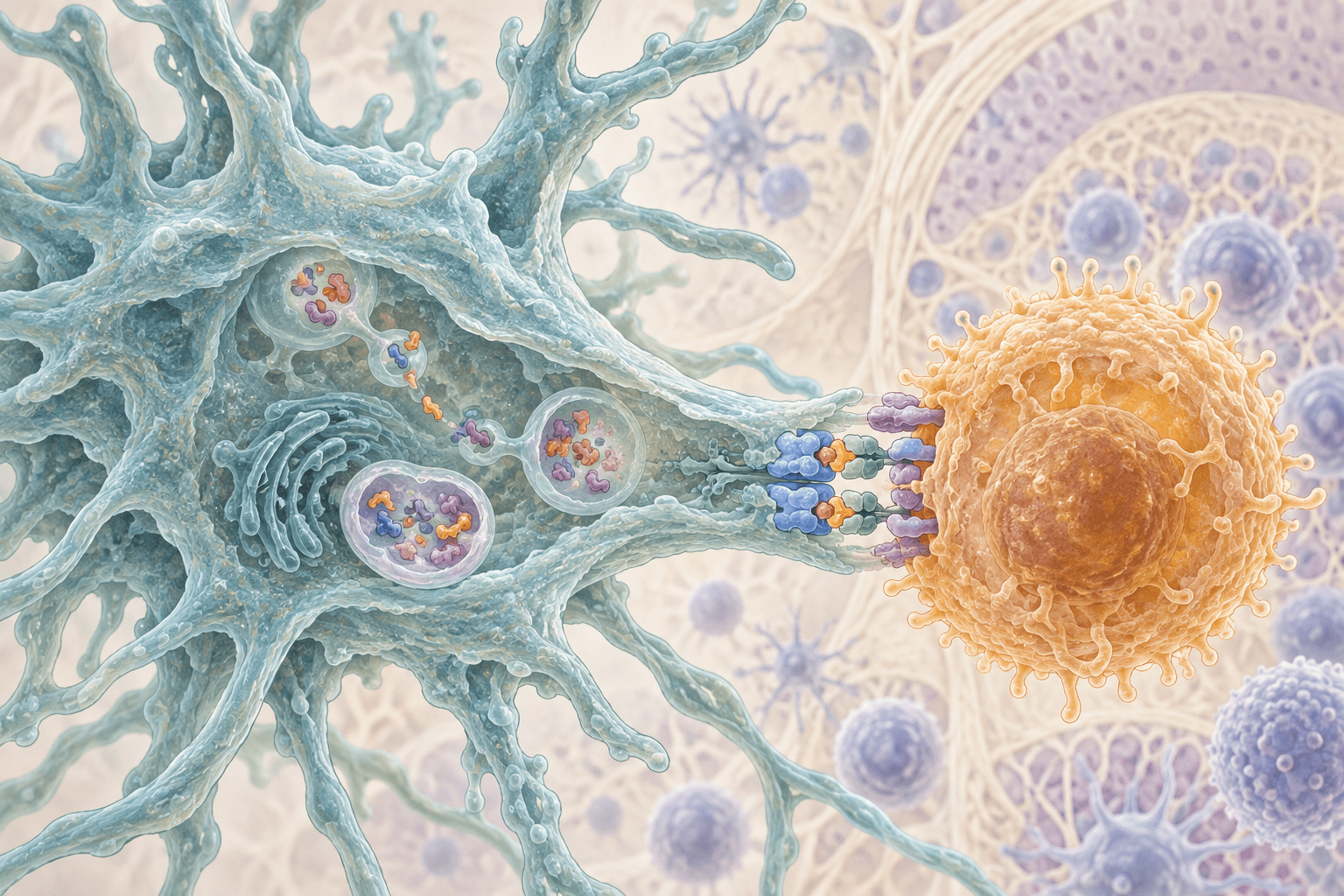
A dendritic cell performs two connected but distinct tasks: it shows a T lymphocyte a fragment of a protein and communicates the circumstances in which that protein was encountered. The peptide–MHC complex determines antigen specificity, while costimulation, cytokines and the tissue environment influence activation, differentiation or tolerance. Toll-like receptors, TLRs, contribute to the second task. They neither replace the T-cell receptor nor provide the dendritic cell's only source of information.
This distinction explains why a protein can elicit protection in a vaccine yet be tolerated without inflammation in food. It also explains therapeutic limitations: strengthening one signaling pathway cannot guarantee protection, and lowering a laboratory inflammatory marker cannot establish clinical benefit. Normal function involves an appropriately scaled response followed by resolution, rather than continuous maximal cell stimulation.
Cell origins and the limits of classification
From progenitors to tissue specialization
Following Steinman and Cohn's description of dendritic cells in 1973, these cells came to be recognized as specialized initiators of primary T-cell responses. Modern classification uses origin, transcriptional programs and function, rather than branching shape or CD11c expression alone. No single marker reliably distinguishes every dendritic cell from monocytes, macrophages and activated cells of other types.
FLT3 and its ligand FLT3L support conventional dendritic-cell development. The receptor tyrosine kinase connects a growth signal to protein phosphorylation and survival and differentiation programs. However, the familiar sequence from a common myeloid progenitor through a common DC progenitor to all mature DCs is a useful model, not a universal route followed by every cell. Clonal lineage and single-cell studies refine contributions from different progenitors; pDC origin is particularly poorly represented by one rigid hierarchy.
cDC1, cDC2, pDCs and monocyte-derived cells
cDC1 cells are associated with IRF8, BATF3 and ID2 programs; human cells are characterized using combinations of XCR1, CLEC9A and CD141. CLEC9A contributes to sensing material from damaged cells, while XCR1 supports cellular interactions and migration. cDC1 cells are especially effective at cross-presenting external antigens through MHC-I and initiating CD8+ responses. This is a specialization, not an exclusive entitlement: other antigen-presenting cells can cross-present under particular conditions.
Human cDC2 cells are usually identified using CD1c together with additional features that distinguish them from monocyte-derived cells. IRF4 and other regulators contribute to development and function, but cDC2 populations are heterogeneous. They support MHC-II presentation and CD4+ responses, including different helper programs. They cannot be assigned exclusively to Th2 or Th17: tissue, antigen, cytokines and cell state determine response direction.
pDCs have a TCF4-associated program, express CD123 and CD303, and can secrete abundant type I interferons following particular stimuli. Endosomal TLR7 and TLR9 and the transcription factor IRF7 are particularly important for sensing. pDCs can also present antigen, but both presentation and interferon output are context dependent. Universal claims of a thousand-fold higher interferon production do not describe every population or state.
Monocyte-derived DCs arise in inflammatory environments and are widely used in cell-therapy research. They are not automatically equivalent to tissue cDC1 or cDC2 cells. Products with similar surface markers can differ in origin, metabolism and migration; findings from cultured monocyte-derived cells require separate verification in primary human cells.
Where TLRs encounter ligands
Architecture and surface receptors
TLRs are single-pass membrane proteins with leucine-rich repeats in their ligand-binding region and a cytoplasmic TIR domain. An appropriate ligand changes receptor-complex geometry and brings signaling domains together. A TLR itself is not a protein kinase: adapters and enzymes assembled on the cytoplasmic face of the membrane transmit the signal.
At the surface, TLR2 pairs with TLR1 or TLR6 to recognize different lipopeptides. TLR5 recognizes flagellin. TLR4 works with MD-2; LBP and CD14 assist delivery of lipopolysaccharide to the complex. Structural studies explain how accommodation of a lipid ligand in MD-2 promotes active-complex formation. This physiological model does not mean that every inflammatory substance binds directly to TLR4.
Endosomes and nucleic acids
TLR3 recognizes double-stranded RNA, TLR7 and TLR8 recognize RNA degradation products, and TLR9 recognizes particular DNA motifs. Delivery to endosomal compartments involving UNC93B1, an acidic environment and receptor proteolysis help restrict responses spatially. These receptors can also respond to self nucleic acids when normal localization and clearance are disrupted. Distinguishing self from nonself is not an infallible reading of a single chemical feature.
TLR expression differs between species and cell populations. TLR7/9 are particularly important in human pDCs, and TLR8 in several human myeloid populations; mouse findings cannot be transferred mechanically to humans. Contextual regulatory effects have been described for TLR10, but its physiological ligands and universal function remain less settled than those of better-characterized receptors. Calling it the only inhibitory TLR conceals this uncertainty.
Tissue damage and quality of evidence
A PAMP is a characteristic microbial structure, while a DAMP is a molecular damage signal. HMGB1 activity depends on redox state, interacting proteins and receptors; different oxidized forms are not interchangeable. For several proposed endogenous TLR ligands, microbial contamination, isolation methods and experimental models must be considered. Hsp70, hyaluronan, saturated fatty acids and cholesterol crystals therefore cannot simply be listed as established direct agonists of one receptor.
Crystalline particles and metabolic changes can cause inflammation through organelle damage and other sensors. TLR signaling may prepare cells for these responses, but preparation and direct damage recognition are different stages. This distinction prevents the incorrect inference that association with inflammation establishes direct TLR binding.
Signaling from membrane to nucleus
MyD88, ubiquitin and kinases
Most well-characterized TLRs use MyD88; TLR3 is an important exception, while TLR4 uses two adapter routes. TIRAP helps organize MyD88 signaling from TLR2 and TLR4 at the membrane. Death domains of MyD88, IRAK4 and IRAK1/2 form the myddosome signaling assembly. A crystallographic model of MyD88–IRAK4–IRAK2 death domains showed a 6:4:4 stoichiometry: this describes a particular structural preparation, not an invariant size of every assembly in a living cell.
IRAK4 and related kinases use ATP and Mg²⁺ to transfer phosphate groups to proteins. TRAF6, together with the ubiquitin-conjugating machinery, contributes to signaling polyubiquitin chains. K63-linked chains provide assembly platforms, a different use of ubiquitination from K48-linked targeting for proteasomal degradation. TAK1 and regulatory TAB proteins connect these platforms to IKK and MAP kinases.
Phosphorylation of IκB promotes ubiquitination and destruction, allowing NF-κB access to the nucleus. MAP kinases, including p38 and JNK, contribute to AP-1 activation. The result depends on transcription-factor cooperation, chromatin accessibility and feedback. IL-12p70 contains p35 and p40, whereas IL-23 contains p19 and the same p40; measuring p40 does not establish exclusive production of IL-12.
TRIF and the interferon response
TLR3 uses TRIF, while endosomal TLR4 recruits TRIF with TRAM. This route connects receptor activation to TBK1 and IKKε, IRF3 phosphorylation and IFN-β induction. TRIF can also activate inflammatory branches, so the division of MyD88 into inflammation alone and TRIF into protective interferon alone is too simple. In pDCs, TLR7/9 can strongly activate IRF7 and interferon production through MyD88.
Secreted type I interferon binds IFNAR and activates STAT1, STAT2 and IRF9 through JAK1/TYK2, forming ISGF3. This changes interferon-stimulated gene expression in the cell and its neighbors. The response is useful during infection, but prolonged activation can contribute to autoimmune damage. Timing, localization and duration determine its meaning, rather than the cytokine's name alone.
How a protein becomes peptide–MHC
MHC-II: endolysosomal processing
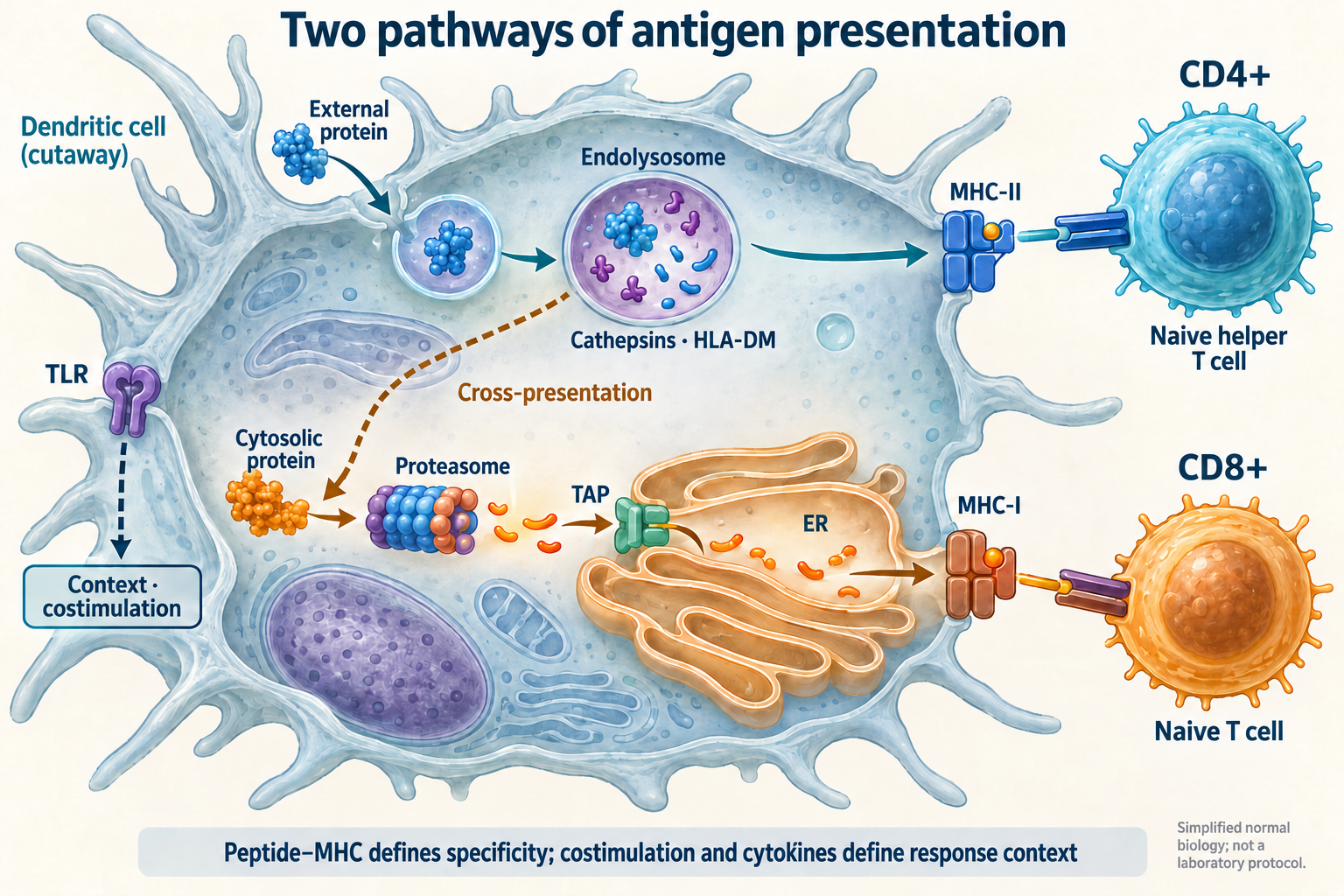
External proteins enter through receptor-mediated endocytosis, phagocytosis or macropinocytosis. Endosomal maturation involves V-ATPase: ATP hydrolysis drives proton transport into the lumen. Early endosomes at approximately pH 6–6.5 lead toward more acidic late endosomes and lysosomes, often around pH 4.5–5.5. These are approximate, cell- and stage-dependent values, not universal settings for all antigen compartments.
Cathepsins S, L and B are cysteine proteases, while cathepsin D is an aspartyl protease; the active enzyme repertoire varies with cell type. GILT facilitates disulfide reduction and accessibility for processing. Proteolysis must produce suitable epitopes rather than necessarily destroy the antigen completely. MHC-II has an open-ended peptide-binding groove, frequently containing peptides of roughly 13–25 amino acids, although longer sequences are possible.
In the endoplasmic reticulum, MHC-II α and β chains assemble with the invariant chain CD74, which protects the groove and directs the complex toward endosomes. Its proteolysis leaves CLIP. HLA-DM catalyzes exchange of CLIP for antigenic peptides and repertoire editing in the specialized MIIC compartment. HLA-DM is neither a protease nor an ATP-driven pump: it acts through MHC-II conformational changes. Loaded complexes reach the surface for recognition by CD4+ T cells.
MHC-I: cytosol, TAP and the endoplasmic reticulum
The ordinary MHC-I pathway displays peptides from proteins synthesized within the cell. Proteasomes cleave proteins using catalytic N-terminal threonines of β subunits; an ATP-dependent regulatory component helps recognize and unfold certain substrates. Interferons alter catalytic-subunit composition to include PSMB9/LMP2, PSMB10/MECL-1 and PSMB8/LMP7. Immunoproteasomes change the peptide repertoire, but do not guarantee cleavage of every protein into one fixed length.
The TAP1/TAP2 heterodimer transports suitable cytosolic peptides into the ER using ATP. ERAP1 and ERAP2 are zinc-dependent aminopeptidases that can trim N-terminal residues; trimming can create an epitope or destroy it by excessive cleavage. The MHC-I heavy chain and β₂-microglobulin interact with a loading complex containing tapasin, calreticulin and ERp57. ERp57 contributes to thiol–disulfide control of protein assembly.
MHC-I grooves often contain peptides of 8–10 amino acids, but length and acceptable anchor residues depend on the HLA allele. After stable-peptide selection, complexes pass through the Golgi to the surface for CD8+ recognition. HLA polymorphism means that two people may present different peptides from the same protein. Chemical identity of an antigen therefore does not guarantee identical T-cell responses.
Cross-presentation and unresolved questions
Cross-presentation loads an external antigen onto MHC-I. In cytosolic variants, some captured protein accesses the cytosol, undergoes proteasomal processing and uses TAP-dependent steps. In vacuolar variants, processing and loading occur in an endosomal environment and may not require proteasomes or TAP. Transfer of preformed peptide–MHC complexes between cells, termed cross-dressing, is a separate process rather than independent antigen processing.
NOX2 uses NADPH to transfer electrons to oxygen, generating reactive oxygen species. In particular DC models this limits phagosomal acidification and proteolysis, preserving antigen for cross-presentation. Other studies found effects on proteases that are not explained by pH alone. A cDC1 phagosome is therefore not an obligatory reservoir held permanently at pH 7.2–7.5. Oxidation, acidification and proteolysis change over time.
The role of Sec61 and the precise route of protein escape from endosomes to cytosol remain debated; one pore model cannot be presented as a universally established mechanism. Cross-presentation also differs from cross-priming: the first describes complex formation, the second productive activation of a naive CD8+ cell. Without an appropriate context, presentation may produce tolerance rather than a cytotoxic response.
Maturation, migration and meeting a T cell
CCR7 and surface-complex stability
Maturation changes costimulator expression, cytokine secretion, MHC trafficking and migration. Reduced MARCH1 activity, a ubiquitin ligase involved in MHC-II and CD86 turnover, helps preserve these molecules at the surface. Complex lifetime nevertheless depends on peptide, cell and conditions; a universal lifetime above 100 hours is too broad a claim. Antigen uptake need not cease completely after maturation either.
CCR7 helps DCs follow CCL19 and CCL21 signals toward lymphatic vessels and lymph-node T-cell zones. Cytoskeleton and adhesion adapt to three-dimensional tissues, rather than just a culture-dish surface. Migration can support tolerance without marked inflammation. CCR7 or a mature marker therefore does not independently establish an immunogenic outcome.
Three signals and a dynamic synapse
The first signal is TCR recognition of peptide–MHC, with CD4 or CD8 helping organize contact. The second involves costimulation, including CD28–CD80/CD86; the third is the cytokine environment. DC CD40 and activated-helper-cell CD40L can reinforce licensing of a CD8+ response. Inhibitory interactions, including CTLA-4 and PD-1, limit activation, and cannot be described by one universal affinity ratio.
LFA-1 and ICAM-1 support contact, while actin structures and receptor microclusters move and reshape signaling. The classic central and peripheral SMAC diagram is useful, but DC contacts are often dynamic and do not form perfect concentric rings. Local exclusion of large phosphatases and Lck/ZAP-70 activity support TCR signaling. The connection with differentiation is explored further in the article on Th1, Th2, Th17 and Treg programs.
The metabolic cost of activation and tolerance
ATP, reducing equivalents and species differences
In experimental models, early DC activation increases glucose flux through glycolysis; TBK1/IKKε and Akt link receptor signals to metabolic changes and hexokinase localization. The hexokinase reaction, glucose + ATP → glucose-6-phosphate + ADP, requires Mg²⁺. The pentose phosphate pathway generates NADPH for reductive reactions and lipid synthesis. Mitochondrial citrate export and ATP-citrate lyase provide cytosolic acetyl-CoA for synthesis of ER and Golgi membranes.
A later reliance on glycolysis and nitric-oxide-mediated respiratory inhibition are well demonstrated in some mouse inflammatory DC models. iNOS uses arginine, oxygen and NADPH and requires heme, tetrahydrobiopterin, FAD and FMN. NO affects mitochondrial enzymes, including cytochrome-c oxidase. However, NOS2 responses differ in human DCs and primary populations: the claim that every cell completely shuts down respiration after 12 hours is incorrect. Metabolic features alone do not classify immunogenicity.
Tolerogenic states and the intestinal barrier
A tolerogenic DC is a functional state, not one immutable lineage obligatorily lacking CD80/CD86. Presentation of self or food antigens without appropriate costimulation may support anergy, removal of reactive clones or regulatory programs. IL-10, TGF-β and inhibitory interactions contribute; none guarantees complete suppression of every inflammatory response. Tolerance preserves infection defense and is not synonymous with globally switching immunity off.
IDO1 is a heme-containing dioxygenase that converts tryptophan and oxygen into N-formylkynurenine, followed by kynurenine formation. Changes in amino-acid availability and AhR signals can affect T cells. A single tryptophan concentration cannot be declared a universal arrest threshold for every lymphocyte, nor can every kynurenine effect be equated with direct, inevitable FOXP3 induction. Metabolic conditions and interacting cells determine the result.
Intestinal antigens arrive through M cells and other epithelial transport routes; direct dendritic sampling depends on anatomical region and model. Retinaldehyde dehydrogenases, including ALDH1A2, oxidize retinal to retinoic acid using NAD⁺. Retinoids and local signals support intestinal homing and some regulatory responses. These mechanisms do not establish that high vitamin A or D doses safely generate tolerogenic DCs in humans.
Sensors work together
TLRs interact with lectin receptors and intracellular sensing systems. Dectin-1 and other CLRs can signal through Syk and CARD9-associated complexes. NLRP3 responses involve preparation, including altered expression of inflammatory components, and subsequent assembly with caspase-1 activation. These stages vary between cells; IL-18 does not always require the same transcriptional induction as IL-1β.
Negative feedback includes A20/TNFAIP3, IRAK3 and DUSP phosphatases. Ubiquitin editing, kinase restraint and chromatin changes reduce responses during sustained stimulation. Aging alters population abundance, tissue signals and functional reserve, but does not establish that every DC ages identically. Blood is not an exact representation of the cell composition of intestine, skin or tumor.
What clinical trials demonstrated
An adjuvanted vaccine: ZOE-50
The double-blind, placebo-controlled ZOE-50 trial evaluated a recombinant shingles vaccine in adults aged at least 50 years. The analysis included 15,411 participants receiving two intramuscular doses two months apart. The vaccine contained 50 μg glycoprotein E with AS01B, combining MPL and QS-21 in a liposomal formulation. The primary objective was prevention of confirmed herpes zoster; mean follow-up was 3.2 years.
Herpes zoster occurred in 6 vaccine recipients and 210 placebo recipients, giving 97.2% efficacy, 95% CI 93.7–99.0. Marked local or systemic reactions were more frequent after vaccination. This demonstrates benefit of the complete vaccine in the studied population, not isolated MPL or selective cDC1 reactivation. It was not a direct comparison with a live vaccine. Older populations and protection over longer periods require their own evidence, rather than extrapolation of one number to every time point.
Cellular immunotherapy: IMPACT
IMPACT was a double-blind randomized phase III trial in 512 men with asymptomatic or minimally symptomatic metastatic castration-resistant prostate cancer. Participants received sipuleucel-T or control in a 2:1 ratio, 341 versus 171. Three autologous cellular infusions were given intravenously two weeks apart; overall survival was the primary endpoint. The product contains antigen-presenting and other cells, rather than a purified homogeneous cDC1 population; its dose cannot be described by one protein mass.
At a median follow-up of 34.1 months, median overall survival was 25.8 versus 21.7 months. The death hazard ratio was 0.78, 95% CI 0.61–0.98, p=0.03. Time to objective progression was not significantly improved; infusion reactions included chills and fever. Benefit from this particular cellular product in this setting does not establish efficacy of every dendritic-cell vaccine in every tumor, nor does an increase in antigen-specific T cells equal longer survival.
TLR4 blockade: the negative ACCESS result
In the randomized double-blind ACCESS trial, 1,961 patients with severe sepsis received eritoran or placebo, 1,304 versus 657. Eritoran, an MD-2–TLR4 antagonist, was administered intravenously over six days, 105 mg total, beginning within 12 hours of the first organ dysfunction. The primary endpoint was all-cause mortality at day 28. Both groups continued receiving sepsis treatment.
Mortality was 28.1% versus 26.9%, with no significant benefit. A molecularly rational receptor blockade did not establish reduced mortality in this population. Infection heterogeneity, timing and the coexistence of inflammation with immune dysfunction limit translation of a simple signaling diagram into clinical trajectories. This negative result must not be concealed behind promises of precisely suppressing a cytokine cascade.
Experimental cellular approaches and supplements
Early studies of personalized or tolerogenic cellular products may assess tolerability, migration and antigen-specific responses. A small cohort without suitable controls cannot establish longer survival or universal autoimmune remission. Differences in antigens, cell origins, tumors and concurrent therapy prevent all such products being treated as one class with proven efficacy. In early studies, immunogenicity must be distinguished from demonstrated clinical efficacy.
Vitamin D, omega-3 fatty acids, sulforaphane and lactoferrin have experimental effects on inflammatory and cellular measurements. Effects depend on chemical form, concentration, availability and model. Calcitriol in culture cannot be equated with an ordinary cholecalciferol supplement, and binding an inflammatory component in a test tube cannot be equated with treating sepsis. Recommendations require human outcomes and assessment of harm; TLR modulation alone is not an established indication for supplement use.
Reading results without false precision
Research must distinguish cell number, phenotype, signaling activity, antigen-presentation ability and actual clinical outcomes. CD80 expression, interferon concentration or T-cell clone expansion addresses only part of this picture. Tissue, HLA, sampling time and method affect results; no universal blood test measures the correct activity of all dendritic cells.
A dendritic cell integrates molecular recognition, enzymatic protein processing, peptide transport, metabolism and cellular contact. Protection emerges from coordination of these processes while retaining mechanisms that limit responses. A convincing mechanistic explanation identifies compartments, causal links and uncertainty; a convincing therapeutic claim additionally requires a controlled clinical outcome.
FAQ
Do TLRs determine T-cell antigen specificity?
Specificity comes from T-cell receptor recognition of peptide–MHC; TLRs help convey context.
Are cDC1 the only cells able to cross-present?
No. This is a strong cDC1 specialization, but other cells can do it under particular conditions.
Does lowering a marker prove supplement efficacy?
No. Human clinical outcomes and assessment of harm are required.
Sources
- Wilson KR, Jenika D, Blum AB, et al. MHC Class II Ubiquitination Regulates Dendritic Cell Function and Immunity.
- Lawand M, Evnouchidou I, Baranek T, et al. Impact of the TAP-like transporter in antigen presentation and phagosome maturation.
- Luchner M, Reinke S, Milicic A. TLR Agonists as Vaccine Adjuvants Targeting Cancer and Infectious Diseases.
- Rodrigues PF, Trsan T, Cvijetic G, et al. Progenitors of distinct lineages shape the diversity of mature type 2 conventional dendritic cells.
- Cui Q, Li W, Wang D, et al. Immune signature and phagocytosis of circulating DC subsets in healthy adults during aging.
- Reitsema RD, Kumawat AK, Hesselink BC, et al. Effects of ageing and frailty on circulating monocyte and dendritic cell subsets.
- Feng J, Pucella JN, Jang G, et al. Clonal lineage tracing reveals shared origin of conventional and plasmacytoid dendritic cells.
- Balan S, Saxena M, Bhardwaj N. Dendritic cell subsets and locations.
- Collin M, Bigley V. Human dendritic cell subsets: an update.
- Deifl S, Kitzmüller C, Steinberger P, et al. Differential activation of dendritic cells by toll-like receptors causes diverse differentiation of naïve CD4+ T cells from allergic patients.
- Park et al. The structural basis of lipopolysaccharide recognition by the TLR4–MD-2 complex.
- Lin et al. Helical assembly in the MyD88-IRAK4-IRAK2 complex in TLR/IL-1R signalling.
- Pos et al. Crystal structure of the HLA-DM–HLA-DR1 complex defines mechanisms for rapid peptide selection.
- Savina et al. NOX2 controls phagosomal pH to regulate antigen processing during crosspresentation by dendritic cells.
- Phagosomal proteolysis in dendritic cells is modulated by NADPH oxidase in a pH-independent manner.
- Everts et al. TLR-driven early glycolytic reprogramming via the kinases TBK1-IKKε supports the anabolic demands of dendritic cell activation.
- Lal et al. Efficacy of an adjuvanted herpes zoster subunit vaccine in older adults.
- Kantoff et al. Sipuleucel-T immunotherapy for castration-resistant prostate cancer.
- Opal et al. Effect of eritoran, an antagonist of MD2-TLR4, on mortality in patients with severe sepsis: the ACCESS randomized trial.