Berberine and the Intestinal Barrier: AMPK, Tight Junctions, and the Limits of Clinical Evidence
How berberine affects AMPK, MLCK, ZO-1, and occludin in epithelial injury models; pharmacokinetics, separate IBS-D and ulcerative-colitis findings, and limits of clinical inference.
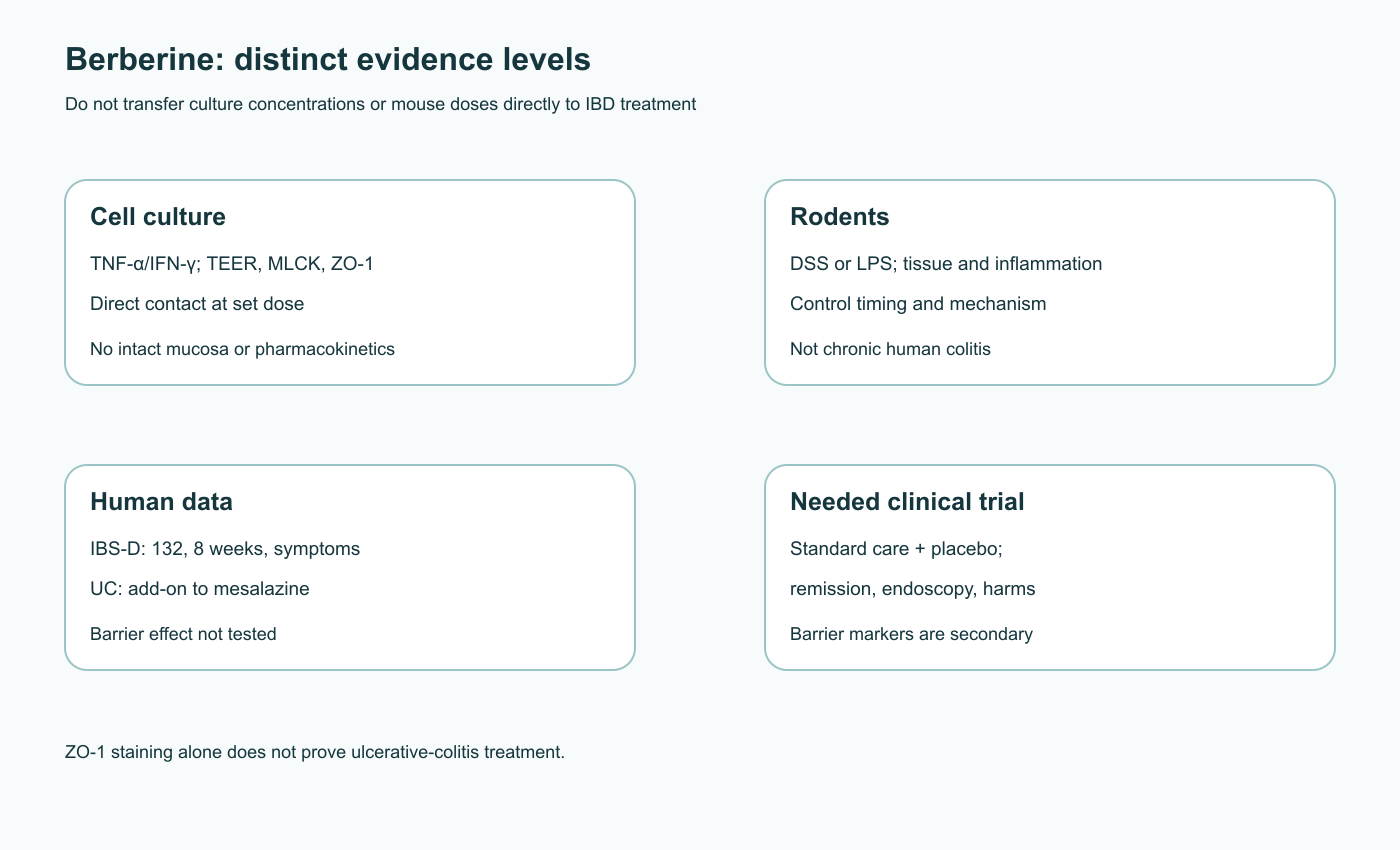
Berberine changes intestinal epithelial readouts in several experimental systems. Under inflammatory cytokines, endotoxin, and chemical colitis inducers, studies have observed preservation or recovery of some barrier measures. Mechanistic work involves the energy sensor AMPK, myosin light-chain kinase (MLCK), actomyosin tension, tight-junction proteins, and the microbiota. These findings do not show that berberine supplements treat “leaky gut” in people. A laboratory concentration, a rodent dose, human tolerability, and an ulcerative-colitis outcome are different levels of evidence.
1. The molecule and where it acts after oral intake
Berberine is a quaternary protoberberine alkaloid with a permanent positive charge. It occurs in plants of the genera Coptis and Berberis, but its content varies among roots, extracts, and finished products. Its planar aromatic core and cationic properties affect solubility and interactions with membranes, transporters, and microbial enzymes. “Plant-derived” does not define pharmacokinetics or safety: purified berberine, mixed alkaloids, and formulations designed to increase absorption can deliver different amounts to the mucosa and circulation.
After oral intake, systemic exposure to unchanged berberine is generally low relative to the administered amount. Limited passage across intestinal epithelium, efflux by P-glycoprotein (ABCB1), intestinal and hepatic metabolism, and microbial transformation all contribute. Rat transport and pharmacokinetic studies help examine these pathways, but “less than 5% bioavailability” is not a universal constant for every person, formulation, or analytical method. Nor does a low plasma concentration establish a specific micromolar concentration of free berberine at the basolateral membrane of a colonocyte: the compound may bind luminal contents, be transformed by bacteria, and distribute unevenly along the gut.
Microbial metabolism can produce reduced and demethylated derivatives; some metabolites are conjugated with glucuronic acid or sulfate. Deconjugation in the lumen could release free forms again and change contact time. Comparisons should state whether they examine parent berberine, a metabolite, or a complex botanical mixture. A phytosome or other delivery system that increases systemic exposure may also increase extraintestinal actions and interaction risks. “More absorption” does not automatically mean “better barrier treatment.” Likewise, inhibiting an efflux transporter in Caco-2 cells does not justify adding a P-gp inhibitor to a patient’s regimen to raise berberine exposure.
System
Where exposure is measured
What is needed before applying it to patients
Caco-2 or HT-29/B6 culture
Free compound concentration in the medium over cells
Active mucosal concentration after a real dose and digestion
LPS- or DSS-treated mouse
Dose per kg and local/systemic response
Comparable human exposure and safety; different disease context
Animal pharmacokinetics
Plasma, intestinal contents, metabolites
Human profiles accounting for formulation and microbiota
Person with IBD
Symptoms, endoscopy, inflammation, safety
Comparator, standard therapy, and adequate follow-up
Exposure, formulation, and chemical balance
Berberine’s permanent positive charge favors interactions with negatively charged molecules while making passive passage through lipid membranes more difficult. Systemic pharmacokinetics depend on dissolution of the salt, intestinal transport, biotransformation, and hepatic extraction; these steps cannot be reduced to “it is not absorbed.” Studies may measure parent compound or parent plus metabolites, plasma concentration or tissue accumulation, so reported “absorption” percentages are not always comparable. Free dissolved molecule, material bound to food or mucus, and total analytical signal in intestinal contents are also distinct. The fraction available at the apical epithelial surface is the relevant one for a direct epithelial effect.
Microbial reduction of berberine to dihydroberberine may alter membrane passage and subsequent reoxidation, and its rate depends on community composition. The comparison of berberine and dihydroberberine discusses pharmacokinetic and AMPK-related differences between the forms. Pharmacokinetic comparisons in conventional and partly germ-free rats indicate a microbial contribution to metabolism. They do not establish the same transformation in a person with IBD who may take antibiotics, mesalazine, or other drugs. P-gp findings also require care: berberine can be a transporter substrate and, under some conditions, an inhibitor. Direction and clinical magnitude depend on concentration and co-medication. This is a reason to study interactions, not to combine substances independently to “boost” berberine.
2. What does it mean to “restore tight junctions”?
The intestinal barrier is more than junctions. MUC2-containing mucus, antimicrobial molecules, renewing epithelium, immune cells, and microbiota all affect molecular flux. At the apical region between neighboring epithelial cells, claudins, occludin, tricellulin, and other transmembrane proteins create selective pathways. Scaffolding proteins ZO-1, ZO-2, and ZO-3 link some of these components to actin. E-cadherin and catenin adherens junctions support monolayer mechanics. A protein’s position along the membrane, phosphorylation, and turnover can matter more than its total concentration in a tissue homogenate.
Claudins are not simply “good” or “bad.” Claudin-2 forms a cation-selective paracellular pathway and can rise during inflammation, yet its function depends on gut region and regenerative state. A reduction in claudin-2 in one experiment does not mean all permeability routes have been corrected. Occludin moves between membrane and endosomes; loss of continuous ZO-1 staining indicates disorganized junctions, not necessarily complete protein destruction. Claims that any decrease in ZO-1 causes immediate total breakdown or obligatory proteasomal destruction of every component are too categorical.
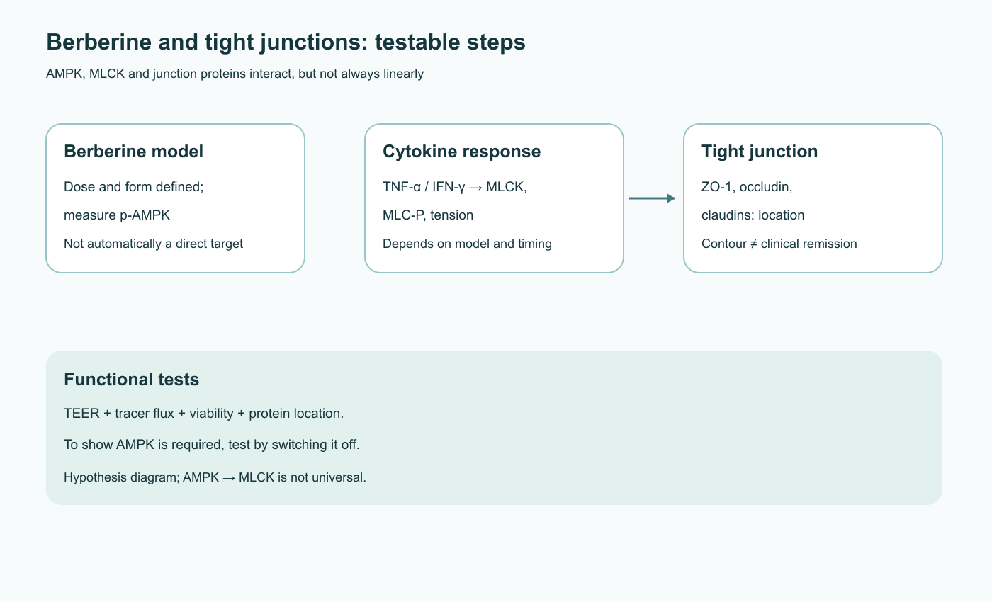
Inflammatory TNF-α and IFN-γ can increase expression of the long epithelial MLCK isoform and phosphorylation of the regulatory myosin-II light chain. MLCK transfers phosphate from ATP to MLC, primarily at Ser19, increasing actomyosin activity and changing tension in the perijunctional ring. Junctions may reorganize and occludin may enter endosomes. Endocytosis need not mean irreversible destruction; recycling can occur when the stimulus stops. Studies by Shen and Marchiando support roles for MLCK and caveolin-dependent occludin endocytosis in specific models, but berberine’s response must be tested in the model at issue.
A fall in transepithelial electrical resistance (TEER) reflects altered total ionic current across a monolayer; it can also depend on cell death or electrogenic transporters, not only tight junctions. Fluorescein or dextran flux reports permeability to a tracer of a chosen size. ZO-1/occludin microscopy shows protein localization. Viability, proliferation, and cell counts help distinguish repair from simple restoration of a monolayer. Human sugar tests and biopsy findings do not automatically identify berberine as the cause of an improvement. A barrier marker is an intermediate measure; clinical treatment requires symptom, endoscopic, and safety outcomes.
Pore and leak pathways
Paracellular transport includes regulated, narrow ion pores and a larger leak pathway that can increase with inflammation. Claudin-2 is especially associated with cation conductance; changing its expression may substantially alter TEER without necessarily changing flux of large dextran. Claudin-1 and other barrier proteins have different effects depending on epithelial context. A test using two tracer sizes plus resistance is therefore more informative than one readout alone. Secretory diarrhea can also result from altered ion transport without a “break” in tight junctions, so reduced diarrhea after berberine could reflect a transport mechanism.
Cell lines often form monolayers with a different claudin and mucus-cell profile from intact colon. Immunofluorescence, biochemical separation of membrane and cytosolic fractions, and live observation of recycling can distinguish protein loss from redistribution. Recovery of a continuous ZO-1 contour after treatment could follow cell survival or a weaker inflammatory stimulus. To establish a direct berberine effect on junction assembly, cells should be compared at similar viability and injury intensity. Timing matters too: administration before TNF-α tests prevention, while treatment after established colitis asks a different question.
3. AMPK: energy chemistry and the limits of molecular inference
AMPK is a heterotrimer with catalytic α and regulatory β and γ subunits. The γ subunit binds AMP, ADP, and ATP; when cellular energy falls, AMP/ADP compete with ATP and favor retention of activating phosphate on α-subunit Thr172. LKB1 in a STRAD/MO25 complex and CaMKK2 under calcium-dependent conditions can phosphorylate this site. The adenylate-kinase reaction, 2 ADP ⇄ ATP + AMP, links small ATP changes to potentially larger AMP changes. But the source manuscript’s numerical rule that a 5–10% ATP fall must produce a 100–300% AMP rise is not universal: concentrations depend on the starting nucleotide pool, compartment, and measurement.
In cell studies, berberine can restrict mitochondrial respiration, including complex I, changing adenylate ratios and AMPK activity. Complex I transfers electrons from NADH to ubiquinone while coupling this to proton transfer. Restricting the flow may reduce ATP production, but a higher exposure may also injure cells. Increased AMPK phosphorylation is therefore not unconditionally “cytoprotective”; exposure range and viability controls matter. Specific binding of berberine in an ND1/PSST channel or “hundreds-fold” accumulation inferred from one membrane-potential measurement has not been established in enterocytes at dietary exposure. AMPK can also respond to inflammatory, calcium, or metabolite signals, so p-AMPK after berberine does not identify the primary target.
To show that AMPK is necessary for barrier protection, experiments should combine changes in p-Thr172 and kinase activity with TEER, tracer flux, protein localization, and viability. AMPK inhibition or genetic depletion should remove the effect, and a rescue should restore it; inhibitor off-target effects must be considered. If berberine still improves barrier readouts when AMPK is blocked, AMPK activation may accompany the response without being required. Nor is AMPK necessarily a direct upstream regulator of MLCK in every cell type. Cytokine signaling, energy state, and junction remodeling may intersect without forming one universal linear chain.
AMPK also affects lipid synthesis, autophagy, mitochondrial turnover, and inflammatory signaling. An effect on one of these processes may indirectly alter barrier readouts. The key distinction is between “berberine changes AMPK phosphorylation” and “AMPK mediates the barrier effect.” The latter requires a causal perturbation, appropriate controls, and replication in a relevant epithelial or animal model.
4. The injury model determines what a result means
DSS in rodents’ drinking water causes mucosal injury and inflammation that depend on polymer concentration, duration, microbiota, sex, and animal strain. It is not simply “detergent lysis” of one membrane: primary injury and the subsequent immune response together shape the phenotype. TNBS and oxazolone use chemical haptenization and different immune contexts. LPS endotoxemia is often induced by injection, bypassing the usual route from the intestinal lumen. Protection after intraperitoneal LPS therefore does not show that berberine prevents ulcerative colitis after oral exposure. A rat irritable-bowel model, Caco-2 culture, and acute DSS colitis also test different mechanisms and outcomes.
Gu and colleagues used mouse endotoxemia and assessed tight junctions and MLCK; this supports a mechanism experimentally, but the original bibliography misattributed the journal and identifier. Amasheh and colleagues studied TNF-α-dependent barrier loss in an epithelial monolayer and rat tissue, including claudin-1/2; again, this concerns direct exposure to specified concentrations, not controlled treatment in people. Yan and colleagues described recovery after DSS colitis and lower inflammatory signals in mice. These studies are consistent with possible local protection, but do not show that it is always mediated by one AMPK mechanism.
In chemical colitis, a smaller pathological area and preservation of goblet cells may accompany more even ZO-1 staining, more MUC2, and changes in claudins. The causal sequence is ambiguous: berberine may have reduced the initial inflammation, affected bacteria, or improved cellular nutrition, after which the barrier recovered secondarily. To establish a direct effect on junctions, investigators need separate time points and a control with comparable inflammatory severity. A single terminal biopsy cannot establish that berberine “blocked the proteasome,” “activated FAK,” and “prevented EMT”; each proposed node needs its own intervention and measurement.
Separating outcomes is especially important when translating from a model to IBD. In mice, researchers assess weight, colon length, histology, and sometimes local cytokines; in patients, relevant outcomes include clinical and endoscopic remission, relapse, steroid need, quality of life, and harm. An improvement in TEER in a dish is equivalent to none of these outcomes.
Inflammatory networks and feedback with the barrier
TNF-α can increase MLCK through transcriptional pathways, while IFN-γ amplifies the epithelial response; IL-1β and other mediators act through their own receptors. NF-κB regulates a broad set of inflammatory genes and cell survival, so reduced activation is not necessarily the first step in berberin’s protective effect. In some models berberine lowers inflammatory signaling; in others MLCK/MLC changes without measurable NF-κB suppression. Oxidative stress and iNOS/NO pathways are also context-dependent: nitric oxide can be a signaling molecule, whereas excessive reactive nitrogen species can damage tissue. A universal claim that “berberine blocks all cytokines and radicals” does not follow from separate experiments.
The barrier and immune response form a feedback loop. Injured epithelium allows more microbial products to reach mucosal receptors; immune cytokines then further alter junctions. A drug may interrupt the loop at any node, after which several markers improve together. This explains why terminal improvement in ZO-1, MUC2, calprotectin, and microbiota does not rank the primary targets in sequence. Early time points and blockade of each proposed pathway are needed to determine order. Direct antimicrobial effects are especially difficult to distinguish from epithelial effects in DSS colitis because both change exposure to bacterial products.
Prevention, recovery, and chronic control are different questions
Many preclinical protocols give berberine before injury is induced or at the same time as DSS. This tests possible preventive protection at a known time, whereas people usually seek care after inflammation has developed. A treatment study that starts after disease onset needs different timing, control of baseline severity, and relapse assessment. Acute DSS colitis is relatively short; human ulcerative colitis can last for years, with remissions, different affected regions, and prior medicines. Even a favorable short-term mouse result does not predict maintenance of remission or cancer prevention over years.
Oxidative stress, cell survival, and fibrosis are three different tasks
Nrf2 is released from KEAP1-dependent control in response to appropriate redox signals and regulates antioxidant genes, including HMOX1, which encodes HO-1. SIRT1, an NAD⁺-dependent deacetylase, may intersect with energy and inflammatory pathways. These links allow researchers to test whether berberine changes epithelial sensitivity to oxidative injury. But high HO-1 in inflamed tissue may be a response to stress, not necessarily proof of protection. A direct antioxidant effect, signaling-mediated induction of a protective enzyme, and reduction of the initial inflammation are different explanations for a lower ROS signal. They require a time course, genetic loss of Nrf2/HO-1, and measurement of oxidative injury, not expression alone.
Caspases-3/7 participate in the effector phase of apoptosis; the balance between anti-apoptotic and pro-apoptotic Bcl-2/Bax family members changes the likelihood of mitochondrial cell death. If a model shows less active caspase and more intact colonocytes after berberine, that could indicate direct cytoprotection or simply less severe primary colitis. Apoptosis measurements should be combined with proliferation and crypt regeneration: blocking all cell death is not the same as healthy epithelial renewal. During prolonged inflammation, the balance between survival and removal of damaged cells may also matter for cancer risk, so a short mouse experiment does not establish chronic safety.
Epithelial–mesenchymal transition (EMT) and intestinal fibrosis are not reducible to a fall in E-cadherin. Chronic injury involves stromal cells, TGF-β, matrix enzymes, and collagen accumulation. Changing one marker in culture does not show that berberine will prevent Crohn’s strictures. An antifibrotic claim requires an appropriate long-term disease model, tissue architecture, and mechanical outcomes. Thus, Nrf2/HO-1, Bcl-2/caspase, and EMT branches are useful research directions, but cannot be combined into a required “repair cascade” after every berberine dose.
The MLCK–MLC pathway is one plausible route from inflammatory signaling to increased paracellular permeability. Cytokine receptors can increase long MLCK expression or activity; phosphorylated MLC promotes perijunctional actomyosin contraction. This can alter junctional tension, redistribute occludin, and change leak-pathway flux. Berberine attenuated MLCK/MLC-associated changes in some endotoxin and cytokine models. Yet an IFN-γ/TNF-α Caco-2 model reported reduced MLCK/MLC signaling without suppressing NF-κB. That result argues against treating NF-κB suppression as a required universal explanation.
Tight-junction readouts answer different questions. Total protein abundance does not show whether a protein reaches the junction. Continuous staining does not prove selective barrier function. TEER is sensitive to small-ion conductance and cell health; tracer flux samples permeability for a particular molecular size. Combining localization, multiple tracer sizes, resistance, viability, and relevant inflammatory readouts helps avoid interpreting one surrogate as the whole barrier. In vivo, stool or serum markers also reflect more than epithelial tight junctions and cannot by themselves establish a berberine mechanism.
5. Microbiota, metabolites, and the barrier: alternative pathways
Oral berberine can meet microbes at high local concentrations even when plasma exposure is low. It may alter microbial growth or metabolism, while microbes transform berberine and affect its absorption. These directions can coexist. An antimicrobial effect may help under one starting condition and disrupt ecological balance under another. Strains, diet, and colitis model can substantially change the result.
Akkermansia muciniphila uses mucin substrates and has been associated with metabolic benefits in animal experiments. A rise in its relative abundance after berberine does not by itself prove causal restoration of MUC2 or tight junctions. Absolute abundance, mucus production and thickness, inflammation, and epithelial fate would need measurement. Studies of purified Akkermansia proteins or pasteurized bacteria do not clinically establish that berberine “grows a protective symbiont” in every person with ulcerative colitis. Butyrate-producing bacteria likewise depend on substrate and local metabolite concentration; taxonomic composition alone is not enough.
Microbes may transform berberine and alter absorption; berberine may in turn affect bacterial bile-acid enzymes and FXR signaling. A frequently cited study of microbiota and intestinal FXR in people examined metformin, not berberine; its citation was misattributed in an earlier manuscript. It does not establish berberine-mediated barrier healing in IBD. Short-chain fatty acids, indoles, and bile acids can affect epithelium and immunity, but the chain “berberine → one microbe → one metabolite → ZO-1 → remission” requires direct intervention evidence. Transfer of microbiota from a treated animal can support a role for the transferred community in those conditions, not identify a human strain or dose.
New formulations: more exposure is not always better for the gut
Phospholipid complexes, emulsions, nanoparticles, and enteric-release products address different delivery goals. A phytosome may raise plasma exposure compared with conventional berberine in a pharmacokinetic comparison in healthy volunteers; that is a delivery result, not proof of reduced ulcerative colitis. Enteric or delayed-release products instead aim to change release location and local contact. Distribution may differ in inflamed mucosa. Evaluation should measure active alkaloid content, release profile, plasma and, if feasible, local concentrations, metabolites, and tolerability. A dose of a conventional salt cannot be mechanically transferred to a more absorbable formulation.
Claims of pharmacokinetic “synergy” with a P-gp/ABCB1 inhibitor need particular caution. This efflux transporter handles many drugs as well as berberine. Blocking it may increase exposure to a co-administered medicine. Greater plasma berberine does not guarantee concentration in an inflamed crypt, and deliberately changing transporter activity to boost a nutraceutical may raise interaction risk. Caco-2 and rat studies explain a research direction; they do not provide a safe combination regimen for people with IBD.
6. Human evidence, safety, and decisions about use
Early studies of berberine with mesalazine in ulcerative colitis examined tolerability and possible add-on effects; a pilot phase I study does not establish efficacy as standard treatment. A later review of randomized studies of berberine plus 5-aminosalicylic acid reported favorable symptom findings, while noting the need for better-quality studies with lower risk of bias. In combination therapy, an outcome cannot be attributed to berberine alone. Endoscopy, dose, product standardization, remission duration, and adverse events need clear reporting. No established berberine regimen restores tight junctions in Crohn’s disease or an unvalidated “increased permeability syndrome.”
IBS-D is a separate evidence set. Chen and colleagues conducted a randomized, double-blind, placebo-controlled study of berberine hydrochloride in people with diarrhea-predominant irritable bowel syndrome. Of 196 enrolled, 132 were randomized; treatment lasted eight weeks after a two-week run-in, followed by four weeks off treatment. The publication abstract refers both to “400 mg daily” and to dosing “twice daily”; without the full protocol, the dose per administration cannot be stated confidently or treated as a universal regimen. Participants recorded diarrhea frequency, pain, urgency, and adverse events daily; symptom, anxiety/depression, and quality-of-life scales were assessed before and after treatment. Compared with placebo, diarrhea frequency, pain, and urgency declined, and the authors reported good tolerability. The abstract does not make clear which single outcome was prespecified as primary, so the result should not be described as confirmation of a known primary endpoint without the protocol. This is a clinical signal for IBS-D symptoms, not evidence of ZO-1 restoration, benefit in IBD, or long-term remission maintenance.
Human metabolic outcomes cannot be used as ready evidence for colitis treatment. Plasma glucose, lipids, or microbiome changes do not measure intestinal ZO-1 structure. Even if a medicine acts through AMPK in one tissue, its therapeutic window in inflamed mucosa may differ. A mouse dose in mg/kg cannot be converted by simply multiplying by human body weight; species metabolism, formulation, duration, and experimental aim matter. The original manuscript proposed a universal course and “synergy” with butyrate, zinc carnosine, and glutamine without controlled confirmation of that combination, so its dosing protocol is not carried into practical recommendations.
Nausea, abdominal pain, constipation, or diarrhea can be confused with colitis symptoms. Berberine and some formulations or extracts may interact with drug transporters and enzymes. A small repeated-administration human study found changes in activity of some CYP enzymes; this warrants checking concomitant medicines, especially those with a narrow therapeutic range, rather than assuming the same interaction with every drug. Pregnancy, breastfeeding, childhood, significant liver or kidney disease, and multiple medicines call for individual medical review. Lack of a clear hepatotoxicity signal in a small study does not prove absolute safety. A phytosomal formulation with higher absorption may have a different exposure and needs its own safety assessment.
For active IBD, treatment decisions depend on severity, endoscopic and laboratory findings, complication risk, and established standard therapy. Berberine should not delay assessment of bleeding, fever, dehydration, or sudden worsening. A research path remains: test a standardized formulation as an add-on to usual care while measuring clinical remission, endoscopy, barrier markers, and adverse events. Current data support studying the mechanism, not replacing prescribed treatment or routine “AMPK therapy” based on one permeability test.
What a persuasive clinical study would look like
A study of add-on berberine in ulcerative colitis should start with a standardized chemical form and composition analysis. Participants should receive comparable evidence-based background treatment, with concealed allocation to berberine or placebo. The clinical and endoscopic outcomes, assessment times, worsening criteria, and interaction monitoring should be defined in advance. Barrier tests and microbiota can be secondary mechanistic measures, not substitutes for remission. Reports should include the proportion in remission, discontinuations due to adverse events, and durability, not only an average score change. Dedicated pharmacokinetic measurements are needed when participants take medicines with narrow therapeutic ranges.
Such a design could separate symptom relief from mucosal healing and test whether outcomes depend on baseline microbiota or disease severity. A small open study or pooled set of heterogeneous small studies can generate a hypothesis but is poorly protected against expectation effects, changes in standard therapy, and selective outcome reporting. Even a favorable signal from combined treatment with mesalazine therefore does not establish a universal dose for Crohn’s disease, IBS-D, or “food intolerance.”
7. Review method and limits
This is a narrative review of molecular and pharmacokinetic studies, cell and animal experiments, and available human trials, with targeted checking of disputed primary records on October 9, 2026. It compares endotoxemia, cytokine injury, chemical colitis, and separate patient populations with ulcerative colitis or IBS-D. Evidence from different fields does not have equal strength; this was not an exhaustive search, meta-analysis, or formal risk-of-bias assessment.
Targeted checking corrected bibliographic errors in key work on MLCK, DSS colitis, and microbiota-dependent pharmacokinetics. A study of intestinal FXR previously attributed to berberine actually examined metformin. For IBS-D, the design, population, symptoms reported in the abstract, and ambiguous dose wording were checked; no unsupported primary endpoint is asserted without the full protocol. The main evidence limitation is the small number of high-quality human trials with endoscopic remission and standardized barrier measurements, so the mechanism does not translate into a treatment protocol.
8. What is established and what remains to be tested
Berberine can change epithelial barrier measures in cell and animal experiments; the clearest findings concern specific cytokine, endotoxin, and DSS injury conditions. MLCK/MLC, claudin remodeling, occludin and ZO-1, AMPK, inflammatory pathways, and microbes offer complementary but not necessarily sequential explanations. Luminal berberine and low systemic exposure make a local mechanism plausible, yet active concentration in human mucosa and clinical effect depend on formulation and context. Translation to IBD treatment requires reproducible exposure, separation of direct epithelial effects from microbiota-mediated effects, and then well-controlled human outcomes. Until then, AMPK biochemistry and attractive ZO-1 staining are reasons to investigate, not a clinical prescription.
FAQ
Does berberine restore intestinal tight junctions?
Some barrier measures improved in cell and animal models. This does not prove intestinal repair or IBD remission in an individual.
How is berberine linked to AMPK?
In experiments, berberine can restrict mitochondrial respiration and change cellular energy state, after which AMPK may activate. Each model must test separately whether AMPK is required for the barrier effect.
Why are ZO-1 and occludin not diagnoses?
Staining shows protein location in a specific tissue or model. Function requires flux and viability tests; treatment requires a clinical outcome.
Is there an established berberine dose for ulcerative colitis?
The reviewed studies do not establish a single dose to treat disease or maintain remission. Animal doses and different supplement forms cannot be transferred directly.
Has berberine been studied in diarrhea-predominant IBS?
Yes. In a double-blind trial, 132 people with IBS-D received berberine hydrochloride or placebo for eight weeks; diarrhea, pain, and urgency decreased. This concerns IBS-D symptoms, not tight-junction restoration or IBD efficacy.
Can berberine replace mesalazine?
No. Early human studies mainly evaluated adding it to standard therapy; replacing prescribed treatment is unsupported.
Can berberine interact with medicines?
A small human repeated-dose study found changes in CYP activity. Concomitant medicines, especially those with a narrow therapeutic range, warrant a professional interaction review.