
Astaxanthin in membranes: lipid peroxidation, pharmacokinetics and clinical limits
Astaxanthin chemistry and stereoisomers, model bilayer orientation, LPO chain kinetics, mitochondria, Nrf2/NF-κB and critical interpretation of small human studies.
Astaxanthin chemistry and stereoisomers, model bilayer orientation, LPO chain kinetics, mitochondria, Nrf2/NF-κB and critical interpretation of small human studies.
Astaxanthin offers a useful test of how a plausible molecular hypothesis becomes an overly strong therapeutic promise. Its terminal hydroxyl and keto groups interact with the polar membrane region, the conjugated polyene is distributed among acyl chains, and different model systems detect changes in lipid oxidation. Yet orientation in an artificial bilayer depends on composition and concentration; a change in a human biomarker is not equivalent to reduced disease risk. Chemistry, membrane biophysics, chain kinetics, cellular pathways and human experiments are therefore distinguished below by their level of evidence.
Astaxanthin is a C₄₀ xanthophyll with the formula C₄₀H₅₂O₄ and a molar mass of approximately 596.8 g/mol. Its framework consists of a polyene chain and two β-ionone rings, each bearing OH at C3 or C3′ and a keto group at C4 or C4′. The systematic name of one particular natural enantiomer is (3S,3′S)-3,3′-dihydroxy-β,β-carotene-4,4′-dione, whereas “astaxanthin” without a stereochemical qualification also includes other configurations. The often mentioned “13 conjugated double bonds” means 11 C=C bonds and two conjugated C=O bonds: these must not be confused with 13 C=C bonds. Delocalized π electrons determine visible absorption and the capacity to transfer excitation energy or react with radicals. The spectral maximum depends on solvent, aggregation, cis/trans composition and matrix; no single value applies to every membrane. HOMO/LUMO energies calculated for an isolated molecule do not themselves rank the rates of protective reactions in a cell: the particular oxidant, rate constants, competitors and local concentration are required (Woodall et al., 1997).
C3 and C3′ are chiral centers. For the symmetrical skeleton, the mirror-image (3S,3′S) and (3R,3′R) forms are distinguished from the meso configuration (3R,3′S), equivalent to (3S,3′R). In Haematococcus pluvialis, (3S,3′S) usually predominates; in Xanthophyllomyces dendrorhous (Phaffia rhodozyma), the opposite configuration predominates. Chemical synthesis through an appropriate nonselective process produces three stereochemical forms. Their proportions depend on strain, production method and batch analysis, so a fixed percentage table cannot be applied to all natural and synthetic products. In algal material the molecule is often esterified with fatty acids, while a synthetic preparation may contain the free form. This affects extraction and hydrolysis, but a bioavailability comparison requires an identical formulation matrix and dose rather than a “natural” label (Odeberg et al., 2003).
Biosynthesis involves hydroxylation of the β-ionone rings by β-carotene hydroxylase and ketolation by β-carotene ketolase; the order of steps and intermediate xanthophylls depend on the organism's enzymes. Schematically: β-carotene → hydroxy and/or keto intermediates → astaxanthin. This reaction map explains the presence of both OH and C=O, but does not prove that one enantiomer binds an enterocyte receptor better than another: separate comparative studies are needed.
The polyene permits all-E and Z isomers, including 9-Z and 13-Z. Light exposure, heating, oxygen and the matrix can change isomer composition or cause oxidative degradation. A Z bond changes molecular projection, crystal packing and dissolution in lipids, but this is not a universal “shortening to 22–25 Å” or an automatic loss of antioxidant activity. Isomerism and stereochemistry are independent: (3S,3′S) does not specify whether the chain is all-E. Ester forms, the free molecule and oxidation products must also be distinguished analytically when comparing studies. Tissue accumulation depends on starting composition, intestinal transformations, lipoprotein transport and elimination, rather than one conformation in a drawing.
A phospholipid bilayer contains aqueous interfaces with hydrated head groups and a less polar core of acyl chains. Interface spacing, hydrophobic thickness, hydration and lateral order vary with lipids, unsaturation, cholesterol, temperature and phase. Comparing one calculated length of an extended molecule with one “living membrane thickness accurate to tenths of an Å” is therefore methodologically unsound. Gel-phase DPPC, fluid-phase DOPC and a cardiolipin-containing mitochondrial membrane do not share the same geometry. Determining depth and tilt requires a particular model and method, for example spin-label parameters, spectroscopy, scattering or molecular dynamics under specified conditions (Goto et al., 2001; McNulty et al., 2007).
The two oxygen-containing terminal groups make astaxanthin amphipathic: terminal regions can interact with hydrated heads and the polyene with the acyl region. In some artificial bilayers, an orientation with the ends facing opposite surfaces is energetically possible, helping explain changes in local lipid order and radical accessibility at different depths (Goto et al., 2001; McNulty et al., 2007). This is a model hypothesis about an orientation distribution, not a proven fixed pose of every molecule in all cell membranes. Tilt angle is not a universal constant; “membrane cross-linking” here describes noncovalent interactions, not the formation of chemical cross-links between phospholipids.
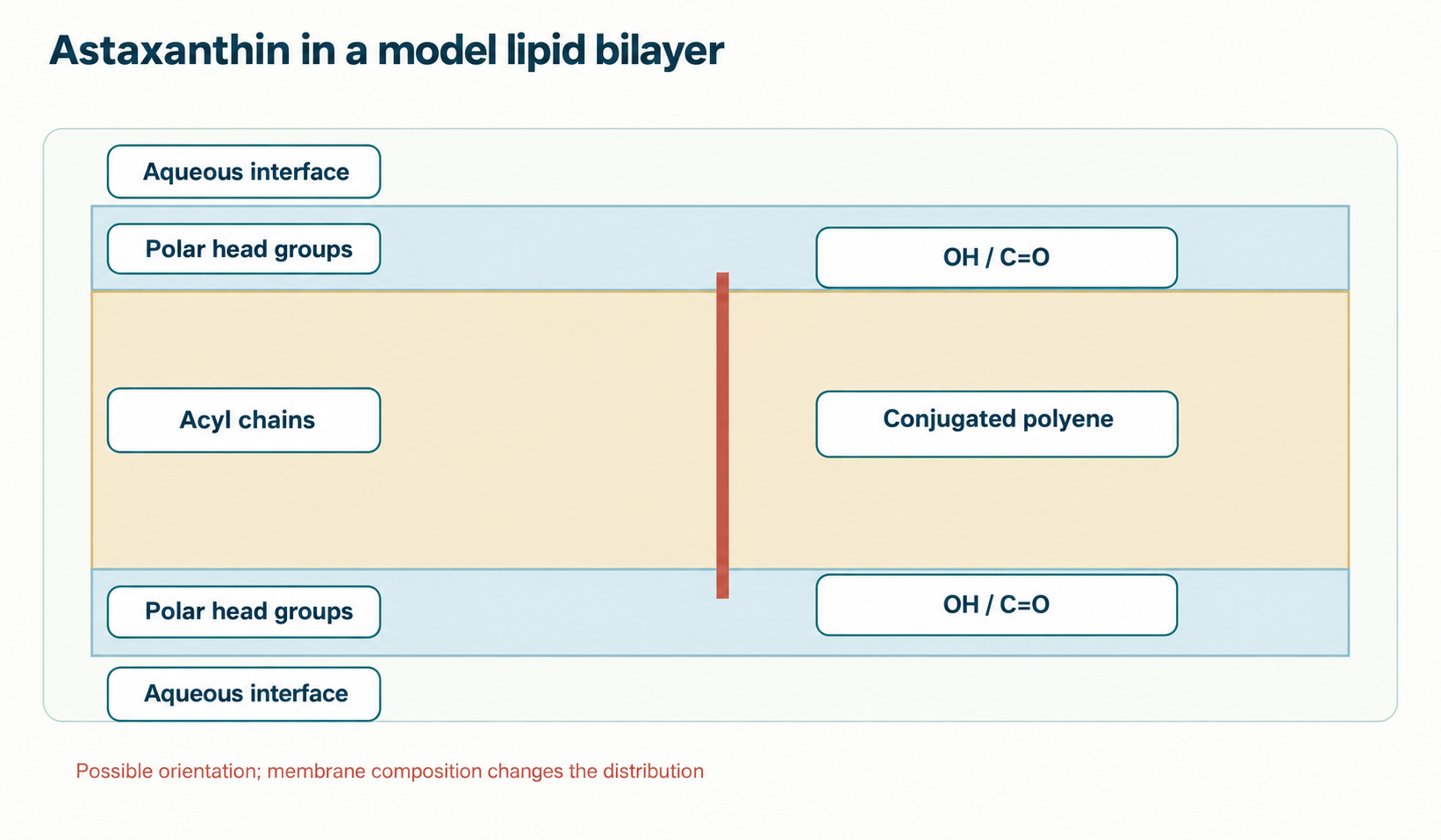
β-Carotene and lycopene lack these polar terminal groups and are consequently distributed differently in bilayers; this does not imply a compulsory horizontal position for every molecule, loosening of every membrane or toxicity at every concentration. Lutein and zeaxanthin also have polar ends and can adopt orientations spanning the bilayer. Astaxanthin's keto groups broaden its interactions, but it is not the only carotenoid with a potential interleaflet orientation. Comparisons require identical membrane composition, molar fraction, temperature and measured property; without these conditions, a table of “absolute superiority” is invalid. Differences in photophysics and radical chemistry do not automatically equal differences in clinical efficacy.
Adding a carotenoid to a model membrane can change order parameters, microviscosity, permeability and the thermal profile of the Lβ → Lα transition. The direction depends on whether the starting lipid is in a gel or liquid-crystalline phase, the astaxanthin fraction, cholesterol and the probe used. Broadening or attenuation of a calorimetric peak indicates altered phase-transition cooperativity in that sample, not universal prevention of fever-related membrane damage. Liposome and erythrocyte observations inform mechanisms, but cannot establish that oral intake prevents osmotic hemolysis, vascular leakage or loss of ion gradients in humans (McNulty et al., 2007). The practical question is whether a comparable molar fraction reaches the relevant membrane after absorption and transport; for most proposed clinical scenarios this has not been established.
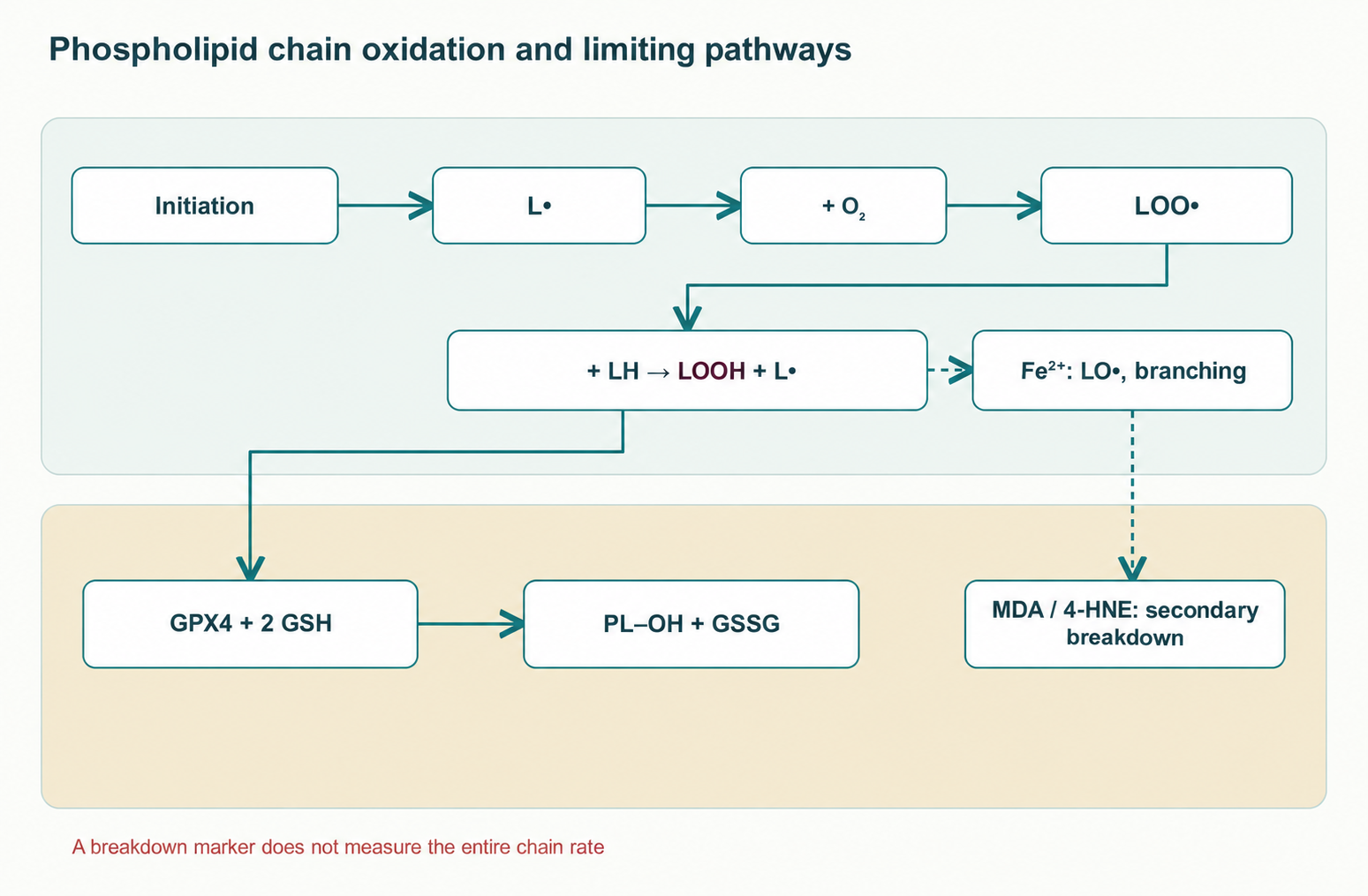
The bis-allylic methylene hydrogen in a polyunsaturated phospholipid acyl residue is vulnerable to radical abstraction. Denote this residue LH. The initial reaction can be written LH + X• → L• + XH; then L• + O₂ → LOO• and LOO• + L′H → LOOH + L′•. The last step regenerates a radical and permits chain propagation. Reaction constants vary with substrate, oxygen, phase and temperature; one number cannot represent the rate in all membranes. LOOH + Fe²⁺ → LO• + OH⁻ + Fe³⁺, when the metal is accessible, produces an alkoxyl radical and promotes branching. Enzymatic hydroperoxide removal competes with this route: GPX4 catalyzes PL–OOH + 2 GSH → PL–OH + GSSG + H₂O when the selenoprotein and reduced glutathione are available.
Termination is a reaction after which the radical chain no longer generates a new L•, for example LOO• + LOO• → stable products + O₂, or sufficiently rapid interception by a chain-breaking donor. Malondialdehyde (MDA), 4-hydroxy-2-nonenal (4-HNE) and other reactive carbonyls arise mainly from secondary breakdown of oxidized polyunsaturated residues: they are damage products, not the mechanism of termination itself. Their concentration reflects formation, further metabolism, protein binding and the analytical method. An increase in one marker does not automatically measure the flux of the entire chain. A separate article on dietary polyphenols and the limits of antioxidant-marker interpretation is useful in this membrane-chemistry context: food polyphenols and safety.
¹O₂ arises through photosensitization and certain other reactions; it can oxidize an unsaturated bond without an initial carbon radical. The carotenoid polyene can accept excitation energy: ¹O₂ + AST(S₀) → ³O₂ + ³AST*; return of ³AST* to its ground state dissipates energy. Physical quenching does not require stoichiometric carotenoid consumption, whereas chemical oxidation of astaxanthin consumes it. These pathways compete, with proportions depending on solvent, oxygen concentration, aggregation and neighboring molecules. Relative constants in solutions and liposomes cannot be translated into “550/800/6000 times better” clinical efficacy by comparing ascorbate, tocopherol or coenzyme Q10 without a shared compartment and an identical analytical assay (Woodall et al., 1997).
AST interaction with LOO• can involve hydrogen-atom transfer, electron transfer and polyene addition; products and relative contributions depend on the environment. The simplified reaction LOO• + AST–H → LOOH + AST• assumes that the resulting AST• is less inclined to propagate the chain, but this must be established kinetically. Radical resonance delocalization does not imply complete chemical inertness. Reduction of oxidized AST by a reductant at the aqueous interface, such as ascorbate, is physically possible; claiming a proven obligatory AST → ascorbate → glutathione “electron relay” in a human cell would be excessive. This requires the rate of each step, encounters of reactants in the same compartment and a demonstrated balance of oxidized/reduced forms, rather than an attractive topological scheme alone. Peroxynitrite and lipid peroxyl radicals are chemically different reactants; the generic label “scavenger” does not make their reactions equally effective.
Large β-carotene trials in smokers and asbestos-exposed populations do not justify mechanically attributing the same risk to astaxanthin; it is equally incorrect to infer guaranteed complete safety at any dose or pO₂ from structural differences. Polyene autooxidation and formation of epoxides, apocarotenoids and radical adducts depend on oxygen, light, matrix and concentration. Experimental differences between carotenoids may exist, but “O₂ addition is thermodynamically forbidden” cannot be inferred from a lowered LUMO without measuring the reaction and products (Woodall et al., 1997). Even a positive oxidation-inhibition test in one model does not exclude adverse effects in pregnancy, smokers or long-term use. Clinical risk assessment must concern a specific formulation, population and regimen, as in the discussion of coenzyme Q10 in food combinations.
Cardiolipin is a four-acyl-chain phospholipid enriched in the inner mitochondrial membrane, contributing to crista organization, respiratory-chain proteins and interactions with cytochrome c. Its acyl composition is tissue-specific; tetralinoleoyl cardiolipin is particularly significant in the heart, but percentages cannot be extrapolated to brain or all animal species. Cardiolipin peroxidation can alter cytochrome c binding and protein-complex function. However, cytochrome c release into the cytosol also requires outer-membrane permeability, generally governed by BAX/BAK or other context-dependent mechanisms. The mPTP mechanism cannot be reduced to an obligatory ANT–VDAC pair; permeability-transition composition and regulation remain under investigation.
In isolated cardiac mitochondria, astaxanthin has been studied using oxidation, calcium-dependent swelling and bioenergetic measures (Belova et al., 2019; Krestinina et al., 2023). Direct addition to a suspension is not equivalent to its inner-membrane concentration after a person takes a capsule. Reduced damage in a particular model does not prove “complete blockade” of cardiolipin peroxidation, a fixed ΔΨm or prevention of myocardial infarction. The fraction of oxygen converted to ROS varies with respiratory-chain state and substrates; a universal “0.2–2%” is not a constant. For a comparison of carotenoid membrane chemistry with another mitochondrial signaling system, see melatonin and mitochondria, which involves a different delivery pathway and different outcomes.
Under basal conditions KEAP1 promotes Nrf2 association with the CUL3–RBX1 complex, ubiquitination and proteasomal degradation. When this control is disrupted, newly synthesized Nrf2 accumulates, enters the nucleus, complexes with small Maf proteins and binds ARE sites; other regulatory pathways may also contribute. Changes in Nrf2 and several downstream genes after astaxanthin have been reported in cellular and animal models (Fakhri et al., 2022). Yet direct covalent AST binding to KEAP1 Cys151/Cys273/Cys288 and selective KEAP1–CUL3 uncoupling cannot be considered established without a target-level experiment: astaxanthin should not automatically be modeled as a classic cysteine-reactive electrophile.
Potential outputs of this axis differ in substance. HMOX1 degrades heme to biliverdin and CO with iron release; ferritin is not a product of this reaction but a separate part of the cellular response to iron. NQO1 reduces quinones through a two-electron NAD(P)H-dependent route; GCLC/GCLM support the first ATP-dependent step of GSH synthesis from glutamate and cysteine, after which glutathione synthetase adds glycine. GPX4 reduces phospholipid hydroperoxides while consuming GSH, but is not the only enzyme capable of detoxifying all membrane peroxides. Matrix SOD2 dismutates O₂•⁻ to H₂O₂, which must subsequently be removed by peroxidases. A change in any enzyme transcript is only one level: functional inference requires protein, activity, substrates and lipid injury in the relevant compartment.
Canonical TNF, IL-1 or TLR signaling activates the IKK complex; IκBα phosphorylation at Ser32/Ser36 promotes ubiquitination and degradation, allowing released RelA/p50 to enter the nucleus. Astaxanthin models have reported lower nuclear NF-κB activity and changes in TNF, IL-1β, IL-6, NOS2/iNOS and PTGS2/COX-2 (Fakhri et al., 2022). This is compatible with indirect effects on oxidation, receptor pathways or the inflammatory stimulus. These observations do not identify AST unambiguously as a direct allosteric IKKβ inhibitor. Concurrent Nrf2 and NF-κB changes likewise do not prove that one axis causally suppresses the other. Cell culture at a specific concentration and exposure is a mechanistic model, not a promise of clinical anti-inflammatory efficacy.
Shared oxidation mechanisms in LDL, skin and myocytes do not allow heterogeneous small experiments to be combined into proven prevention of atherosclerosis, photoaging, dry-eye syndrome or exercise exhaustion. Randomization, matrix control, participants' initial condition, data completeness and prespecified outcomes matter. Studies from the original evidence base are retained below, with every observation tied to the outcome measured.
An LDL particle contains ApoB-100 and oxidizable lipids. Extensive modification alters recognition by the classical LDL receptor, while macrophage receptors, including CD36 and SR-A, can take up oxidized particles; in tissue this process is associated with foam-cell formation. This is a pathogenic context, not evidence that oral astaxanthin changes plaque or event frequency. In Iwamoto's study volunteers took astaxanthin, after which isolated LDL was oxidized with copper ex vivo and the lag period of diene formation was measured (Iwamoto et al., 2000). A longer lag phase in this laboratory system means resistance of an isolated particle to a specified oxidant, not fewer infarctions or proven “endothelial restoration.” Choi's experiments in overweight people or another selected population measured MDA, isoprostanes, SOD activity and total antioxidant capacity (Choi et al., 2011; Choi et al., 2011). Population composition, matrix and analytical method limit generalizability; original percentages without reproduction of the primary table are not declared universal effects here.
The mechanistic chain O₂•⁻ + NO• → ONOO⁻ and BH₄ oxidation are important for understanding eNOS uncoupling: with insufficient functional cofactor, the enzyme can produce superoxide instead of NO. But a lower LPO marker alone does not prove that astaxanthin restored BH₄, eNOS coupling and flow-mediated dilation (FMD) in that participant. Direct measurements and a controlled intervention are required. FMD and pulse-wave velocity are functional surrogates, not established hard cardiovascular outcomes here.
In a small randomized study Yoshida and colleagues examined astaxanthin in mild hypertriglyceridemia, comparing several dose groups with placebo over twelve weeks (Yoshida et al., 2010). The authors discussed triglyceride, HDL-C and adiponectin changes. These cannot be described as established treatment of “metabolic syndrome”: lipid-based inclusion does not necessarily imply the syndrome, and adiponectin is a hormonal marker rather than an independent clinical outcome. Correlation of adiponectin and HDL-C changes does not establish causality through AdipoR1/R2, AMPK, PPARα and ABCA1; interventions at these nodes and verification in relevant tissues would be needed. The original article's table assigns exact group changes and p-values without a checked primary table; they are deliberately not reproduced here. Clinical decisions require independent trials controlling diet, medication, multiple comparisons and prespecified endpoints. Comparison with pharmacological lipid lowering calls for particular caution; niacin, lipids and safety explains why a change in HDL-C does not guarantee an outcome.
Photooxidation can activate MAPK–AP-1 and increase matrix metalloproteinase expression; MMP-1 cleaves fibrillar collagen, while MMP-2/9 act on certain matrix components. The original text incorrectly calls MMP-13 “stromelysin”; it is collagenase-3. Tominaga's female cohort received both an oral agent and a topical preparation, so changes in elasticity, moisture or wrinkles cannot be attributed specifically to swallowed astaxanthin; the male group was a separate study (Tominaga et al., 2012). Ito's randomized experiment in 23 healthy people used an oral supplement and controlled local UV exposure, measuring minimum erythema dose (MED), hydration and TEWL; MMP-1 cell experiments represent another evidence level (Ito et al., 2018). A favorable experimental MED does not replace sun protection, prove skin-cancer prevention or define a therapeutic dose for skin diseases.
Intense muscular work involves mitochondrial redox flux and, in certain conditions, NOX2-dependent signals. However, membrane injury cannot be reduced to “CPT1 destruction,” and altered CPT1 activity does not automatically explain improved cycling time. In Earnest's primary study, 21 competitive cyclists were randomized for 28 days, but 14 participants completed the entire test: 7 in each group. After two hours of exercise the authors observed improved 20-km trial time in the astaxanthin group versus placebo and a power change; there was no effect on measured fat/carbohydrate oxidation or fuel-mobilization markers, and the mechanism was described as unclear (Earnest et al., 2011). Thus the original formula “21 elite athletes, +15% power through CPT1 preservation” exaggerates the analyzed sample and substitutes a mechanism despite a negative finding. Baralic and Brown studies using other training protocols must be assessed as independent datasets; they do not establish universal acceleration of recovery, lactate clearance or prevention of rhabdomyolysis.
Saito's study included 20 healthy volunteers and four weeks of astaxanthin or placebo; laser speckle flowgraphy measured a relative index of choroidal blood-flow velocity (Saito et al., 2012). Even if the between-group index change is reproducible, this is a hemodynamic surrogate in healthy eyes, not evidence of blood–retinal barrier passage, improved visual acuity or treatment of retinal degeneration. Computer-related visual fatigue and accommodation studies define different outcomes and must not be merged with Saito into one result. A subjective asthenopia change likewise does not confirm a particular ciliary-muscle mechanism without separate physiology.
H. pluvialis accumulates astaxanthin mainly as fatty-acid esters; extraction, drying and oil determine free-form content, geometric isomers and associated lipids. Yeast and synthetic products may differ in stereochemical composition. The source should be described through verifiable features: mass fraction, stereoisomer profile, esterification degree, oxidative impurities, stability and formulation. Neither “natural” origin nor “petrochemical” raw material itself measures absorption or tissue benefit. A claim that (3S,3′S) has the highest proven enterocyte-receptor affinity and human accumulation requires direct study of identically prepared isomers; the original evidence base does not provide it. Fish and crustaceans obtain pigment through food and do not establish the optimal supplement.
The esterified form requires liberation of free astaxanthin for efficient absorption; bile and hydrolases, including carboxyl ester lipase under appropriate conditions, can contribute in the intestine. Dietary lipids undergo emulsification and lipolysis, and mixed micelles deliver hydrophobic molecules to the brush border. Carotenoid uptake can involve SR-BI/SCARB1, CD36 and passive components, but one scheme cannot specify each route's contribution for every astaxanthin form. In enterocytes the molecule enters lipid structures and can leave with ApoB-48-containing chylomicrons through lymph; chylomicron remnants reach the liver, after which pigment is distributed among lipoproteins. Metabolic transformations and elimination can occur. “The lymphatic route completely excludes first pass” is too strong: this concerns part of the absorbed material and subsequent hepatic handling. Circulating astaxanthin fractions in LDL, VLDL and HDL depend on time after dosing and lipid profile; a uniform 70–80% for everyone is unsupported.
A lipid matrix can enhance dissolution and micellar delivery. In Odeberg's human pharmacokinetic study, different lipid formulations produced different plasma astaxanthin curves (Odeberg et al., 2003). Cmax reflects peak concentration and AUC integrated exposure over the measurement period; both depend on food, product composition and sampling times. The AUC ratio of two test products cannot automatically become a universal “four times better” claim for all SMEDDS, or fourfold improvement in organ function. Liposomes, nanoemulsions and NLC use different physical mechanisms of protection and dispersion; absorption of an intact nanoparticle in humans requires separate proof. BCS classification of a dietary carotenoid likewise cannot be assigned without a formally defined dosage form and solubility/permeability measurements.
The original table converts disparate small-study regimens into diagnosis-specific prescriptions: “antiaging,” sport, metabolic syndrome, asthenopia and endothelial dysfunction. This transition is unjustified. A dose should instead be compared with formulation, duration, group composition and measured outcome within each study: Ito — local UV challenge of healthy skin; Earnest — a cycle-ergometer test in 14 completers; Yoshida — lipids and adiponectin; Saito — relative choroidal blood-flow velocity. None establishes a permanent preventive course, a therapeutic dose for disease or laboratory monitoring of “TAC for antioxidant protection.” A regimen must be justified by evidence quality for the relevant clinical goal rather than extrapolated molecular activity.
Astaxanthin is lipophilic, so food and lipid formulations can change plasma exposure, consistent with Odeberg's experiment. But “absorption is zero when fasting” is incorrect: bile is secreted without an established 15–20-g dietary-fat threshold, and a preparation often already contains oil. Morning intake for energy or the superiority of one particular edible oil has not been established. Krill oil, fish oil and phosphatidylcholine can alter the physical matrix, but their combination need not yield bioavailability synergy, much less synergy of clinical outcomes. Practical interpretation requires finished products to be compared under identical meal conditions with composition controlled.
Astaxanthin, coenzyme Q10, PQQ, lutein, zeaxanthin, DHA, EPA and citrulline have different targets. CoQ10, for example, transfers electrons in the respiratory chain, whereas astaxanthin is studied as a membrane carotenoid; this does not prove that a fixed multicomponent regimen enhances ATP synthesis or protects human cardiolipin. Lutein/zeaxanthin differ from astaxanthin as macular pigments and do not make a combination a retinal treatment. Citrulline contributes arginine availability for NO synthesis but does not automatically correct eNOS uncoupling or turn a mixture into vascular therapy. Assessing interactions requires a factorial design or direct comparison of the combination against each component, pharmacokinetics and adverse events. The distinct rationale of CoQ10 and other lipophilic compounds is discussed in the article on coenzyme Q10 and polyphenols; mechanistic proximity must not be presented as proven synergy.
Astaxanthin is not regarded as a typical provitamin A, so the mechanism of hypervitaminosis A following β-carotene conversion to retinol cannot be transferred to it. This does not establish absolute safety in liver disease, pregnancy or at any dose: relevant data are required for those conditions. Mouse LD50 or NOAEL cannot be linearly translated into “hundreds of grams are safe for humans”; species differences, duration, product composition and endpoint matter. A particular ingredient's status and permitted uses in a regulatory jurisdiction change and do not equal clinical treatment evidence. Original absolute claims that carotenodermia is impossible, genotoxicity is absent under all conditions and laboratory monitoring is mandatory are not supported by the cited clinical studies. Failing to detect an event in limited-duration studies does not exclude rare or delayed effects. Assessment for an individual depends on the registered formulation, medicines, pregnancy/lactation and intended use; no universal course is provided here.
Astaxanthin chemistry explains why researchers place it in model bilayers and study reactions with excited oxygen and radicals. The strongest mechanistic questions concern actual orientation and concentration across different membranes, rates of competing oxidation pathways and the connection between local chemistry and cell function. Human studies provide limited, heterogeneous signals on skin, lipid, flow and exercise-test surrogates; they do not establish cardiovascular-event prevention, treatment of eye disease or universal safety. Moving from molecule to therapy will require reproducible comparisons of specific formulations with prespecified clinical outcomes and careful adverse-event recording.
This is a narrative review of astaxanthin, the membrane environment and lipid oxidation based on a selection of 18 publications. In the editorial version of October 4, 2026, selected key studies of exercise, UV challenge, microcirculation and pharmacokinetics were checked individually. Renewed full-text access was unavailable for some mechanistic papers: unconfirmed exact angles, membrane thicknesses and numerical effects are not asserted. An exhaustive search, meta-analysis and formal study-by-study risk-of-bias assessment were not performed.
Orientation in a model bilayer, a laboratory lipid-peroxidation measure and a clinical outcome represent different evidence levels. Statistical results are assessed within the studied population, formulation, duration and selected endpoint. Small trials do not establish a universal therapeutic effect; greater exposure from one formulation does not prove greater clinical benefit.
It is a model of possible molecular orientation with polar ends at the two bilayer interfaces; there are no covalent cross-links, and orientation depends on the membrane.
Model systems and some biomarker studies support further investigation, but organ protection and disease prevention have not been established.
No. They describe particular protocols and surrogate outcomes, not a validated therapeutic regimen.
Stereoisomers, esterification and matrix may differ; clinical superiority requires direct comparison of specific formulations.