Vinpocetine: PDE1, Brain Perfusion, Evidence and Safety — NutriFit
Vinpocetine: PDE1, Cerebral Blood Flow, and Clinical Evidence
An in-depth account of PDE1/cGMP, ion channels, energy metabolism, rheology, and IKK/NF-κB. Human outcomes are separated from experiments, with food effects, safety, and limitations explained.
Vinpocetine is studied as a compound that can influence cyclic nucleotides, vascular tone, neuronal excitability, and inflammatory signalling. Evidence includes biochemical experiments, cell and animal models, small brain-imaging studies, and clinical trials. Several independent steps nevertheless separate a change in enzyme activity from restored independence after a stroke. Understanding vinpocetine therefore requires following the molecule, the tissue response, and the clinical outcome separately.
1. The neurovascular unit: linking blood flow to energy
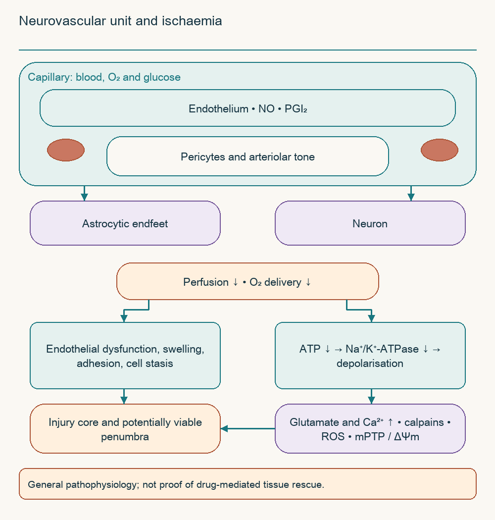
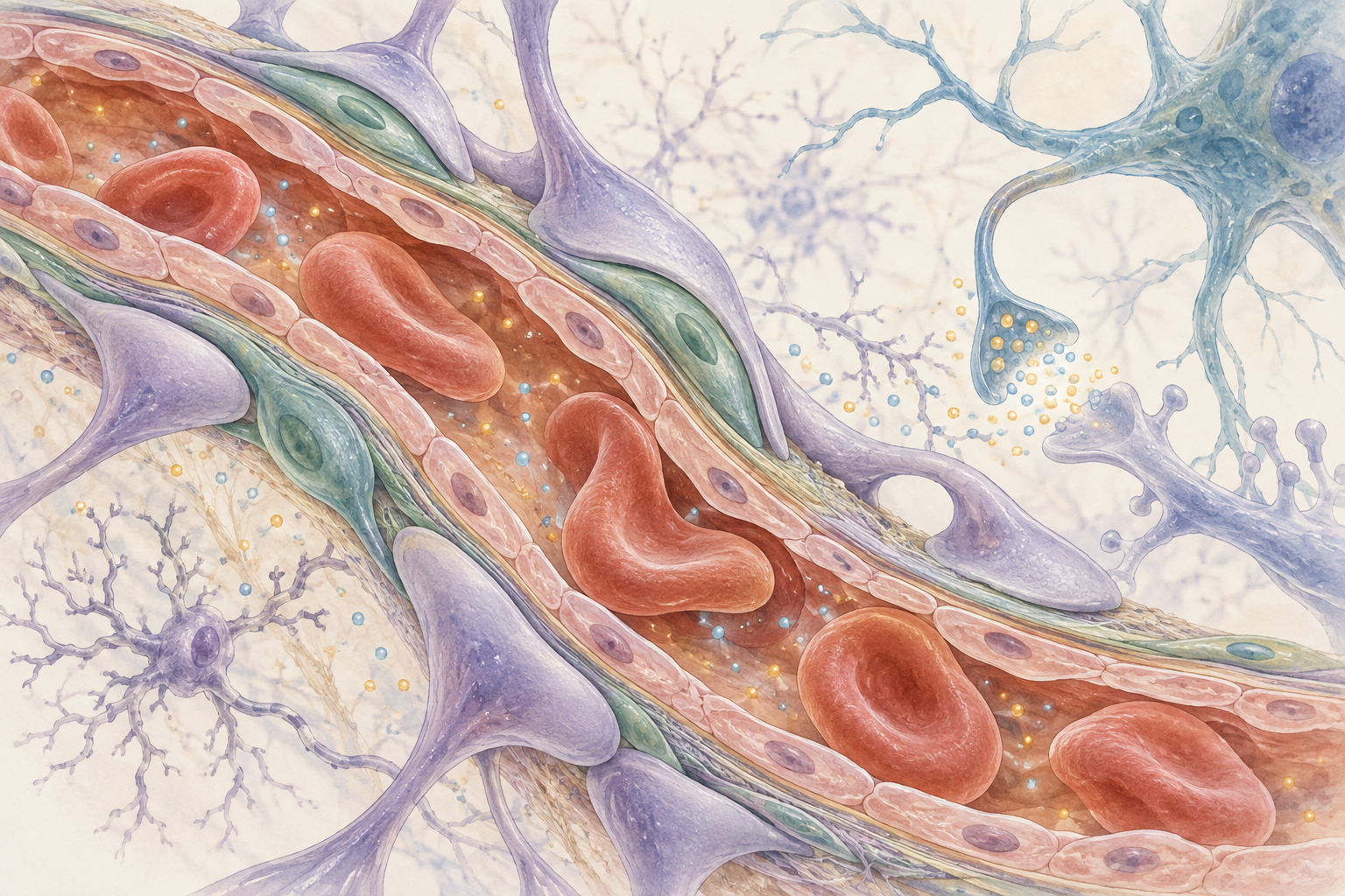
Neurons continuously consume ATP to maintain ion gradients, transmit signals, and remove transmitters from the synaptic cleft. Brain energy reserves are limited, although astrocytic glycogen provides a local buffer. Neurovascular coupling connects neural activity with oxygen and substrate delivery, while the neurovascular unit includes endothelium, pericytes, arteriolar smooth muscle, astrocytic endfeet, and neurons. This system cannot be reduced to a single vessel whose dilation resolves every form of ischaemia.
Reduced perfusion limits oxidative phosphorylation and compromises the Na⁺/K⁺-ATPase. Membranes depolarise, glutamate transport and release change, and calcium loading increases. Endothelial dysfunction, swelling, inflammatory adhesion, and microcirculatory disturbances can simultaneously obstruct delivery even after a large artery reopens. Experiments involving pericytes demonstrated persistent capillary constriction after ischaemia, but the contribution of each vascular segment depends on the model and how the cells are defined.
Excess Ca²⁺ activates calpains, affects phospholipases, and increases mitochondrial stress. Loss of mitochondrial membrane potential, ΔΨm, reactive oxygen species production, and increased membrane permeability can contribute to cell death. Caspases, cytochrome c release, and AIF translocation nevertheless belong to different injury branches rather than one mandatory sequence. There is no universal patient threshold of “ATP below 20%” that automatically triggers the same events.
The penumbra is dysfunctional but potentially viable tissue surrounding the ischaemic core. Its condition depends on time, collateral flow, oxygen, temperature, and other factors. Increased Ca²⁺ can enhance Ca²⁺/calmodulin-dependent enzymes, but this does not demonstrate universal PDE1 overexpression in every ischaemic cell. For vinpocetine, a plausible molecular hypothesis therefore remains distinct from demonstrated penumbral rescue in humans.
2. Origin, chemical identity, and access to the brain
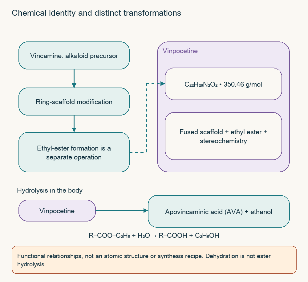
Vinpocetine is a semisynthetic derivative of the vincamine alkaloid family associated with lesser periwinkle. Its molecular formula is C₂₂H₂₆N₂O₂ and its molar mass approximately 350.46 g/mol; a functionally important feature is the ethyl ester of apovincaminic acid. Stereochemistry and the fused ring system are part of its identity, so a few symbolic rings must not be presented as an accurate atomic structure. The plant origin of its precursor also does not make vinpocetine a dietary nutrient or an equivalent of periwinkle extract.
The conversion of vincamine cannot be written simply as “remove H₂O → free apovincaminic acid”. Dehydration and modification of the ester group are separate operations, and obtaining the free acid requires a separate ester transformation. The conceptual diagram therefore distinguishes scaffold modification from ethyl-ester formation rather than offering a laboratory synthesis recipe. The reverse hydrolysis is important for pharmacokinetics: vinpocetine yields apovincaminic acid and ethanol, not a new supply of free parent drug.
Lipophilicity favours membrane interactions but does not by itself determine absorption speed, unbound concentration, or target occupancy in the brain. Ionisation, protein binding, tablet dissolution, and metabolism also matter. PET with radiolabelled vinpocetine demonstrates access of radioactivity to the brain, but tracer signal is not a measurement of unchanged, unbound drug inside a particular cell. A guaranteed two-to-four-minute crossing of the blood–brain barrier cannot therefore be assigned to every tablet and every person.
3. PDE1: connecting calcium to cyclic nucleotides
Enzyme regulation and isoforms
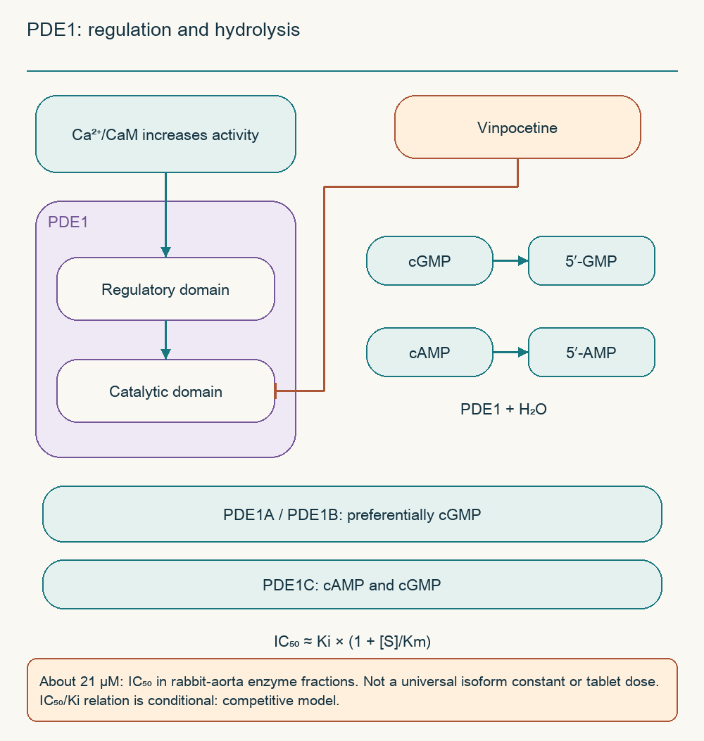
Phosphodiesterases terminate cyclic-nucleotide signals by hydrolysing their cyclic phosphodiester bond. PDE1 is characterised by activation through Ca²⁺/calmodulin: calcium changes calmodulin conformation, and interaction with the enzyme's regulatory region enhances catalytic activity. Regulatory and catalytic domains belong to the same protein, with phosphorylation and alternative splicing further modifying its properties. Calmodulin calcium occupancy should not be depicted as a universally binary switch requiring exactly four ions.
cGMP + H₂O → 5′-GMP; cAMP + H₂O → 5′-AMP.
Water participates in these hydrolytic reactions, and the products no longer provide the same signal as the cyclic molecules. cGMP abundance depends on guanylate-cyclase synthesis and degradation by several PDEs. Blocking one degradation branch therefore does not guarantee the same signal increase in every tissue. Local protein complexes and isoform distribution matter more than assuming a single uniform cellular cGMP pool.
Isoform
Substrate characteristic
Interpretation
PDE1A
Usually preferential affinity for cGMP
Splice variant and assay conditions affect kinetics
PDE1B
Also preferential affinity for cGMP
Present in neural tissue but not the only brain PDE
PDE1C
High affinity for both cAMP and cGMP
Effects depend on cell type and substrate availability
Cloning human PDE1A and PDE1C established differences in their sequence and properties. The table describes the family rather than assigning a fixed percentage of brain nucleotide hydrolysis. Km values require a specified protein, substrate, and assay environment. No universal constant is assigned to the names PDE1A, PDE1B, or PDE1C without identifying the variant.
What vinpocetine experiments actually show
Early experiments with rabbit-aorta enzyme fractions found preferential inhibition of the Ca²⁺-dependent phosphodiesterase by vinpocetine. Half-maximal inhibition occurred at approximately 21 µM in that system, and increasing calmodulin did not overcome the inhibition. Vascular strips mainly showed increased cGMP rather than cAMP. This supports a mechanism in a defined model, not a complete modern selectivity panel covering human PDEs.
IC₅₀ is the concentration that halves measured activity in a specified assay. It is neither a tablet dose nor a binding constant nor the minimum effective brain concentration. For competitive inhibition, the conditional relation IC₅₀ ≈ Ki × (1 + [S]/Km) illustrates dependence on substrate concentration [S], but only when the model assumptions hold. Unsupported PDE2–PDE5 values and the resulting guarantee against cardiac or gastrointestinal adverse effects are consequently not retained.
4. Vascular smooth muscle: from NO to relaxation
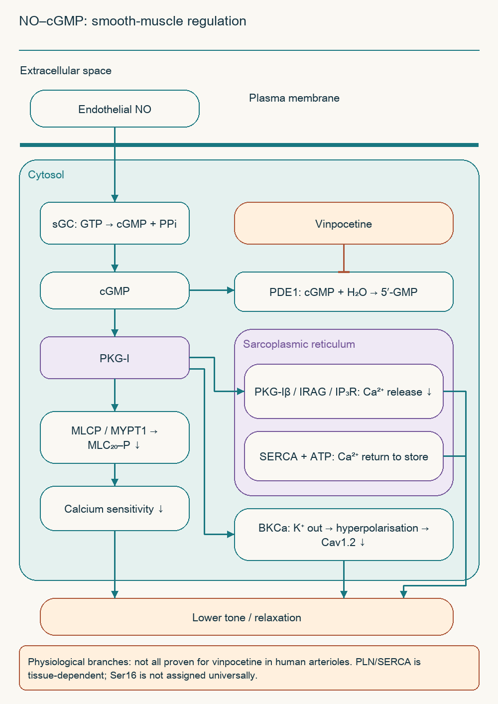
Endothelial NO diffuses into a smooth-muscle cell and activates soluble guanylate cyclase, sGC. The enzyme converts GTP into cGMP, which regulates type-I protein kinase G, PKG-I. Unlike the conventional PKA illustration, PKG regulatory and catalytic regions are in the same polypeptide; activation does not release separate catalytic subunits. PDE1 inhibition can prolong an existing cGMP signal but neither replaces NO formation nor produces it from nothing.
GTP → cGMP + PPi; MLC₂₀–P + H₂O → MLC₂₀ + Pi.
The first reaction represents sGC activity, the second regulatory myosin-light-chain dephosphorylation. MYPT1 directs myosin-light-chain phosphatase, MLCP, to its substrate, and interaction with PKG-Iα contributes to reduced calcium sensitivity of the contractile apparatus. Contraction can therefore weaken at the same Ca²⁺ concentration. Different MYPT1 phosphorylation sites have different functions, so “every MYPT1 phosphorylation activates MLCP” is incorrect.
Other cGMP branches involve calcium stores and membrane excitability. The PKG-Iβ–IRAG–IP₃-receptor complex can limit calcium release from the sarcoplasmic reticulum, while SERCA returns calcium to that store using ATP. In some tissues, phospholamban regulation facilitates SERCA activity, and BKCa opening favours K⁺ efflux, hyperpolarisation, and less Cav1.2 activation. These processes were demonstrated in different experimental systems, not as a proven simultaneous response to oral vinpocetine in a human cerebral arteriole.
Phospholamban requires particular attention to tissue context: well-studied cardiac Ser16 regulation cannot simply become a precise map of vascular drug action. Similarly, a possible cAMP/PKA increase after PDE1C inhibition depends on that isoform and competing PDEs. The diagram consequently distinguishes general physiology from the experimentally supported drug intervention. It explains vasodilation without extending evidence beyond what was measured.
5. Perfusion pressure and vascular steal
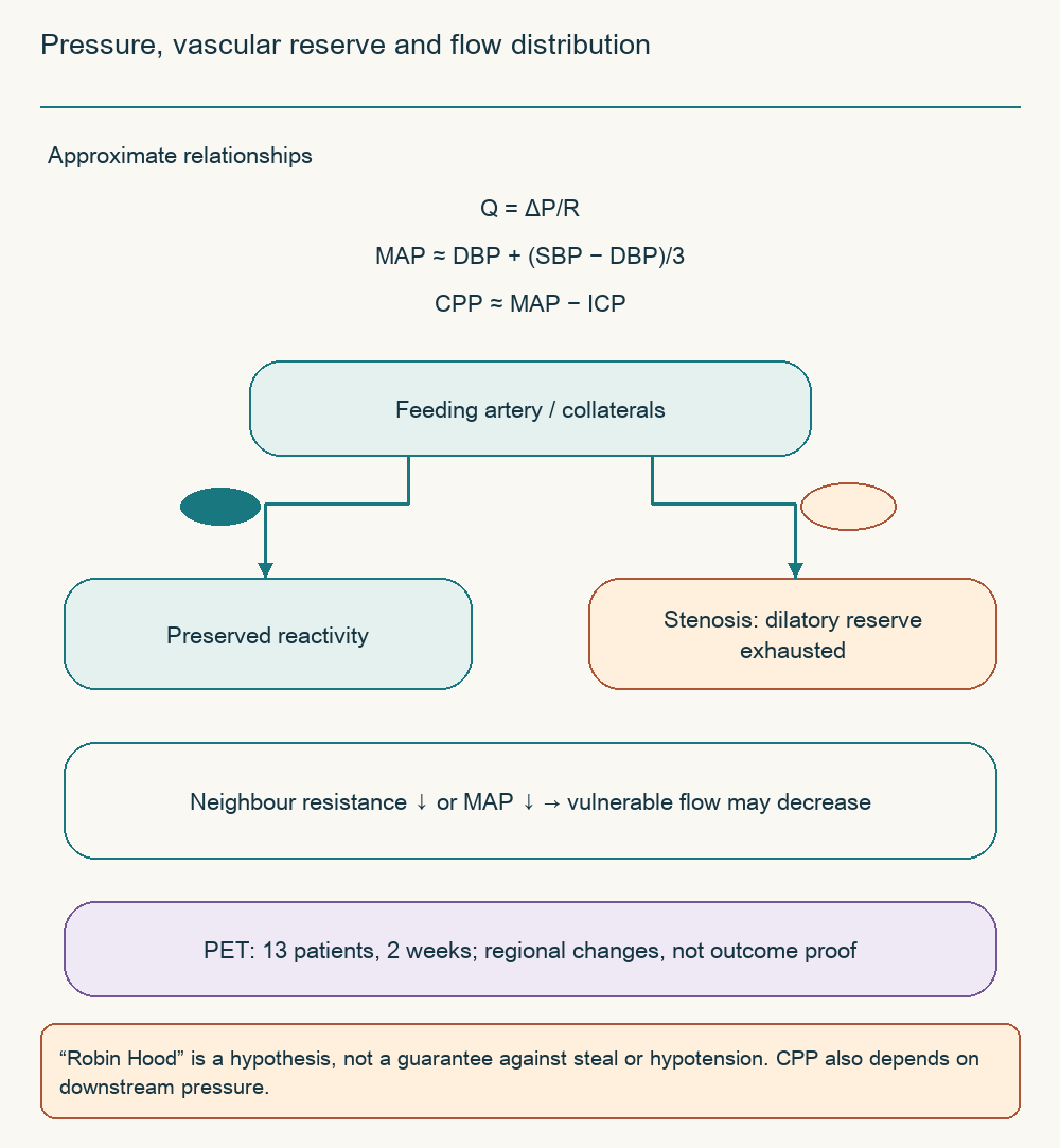
Blood flow depends on pressure, resistance, collaterals, and microcirculatory conditions as well as vessel diameter. In simplified form, Q = ΔP/R, where Q is flow, ΔP the pressure difference, and R resistance. Mean arterial pressure is often approximated by MAP ≈ DBP + (SBP − DBP)/3; SBP and DBP are systolic and diastolic pressures, and the expression is not exact integration of every pulse waveform. Cerebral perfusion pressure is CPP ≈ MAP − ICP when intracranial pressure determines downstream opposition and elevated venous pressure is not dominant.
Beyond a severe stenosis, distal vessels may have exhausted their dilatory reserve. Lower resistance in a neighbouring territory or reduced systemic pressure can then worsen flow to the vulnerable region. Steal nevertheless depends on anatomy and reactivity rather than being inevitable with every vasodilator. A maximally dilated arteriole also cannot simultaneously be described as a vessel rescued solely by relief of spasm.
A small double-blind PET study in chronic stroke included 13 people: six received infusions containing vinpocetine and seven infusions without it. Regional flow changed after two weeks, more prominently with the drug, while global glucose metabolism did not change markedly. This is a physiological observation, not proof of reduced mortality or guaranteed penumbral preservation. The historical “Robin Hood effect” can describe a redistribution hypothesis but cannot promise absence of hypotension or steal.
6. Sodium channels, calcium, and excitotoxicity
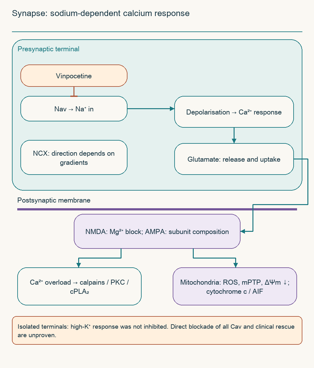
During ischaemic depolarisation, Na⁺ entry and disrupted gradients change nerve-terminal function. Calcium channels, neurotransmitter transport, and the sodium–calcium exchanger NCX are connected to these changes but are not interchangeable mechanisms. NCX can reverse under particular voltage and concentration conditions, rather than obligatorily doing so during every ischaemic episode. Excess glutamate release and impaired clearance increase postsynaptic excitation.
In isolated nerve endings, vinpocetine inhibited responses triggered by sodium-channel activation. With 4-aminopyridine, a smaller intracellular Na⁺ rise was accompanied by a smaller calcium response. However, the Ca²⁺ rise caused by high extracellular K⁺ was insensitive to vinpocetine in the same study. Reduced calcium signalling in that model therefore follows limitation of sodium-dependent excitation, not proven direct blockade of every L-, T-, and N-type calcium channel.
NMDA receptors conduct Ca²⁺ when agonist binding combines with relief of the voltage-dependent magnesium block. AMPA calcium permeability depends on subunit composition, so not every AMPA channel should be drawn as an identical Ca²⁺ source. Excess calcium activates calpains, affects PKC and cPLA₂, and mitochondrial loading can contribute to ROS, ΔΨm loss, and cell death. Limiting excitation can attenuate these processes experimentally but cannot establish complete protease inactivation in a patient.
Clinical translation is complicated by exposure because many cellular effects were obtained at micromolar concentrations. One oral pharmacokinetic study in elderly volunteers found peak parent-drug concentrations in the tens of nanomoles per litre. These values cannot be directly compared without accounting for unbound fraction, tissue, and administration route, but the discrepancy requires explanation. A demonstrable molecular effect is therefore not equivalent to its achievement at a therapeutic dose.
7. Glucose delivery and energy reactions
Barrier and cell-membrane transport
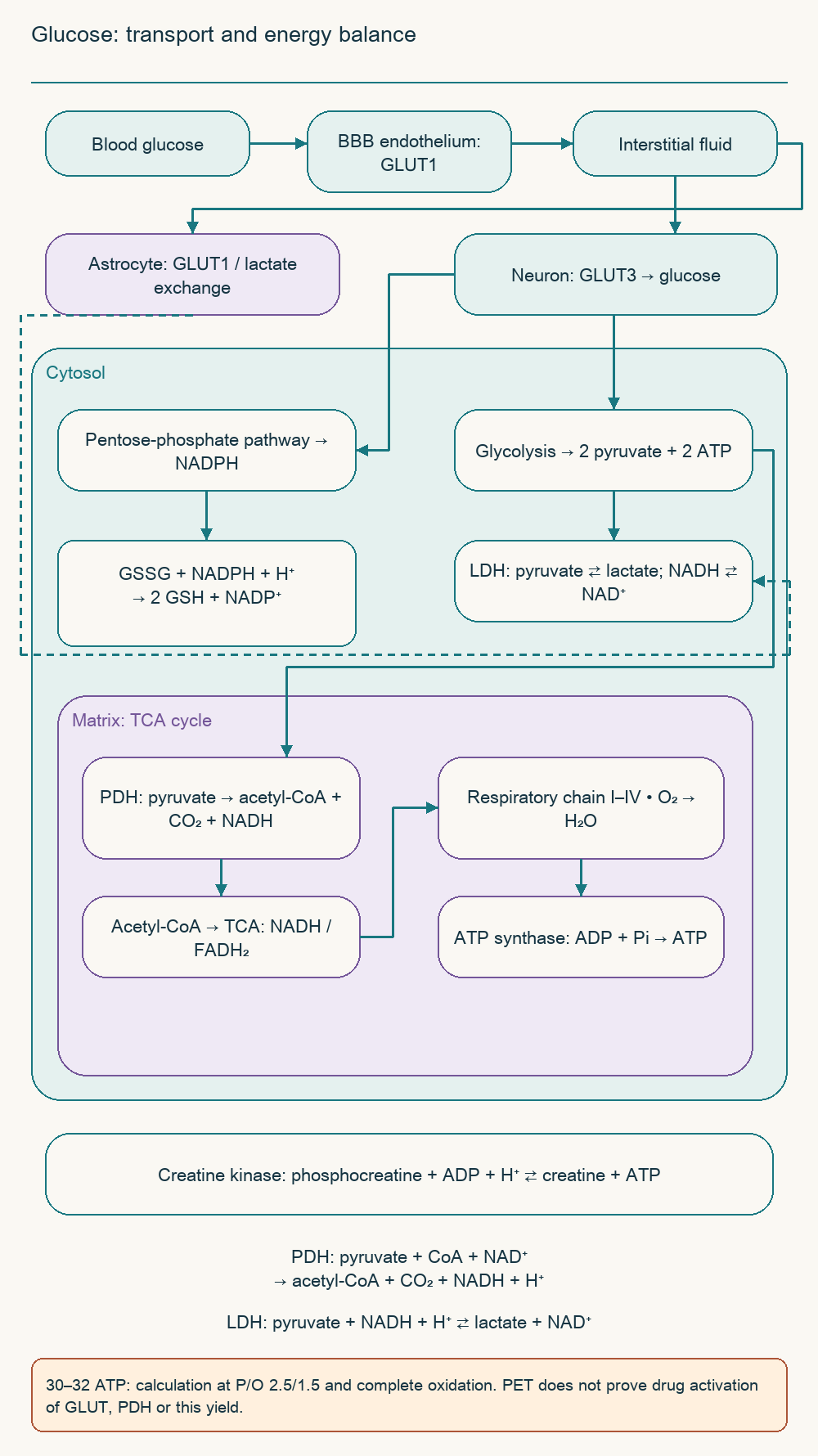
GLUT1 makes an important contribution to glucose passage across blood–brain-barrier endothelium. GLUT3 is principally associated with neuronal uptake, while glia also participate in substrate exchange. Neurons obtain glucose from interstitial fluid rather than necessarily through a serial “astrocytic syncytium → GLUT3” relay. Intercellular lactate exchange complements this system and depends on activity and metabolic conditions.
In a single-dose PET study of vinpocetine, modelled glucose transport across the barrier changed without a significant rise in regional or global metabolic rate. This did not measure GLUT1/GLUT3 vesicle translocation or establish a universal 20–35% transport increase. FDG signal reflects delivery, transport, and phosphorylation of a glucose analogue within a particular model. It does not directly count ATP production or demonstrate PDH activation.
Glycolysis, mitochondria, and antioxidant reserves
Glycolysis converts glucose into two pyruvate molecules with a net yield of two ATP. The LDH reaction pyruvate + NADH + H⁺ ⇄ lactate + NAD⁺ regenerates NAD⁺ and supports glycolysis when oxidation is limited. Lactate is therefore not an unconditional anaerobic dead end: it can be transported and oxidised when conditions permit. During severe ischaemia, glycolytic energy remains insufficient, and acid–base and ionic disturbances aggravate injury.
Mitochondrial PDH catalyses pyruvate + CoA + NAD⁺ → acetyl-CoA + CO₂ + NADH + H⁺. Acetyl-CoA enters the tricarboxylic-acid cycle, reduced cofactors supply the respiratory chain, and oxygen is the terminal electron acceptor. A calculated 30–32 ATP per glucose assumes approximate P/O ratios of 2.5 for NADH and 1.5 for FADH₂ and depends on the shuttle carrying cytosolic reducing equivalents. This is a teaching balance for complete oxidation, not a measured vinpocetine response or a way to bypass oxygen absence.
The pentose-phosphate pathway supplies NADPH for processes including glutathione regeneration. GSSG + NADPH + H⁺ → 2 GSH + NADP⁺ supports antioxidant reserve, while creatine kinase buffers energy through phosphocreatine + ADP + H⁺ ⇄ creatine + ATP. These relationships explain the metabolic hypothesis in the source material. Direct human activation of PDH, creatine kinase, or the pentose-phosphate pathway by vinpocetine is not presented as established.
8. Rheology: red cells, platelets, and endothelium
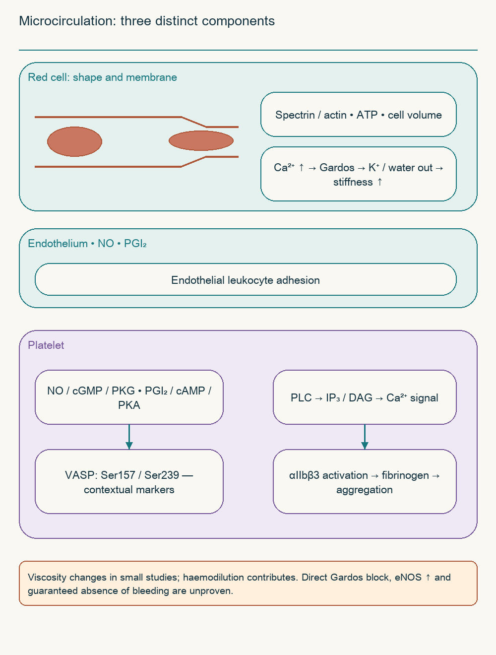
Whole-blood behaviour depends on shear rate, haematocrit, aggregation, and cell deformability. Plasma and whole blood are different measurement objects, and there is no single critical shear rate applicable to all vessels. Erythrocytes traverse narrow capillaries by changing shape; deformability depends on the spectrin–actin framework, cell volume, and membrane condition. Raised intracellular Ca²⁺ can activate the Gardos channel, promote K⁺ and water loss, and impair flexibility.
Small studies in cerebrovascular disease reported rheological changes after vinpocetine. In a 40-patient study, everyone first received parenteral treatment before continued oral drug was compared with placebo. The authors explicitly acknowledged haemodilution as a contributor to early changes. This design does not demonstrate direct Gardos-channel inhibition, increased ATP in every red cell, or prevention of recurrent stroke.
In platelets, cyclic nucleotides can restrain activation through PKA, PKG, and associated proteins. VASP phosphorylation at Ser157 and Ser239 provides context-dependent signalling markers, but is not always equivalent to reduced aggregation or a drug-specific signature. PLC generates IP₃ and DAG, calcium signalling supports αIIbβ3-integrin activation, and fibrinogen binding links platelets. Reduced integrin activation must be distinguished from reduced surface expression.
Endothelial NO and prostacyclin oppose platelet activation, while inflammatory adhesion molecules promote leukocyte interactions. Vinpocetine has been studied at several points in this system, but the clinical chain “PDE1 → VASP → no bleeding” is unproven. A universal 25–40% aggregation reduction with guaranteed haemorrhagic safety is not established. Vinpocetine consequently cannot replace proven antithrombotic treatment.
9. IKK/NF-κB: a separate experimental target
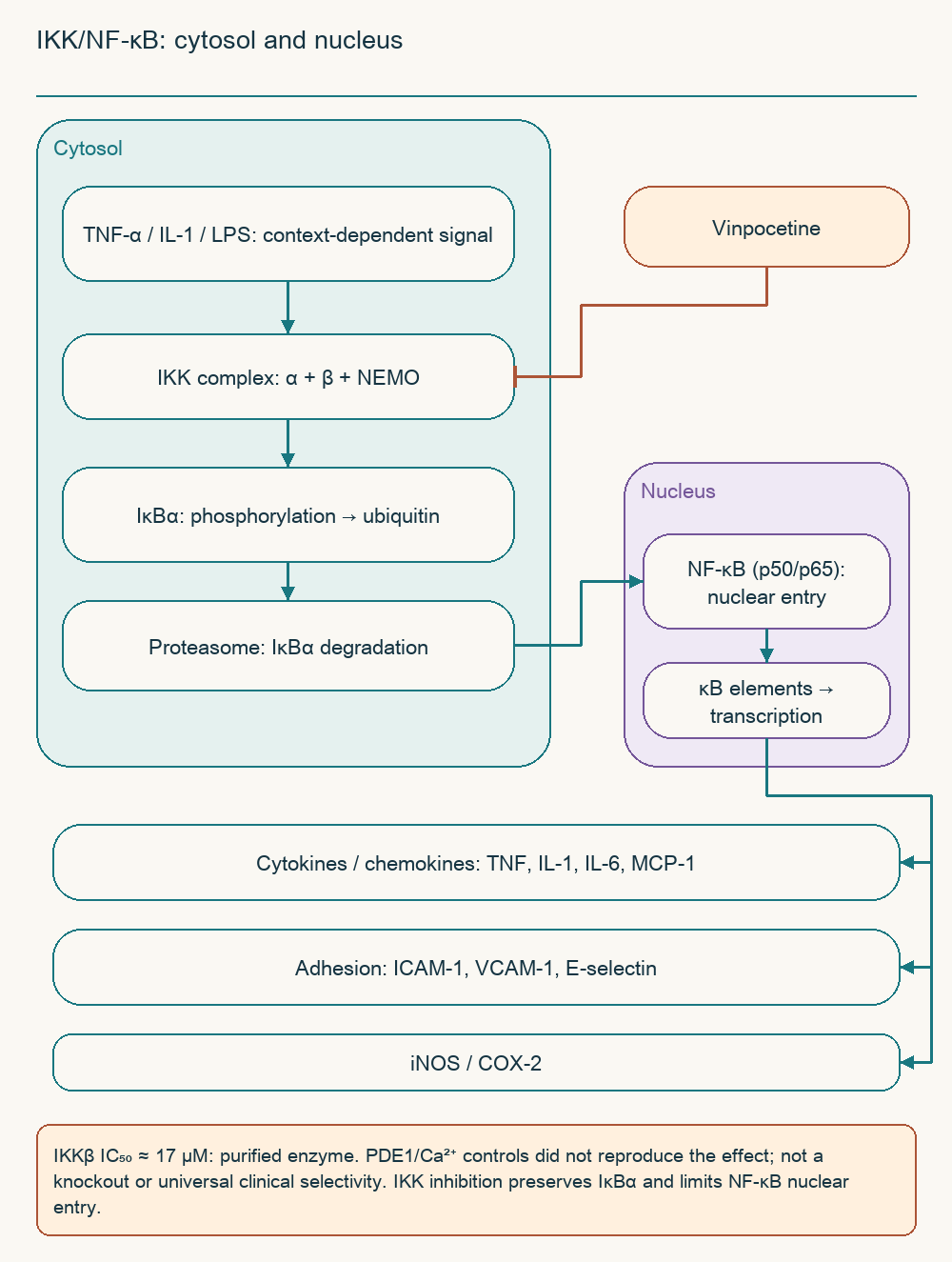
Inflammatory stimuli activate pathways converging on the IKK complex, containing IKKα, IKKβ, and regulatory NEMO. IκBα phosphorylation facilitates ubiquitination and proteasomal degradation. Released NF-κB, including the p50/p65 dimer, can enter the nucleus and alter inflammatory-gene expression. Cytokines, chemokines, adhesion molecules, and enzymes are potential products, but the response differs between cells and stimuli.
Jeon's research group found vinpocetine inhibition of this signalling in several cellular models and reduced lung inflammation in mice. Direct inhibition of purified IKKβ occurred with an IC₅₀ near 17 µM. A separate PDE1 inhibitor and calcium-signalling interventions did not reproduce the principal anti-inflammatory effect. Those pharmacological comparisons, rather than a claimed PDE1 knockout, supported a separate target.
This distinguishes reduced cGMP hydrolysis from restrained NF-κB activation. Nevertheless, an independent pathway does not imply absolute selectivity against all kinases or the same response in every organ. Microglia occupy diverse states, and a binary M1/M2 classification incompletely describes their behaviour after stroke. Experimental marker changes cannot become a promise of clinically proven microglial reprogramming.
A clinical study of 60 patients also reported inflammatory-signalling changes when intravenous vinpocetine was added. It takes the hypothesis towards human investigation but remains small and does not establish IKK inhibition as the sole cause of functional improvement. Its patients must not be merged with CAVIN as though these were the same cohort. Biomarkers, lesion size, and patient independence require separate assessment.
10. Pharmacokinetics: food, hydrolysis, and exposure
Absorption and bioavailability
Oral absorption and absolute bioavailability are different concepts. Absorbed drug may be metabolised before reaching the systemic circulation, so dissolution and intestinal passage do not determine the entire result. Older vinpocetine studies reported substantially different bioavailability estimates depending on methods, formulation, and participants. They cannot simply be combined into “6–7% fasting versus 50–60% with fatty food”.
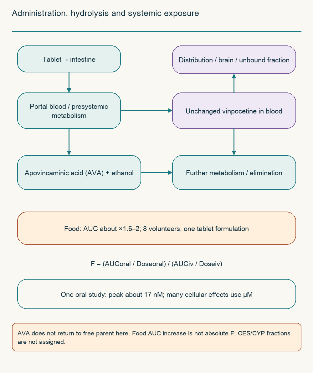
A small crossover study of eight volunteers found approximately 60–100% greater exposure to a particular tablet with food than fasting. This is an approximately 1.6–2-fold AUC increase, not a guaranteed seven-to-nine-fold increase. The experiment does not prescribe a universal dietary fat quantity. When a registered product is prescribed, meal timing follows its instructions rather than an attempt to maximise concentrations independently.
F = (AUCoral / Doseoral) / (AUCiv / Doseiv).
F is absolute bioavailability of unchanged drug, AUC the area under its concentration–time curve, and Dose the administered amount. Comparison with intravenous administration requires compatible kinetics and dose adjustment. A relative food-associated AUC increase does not replace measurement of F. This distinction explains how plausible numbers from separate studies can generate an incorrect dosing rule.
Distribution and metabolites
Vinpocetine ester hydrolysis forms apovincaminic acid, often abbreviated AVA. Parent and metabolite require separate diagram branches: AVA → free vinpocetine would misrepresent metabolism. Protein binding and tissue distribution further alter unbound concentrations available to targets. One study cannot establish universal human clearance fractions for CES1, CYP3A4, and CYP2D6.
In elderly volunteers, peak parent concentration after an oral dose was approximately 17 nM. Comparison with micromolar enzyme experiments helps assess mechanistic plausibility but does not independently demonstrate absence of every effect. Another small repeated-dose study found no significant accumulation in the investigated regimen. This cannot promise no accumulation, interaction, or kinetic change in every disease and prolonged course.
11. Clinical outcomes depend on the question
Cognitive impairment and chronic vascular symptoms
Early trials often enrolled people with “organic psychosyndrome” or “cerebral insufficiency” defined by historical criteria. These groups are not equivalent to modern Alzheimer's disease, vascular dementia, or subjective forgetfulness cohorts. A genuine double-blind trial of 203 outpatients compared two vinpocetine regimens with placebo for 16 weeks. Positive findings on particular scales do not establish dementia prevention or lasting everyday independence.
A systematic review included three older cognitive trials with 583 participants. It noted possible benefit signals alongside diagnostic heterogeneity, limited long-term follow-up, and analytic restrictions. The source's supposedly modern 484-patient study with exact ADAS-Cog and MMSE changes was not substantively identified. Those numbers are not used as evidence, and cognitive complaints require consideration of sleep, medication, mood, vascular factors, and other causes.
Acute stroke: earlier and later trials
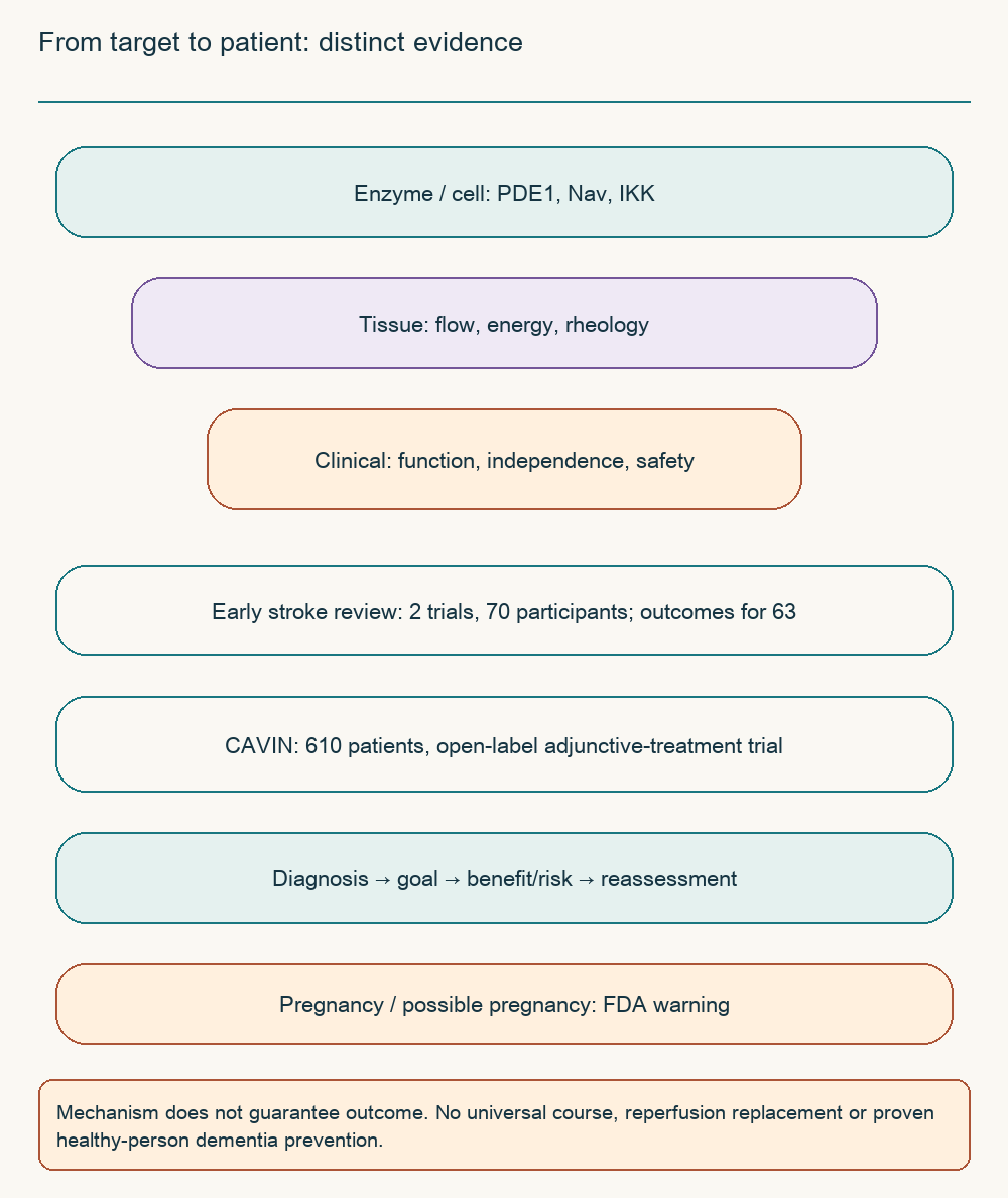
An older acute-stroke review included two small trials totalling 70 participants, with reported outcomes for 63. It could not reliably establish an effect on death or dependency. The later open-label multicentre CAVIN trial enrolled 610 patients and reported advantages of adding vinpocetine on several neurological and functional measures. These are meaningful later data, but lack of blinding and accompanying treatment limit generalisation to every clinical situation.
Later evidence cannot be described using a fictitious meta-analysis of “17 trials and over 1,800 patients”. Small inflammation studies and larger open-label trials ask different questions and carry different bias risks. A favourable scale result does not automatically mean fewer recurrent strokes or improved survival. Suspected acute stroke demands urgent diagnosis and assessment for reperfusion, which discussion of an additional drug must not delay.
Question
Available evidence
Unsupported inference
PDE1 and cGMP
Enzyme and vascular experiments
Identical target occupancy after any tablet
Blood flow and glucose transport
Small PET studies
Guaranteed penumbral rescue or ATP increase
Cognitive symptoms
Older placebo-controlled trials
Dementia prevention in healthy people
Stroke recovery
Pilot and later clinical trials
Replacement of reperfusion or secondary prevention
Inflammation
Cells, animals, and a small human study
Universal safe suppression of inflammation
Hearing, dizziness, and ocular microcirculation
Historical ethyl-apovincaminate studies reported audiometric changes in sensorineural hearing impairment. Their design and diagnostic heterogeneity cannot support a guaranteed 65% recovery rate or a single treatment-start window. Sudden hearing loss, persistent tinnitus, and dizziness need a diagnosis because a vascular explanation is not appropriate for every case. Waiting for a “microcirculation benefit” independently can divert attention from timely specialist care.
Experimental anti-inflammatory activity likewise does not prove treatment of diabetic macular oedema, normal-tension glaucoma, or stabilisation of visual fields. A cellular VEGF reduction, if demonstrated, is not clinical efficacy in retinopathy. These disorders need their own ophthalmic outcomes and comparison with established care. Sudden vision loss requires urgent assessment rather than vinpocetine use by analogy with cerebral blood flow.
12. Practical assessment and safety limits
Use decisions and response assessment
Vinpocetine status differs between countries: some have registered medicinal products, while elsewhere it is discussed as a supplement ingredient. Availability does not demonstrate healthy-memory benefits, and an authorised indication must not be extended to every proposed mechanism. Decisions begin with diagnosis, a defined goal, and benefit–risk assessment. The specific product, local instructions, concurrent drugs, and better-supported alternatives then matter.
The source's universal sequence of “infusions → tablets → twice-yearly repeat courses → tapering” is not an established mandatory protocol. Neither an 8–12-week duration nor a specific dose transition or tapering requirement follows from a single pharmacokinetic study. Research used different regimens, whose description is not a prescription for the reader. If a clinician prescribes the drug, the goal and reassessment date should be clear, with MMSE or MoCA changes interpreted alongside everyday function and repeat-testing effects.
Reproductive toxicity and other risks
A high lethal dose in rodents does not establish a wide human therapeutic window. Animal embryo–fetal development studies found adverse effects including pregnancy loss and developmental abnormalities. FDA advises pregnant people and those who could become pregnant not to take vinpocetine. This significant safety boundary cannot be softened by plant-origin claims or few complaints in a small clinical trial.
Situation
Practical significance
Pregnancy or possible pregnancy
Avoid supplement use; account for official warnings and product instructions
Breastfeeding or childhood
Do not extrapolate adult trials; specialist assessment and product instructions determine applicability
Arrhythmia, prolonged QT, or hypotension tendency
Discuss cardiovascular risk and accompanying medicines
Haemorrhage or high bleeding risk
Do not extrapolate ischaemic-stroke data; formulation and combinations are particularly relevant
Anticoagulants, antiplatelets, or antihypertensives
Individual compatibility assessment is needed; a plausible mechanism does not quantify risk
Nausea, abdominal discomfort, headache, palpitations, and blood-pressure changes should be assessed as possible adverse events rather than obligatory adaptation. Their frequency cannot be declared below 1% for every formulation and patient group. Medicines affecting rhythm, pressure, and haemostasis deserve particular attention. No unsupported guarantee of stable INR with warfarin or automatic adjustment to 15 mg with strong CYP inhibitors is offered here.
The most useful distinction for practice is between a target, a tissue change, and a clinical outcome. PDE1 and IKK explain scientific interest without resolving exposure and trial-quality questions. Vascular-risk management, rehabilitation, and investigation of cognitive complaints remain independently important. The context can be compared with articles on arginine and polyphenols and magnesium, without turning shared pathways into a recommendation to combine products.
FAQ
Does it improve memory in healthy people?
Older cognitive-impairment trials do not establish that benefit. Investigate symptoms, sleep, mood, and medication first.
Why is PDE1 insufficient to prove treatment benefit?
An enzyme assay does not measure unbound brain concentration or patient independence. Those require separate pharmacokinetic and clinical evidence.
Does food increase bioavailability sevenfold?
There is no universal confirmation. A small crossover study found about 1.6–2-fold higher tablet AUC, not a comparison of different absolute F estimates.
Is absence of vascular steal guaranteed?
No. Anatomy, pressure, and vascular reserve matter; a small PET study cannot provide that guarantee.
Can vinpocetine replace stroke care?
No. Adjunctive trials do not replace urgent diagnosis, reperfusion, rehabilitation, or secondary prevention.
Is it safe during pregnancy?
It cannot be assumed safe. Animal reproductive toxicity prompted FDA advice against use in pregnancy or possible pregnancy.
What dose is appropriate for a self-directed course?
No universal course is established here. Trial regimens are not prescriptions; diagnosis, product instructions, and drug combinations determine decisions.