Tributyrin and Sodium Butyrate: Chemistry, Delivery, and Limits of Clinical Inference
How tributyrin and sodium butyrate differ in chemistry, lipolysis, release location, and clinical evidence—and why molecular composition alone does not prove delivery to the colon.
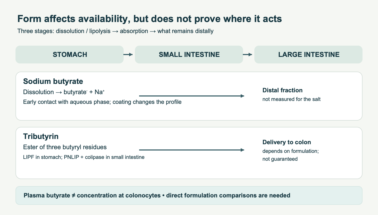
Butyrate is at once a product of microbial fermentation, an energy substrate for colonocytes, and a signaling molecule. That makes the question of its oral form seem straightforward: if three butyric-acid residues are attached to glycerol, could more butyrate reach the large intestine than from a rapidly dissolving salt? Tributyrin does differ chemically from sodium butyrate. Yet the site of release is determined by lipolysis, absorption, and product design, not simply by the number of ester bonds. Guaranteed delivery of ordinary tributyrin to the colon, and clinical superiority over a coated salt, have not been established.
In practice, three goals need to be kept separate: improving tolerance of oral intake, achieving systemic exposure to butyrate, and producing local exposure at the distal intestinal mucosa. A post-dose plasma concentration characterizes the first phase of absorption and presystemic metabolism, but does not prove the concentration at the colonocyte surface. Conversely, fecal butyrate depends on both production and mucosal consumption; it is not a direct measure of the dose delivered by a particular capsule.
1. Molecules and dose equivalents
Butanoic acid, CH₃–CH₂–CH₂–COOH, has four carbon atoms, a molecular mass of about 88.11 g/mol, and a pKa of about 4.8. In physiological aqueous conditions, a substantial fraction is present as butanoate. Sodium butyrate (NaC₄H₇O₂; about 110.09 g/mol) dissociates readily into Na⁺ and butanoate; acidification shifts the equilibrium butanoate⁻ + H⁺ ⇌ butanoic acid. The odor comes from volatile free acid, including when the salt contacts moisture and acid. Organoleptic properties therefore depend on the coating, packaging, and moisture as well as the salt name printed on the label.
Tributyrin, or glyceryl tributyrate, has the formula C₁₅H₂₆O₆ and a molecular mass of about 302.36 g/mol. The three hydroxyl groups of glycerol are esterified with butyric-acid residues. The intact molecule has no free carboxyl group and carries no accompanying sodium. Complete hydrolysis can be written as: tributyrin + 3 H₂O → glycerol + 3 molecules of butanoic acid. By mass, this yields approximately 264.3 mg of free acid per 302.4 mg of tributyrin, or 87.4% of the calculated acid equivalent. For sodium butyrate, the calculated equivalent is about 80.0% of the salt mass. These values help compare moles in product studies, but do not predict bioavailability or the site of delivery. For example, 500 mg of tributyrin corresponds to approximately 4.96 mmol of butyrate residues; 500 mg of NaB corresponds to approximately 4.54 mmol. The latter dose also contributes 4.54 mmol, or about 104 mg, of sodium. Patients limiting sodium should count it within the overall diet without overstating the risk of an arbitrarily selected dose.
The Henderson–Hasselbalch acid–base equation, pH = pKa + log([butanoate⁻]/[butanoic acid]), describes the distribution of forms in an aqueous phase. At pH 5.5, the fraction of undissociated acid is about 17%; at pH 7 it is below 1%. This equilibrium should not, however, be translated directly into a percentage of gastric absorption. The mucus layer, surface area, emptying rate, pH gradients, and transporters make the amount absorbed an empirical question. The original precise claims that NaB almost completely disappears within the first centimeters of the intestine are not supported by direct human measurements.
| Parameter | Uncoated sodium butyrate | Unencapsulated tributyrin | Enteric or microencapsulated form |
|---|---|---|---|
| Active chemical carrier | Soluble butyrate salt | Triacylglycerol with three butyryl residues | Depends on the core: salt or ester |
| What triggers release | Dissolution and dissociation | Lipases at the lipid–water interface | Coating dissolution, erosion, diffusion, and subsequent lipolysis |
| Sodium per mole of butyrate | One mole | None | Depends on the core |
| Distal release | Not implied by the salt composition | Not implied by the ester composition | Possible when the profile of the specific formulation is confirmed |
| What needs measurement | Segmental concentration and tolerability | Hydrolysis products, systemic and luminal kinetics | Dissolution, stability, transit, and clinical endpoints |
2. Lipolysis and the actual site of release
Tributyrin’s ester bonds are more resistant to simple dissolution in the stomach than the salt. That does not mean there is no release in the stomach: gastric lipase LIPF acts in an acidic environment at accessible lipid-droplet surfaces. Then, in the duodenum, pancreatic triacylglycerol lipase PNLIP, with colipase CLPS, hydrolyzes mainly the sn-1 and sn-3 positions. The sequence tributyrin → dibutyrin + butyrate → 2-monobutyrin + another butyrate describes a typical route; other lipases, esterases, and acyl-group migration can release the third residue. The fraction of each intermediate depends on emulsification, meal composition, and digestion conditions. Fixed percentages should not be assigned without measuring the particular product.
Bile salts and phospholipids increase the available interfacial area. Colipase allows PNLIP to work at the droplet surface in the presence of bile, which would otherwise impede lipase adsorption. An emulsion droplet, mixed micelle, free acid, and monobutyrin are different physical states; calling every luminal tributyrin preparation a “micelle” is inaccurate. The hydrolysis rate depends on particle distribution, surface area, lipase and colipase concentrations, bile acids, pH, motility, food, and pancreatic function. A simplified Michaelis–Menten equation without a measured surface concentration of substrate does not give a universal release time.
After release, butyrate and some lipolysis products can be absorbed in the small intestine. Lipolysis during transit therefore does not mean that the same molecule crosses the ileocecal junction. Experimental work on emulsion delivery started from the premise that ordinary tributyrin is predominantly digested and absorbed before the large intestine; special emulsion structures were studied for distal delivery. By contrast, a capsule with protected sodium butyrate may reduce early contact between the salt and the mucosa. A “tributyrin versus NaB” comparison without the coating composition and release conditions is effectively a comparison of unspecified products. The source text’s claims of 15–30% intact ester in the cecum, a six- to tenfold distal AUC, and an obligatory late peak are not supported by an adequate direct comparison in humans.
Evidence for targeted delivery requires information on breakdown of the formulation at physiological pH and in the presence of enzymes, followed preferably by segmental concentration measurements or a validated release marker. Plasma AUC is not a substitute for luminal exposure: a high systemic signal is compatible with early absorption, while a low signal may reflect extensive first-pass metabolism. Finding butyrate in stool after dosing also does not rule out microbial production. Isotope labeling, segment-specific aspirate sampling, and measurement of the release profile would be more informative, but are not available in every study.
Why a salt coating and a lipid matrix are not the same
“Microencapsulated sodium butyrate” can refer to different technologies: salt particles in a solid lipid matrix, granules with a diffusion coating, acid-resistant coatings, or combinations of these. A lipid matrix changes wetting and the rate at which the core dissolves; a pH-threshold polymer should open in a specified medium; a diffusion barrier releases salt gradually after water penetrates. Granule size, active-salt fraction, layer thickness, and surface geometry differ among products. The word “coated” alone does not establish that the entire dose will reach the large intestine. Bile, lipases, pH, mixing, and transit time can change the profile. In addition to laboratory dissolution at several pH values, testing needs conditions with food, enzymes, and a reasonable range of motility. Without such information, lipid coatings cannot be called unstable and polymer coatings cannot be assumed to cause explosive release, as the source text did.
Tributyrin provides chemical protection of the free carboxyl group, but that protection ends when gastric lipase and pancreatic juice act on it. For true targeted delivery, the ester can also be incorporated into a particle or coating. The result then depends on the combination of chemistry and technology and cannot be attributed to free tributyrin alone. Work with tributyrin emulsions examined how changing lipolysis conditions affects colonic release; this is formulation development, not human evidence of cancer prevention. Comparative studies should analyze released butyrate as well as dibutyrin and monobutyrin; otherwise part of the dose may be incorrectly counted as “lost.” Solubility, diffusion through the aqueous unstirred layer, and conversion by bacterial esterases also differ by segment.
For local treatment of distal proctitis, an ordinary oral product is a particularly poor proxy for a rectal dose: the rectum lies beyond a long transit path and multiple sites of absorption. Rectal delivery creates local contact but has its own limitations in convenience, coverage, solution retention, and tolerability. Combining enema studies with capsule studies inflates confidence in an oral strategy. Comparisons should specify the anatomical target—rectal, left-colonic, or whole-colon mucosa—and the method used to assess that target.
3. Colonocyte transport and bioenergetics
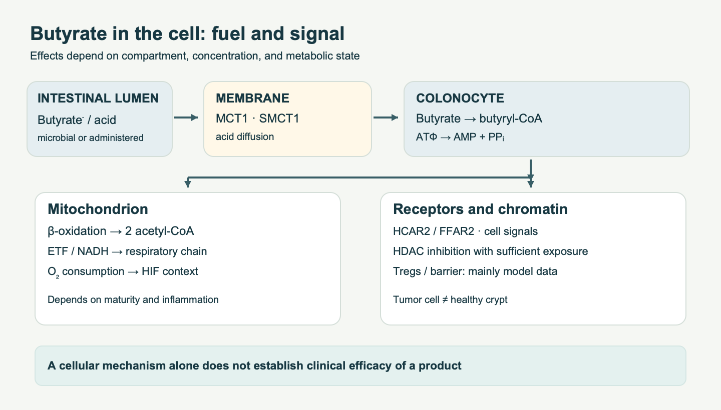
If butyrate reaches the colon, transport across the apical membrane may involve proton-coupled MCT1/SLC16A1, sodium-coupled SMCT1/SLC5A8, and diffusion of the undissociated acid. MCT1 carries a monocarboxylate together with H⁺ and is associated with basigin/CD147; its membrane localization depends on the cellular context. SMCT1 uses the Na⁺ gradient maintained by Na⁺/K⁺-ATPase and can support uptake at lower substrate concentrations. Both transporters also carry other monocarboxylates. Kₘ values reported in isolated systems are therefore not a constant “delivery rate” for a person with intestinal inflammation. Changes in SLC16A1 or SLC5A8 expression may modify the response, but a single gene cannot diagnose butyrate deficiency.
Butanoate is then activated to butyryl-CoA at the cost of ATP converted to AMP and PPᵢ: butyrate + CoA + ATP → butyryl-CoA + AMP + PPᵢ. Several acyl-CoA synthetases may act on short-chain substrates; assigning the entire reaction exclusively to ACSS1 or ACSS2 without a specific cell model is not justified. In mitochondria, ACADS (short-chain acyl-CoA dehydrogenase) converts butyryl-CoA to crotonyl-CoA; electrons from the FAD-dependent reaction pass through ETF and ETF:ubiquinone oxidoreductase to the ubiquinone pool. Crotonase hydrates the double bond, hydroxyacyl-CoA dehydrogenase produces acetoacetyl-CoA and NADH, and thiolase, with CoA-SH, yields two acetyl-CoA molecules. The tricarboxylic acid cycle and respiratory chain then use the acetyl groups and oxygen. This pathway does not mean intact butyryl-CoA freely diffuses across both mitochondrial membranes: access of short-chain substrate and its site of activation depend on compartment and tissue; the carnitine shuttle for long-chain acyl groups should not be mechanically copied here.
In differentiated colonocytes, butyrate is an important and often predominant energy substrate. Experiments with epithelium and microbiota show a relationship between its oxidation and O₂ consumption, but a fixed 70–80% share of ATP for every colonocyte, disease state, or intestinal region is not justified. Crypt stem cells, differentiated cells, and inflamed cells differ metabolically. Early observations of an “energy deficiency” in ulcerative colitis are useful as a hypothesis, but active inflammation itself changes transport, respiration, and microbiota. Butyrate insufficiency cannot explain the whole disease.
Higher epithelial oxygen consumption supports a relatively hypoxic surface and an anaerobic niche. In model experiments, changing the microbiota and losing butyrate producers alters colonocyte respiration and expands ecological opportunities for facultative anaerobes. The metabolite may activate PPARγ-related programs and restrict available oxygen and nitrates; this does not mean one tributyrin capsule prevents Salmonella or C. difficile infection. Local pO₂ is also determined by blood flow, inflammation, and tissue architecture.
Hydroxylation of HIF-1α by prolyl hydroxylases requires O₂, Fe²⁺, and 2-oxoglutarate; when O₂ is sufficient, VHL recognizes the hydroxylated protein and directs it for proteasomal degradation. Reduced O₂ availability stabilizes HIF-1α; its heterodimer with HIF-1β changes expression of adaptation and barrier genes. Work by Kelly and colleagues showed a cross-talk between microbial butyrate and epithelial HIF in experimental systems. The source claim that tributyrin reliably lowers pO₂ below a specified threshold and directly restores all tight junctions in patients would require separate clinical measurements.
4. Receptors, chromatin, and the crypt
Butyrate interacts with short-chain fatty acid receptors, including HCAR2/GPR109A and FFAR2/GPR43; the contribution of FFAR3/GPR41 depends on cell type and concentration, and acetate and propionate are also its ligands. HCAR2 is linked mainly to Gᵢ signaling; FFAR2 can signal through Gᵢ and Gq. Receptor expression differs among epithelial, immune, and enteroendocrine cells. Experiments in Gpr109a-deficient mice support a role for this pathway in anti-inflammatory regulation, but they do not establish one linear cascade—“HCAR2 → complete NLRP3 blockade”—in all human macrophages. Interleukin-18, in contrast, may help maintain epithelial homeostasis; its complete suppression cannot be assumed to be universally desirable.
At millimolar concentrations, butyrate inhibits some zinc-dependent HDACs, shifting the balance of histone and nonhistone protein acetylation. Histone acetyltransferases obtain acetyl groups from acetyl-CoA; butyrate does not simply “add acetyl” directly to a histone. Different cell models have shown changes in p21/CDKN1A, tight-junction genes, and inflammatory responses. Intracellular concentration matters, as does whether the molecule is oxidized as fuel before it accumulates in the nucleus. There is no single IC₅₀ for every class I/II HDAC in living epithelium.
Healthy differentiated colonocytes actively use butyrate, whereas some tumor cell lines with a strong glycolytic phenotype oxidize it less; metabolite accumulation can strengthen HDAC inhibition, alter p21, and slow growth. This is one interpretation of the “butyrate paradox” shown in experimental systems. The real effect varies with tumor genotype, cell location, dose, and crypt state. When the mucosa is injured, excessive exposure of stem cells at the crypt base may suppress their proliferation; protection by the niche and by metabolism in mature cells also matters. Thus, “more butyrate” does not mean “better repair” in every compartment.
In mouse models, microbial butyrate promoted formation of Foxp3⁺ regulatory T cells through a combination of epigenetic and metabolic mechanisms. Parallel studies showed that peripheral Treg differentiation depends on antigen and cytokine context. These findings do not justify promising remission of autoimmune colitis from oral tributyrin. Similarly, reports of changes in MUC2 and tight-junction assembly in cell systems cannot be converted into a known thickness of the human mucus layer after taking a capsule.
5. What disease studies and models show
In diversion colitis, stopping fecal flow deprives a bowel segment of microbial food substrates, including precursors of short-chain fatty acids. Small studies of local irrigation or enemas containing short-chain fatty acids have shown that some patients may respond clinically. For example, a small crossover study in ten people with distal ulcerative colitis examined rectal sodium butyrate at 100 mmol/L and placebo, each for two weeks; this speaks to local exposure, not to the efficacy of oral tributyrin. Results of local therapy differed by design and population, and restoring bowel continuity addresses a different causal problem. Diversion colitis cannot be described as invariably and completely cured “at once” by butyrate.
In ulcerative colitis and Crohn’s disease, microbiota composition, fecal short-chain fatty acids, epithelial ability to use butyrate, and inflammatory activity are often associated. But fecal concentration may fall because production is lower or rise because uptake is impaired. This rules out diagnosing “deficiency” from one number, much less treating it as an independent cause of IBD. Experiments with DSS/TNBS, cell monolayers, and animal diets show mechanisms involving the barrier, immune response, and histology; dose, species, model, and route are not equivalent to treatment in humans. The source’s unsupported claim that tributyrin outperforms coated NaB in crypt repair by specific percentages is not accepted here.
For IBS, it is more informative to discuss specific outcomes than to promise microbiota correction. A study of microencapsulated sodium butyrate as an adjunct to usual care reported a reduction in the frequency of some symptoms over 12 weeks; it did not study tributyrin and did not show a convincing reduction in the severity of the syndrome as a whole. IBS subtypes differ in transit and sensitivity; a change in fecal butyrate in one phenotype cannot be generalized to everyone. Comparative analgesic effects of the two forms remain an open question.
After antibiotics, model data link loss of butyrate-producing anaerobes to changes in colonocyte oxygen consumption and greater substrate availability for facultative bacteria. The extent of loss depends on the drug and community; fixed figures of 90–98% should not be treated as universal. Tributyrin does not reproduce the whole ecosystem of dietary fiber and cross-feeding. It has not been shown to prevent antibiotic-associated colitis, C. difficile disease, or infection.
Dietary fiber is associated epidemiologically with lower colorectal cancer risk, and butyrate is being investigated as one mediator of that effect. Cell-cycle arrest and apoptosis in tumor cell lines do not mean tributyrin prevents APC/KRAS/TP53 mutations or removes micro-polyps in people. Studies of the gut–brain axis, microglia, GLP-1/PYY, liver, muscle, and blood pressure are mainly mechanistic or animal studies. They cannot be extrapolated to prevention of dementia, MASH, sarcopenia, or hypertension without appropriate clinical endpoints. Mouse receptor Olfr78 was studied especially in relation to propionate and acetate, renin, and blood pressure; this is not evidence that tributyrin directly protects human blood vessels.
Particular caution is warranted for premature infants at risk of necrotizing enterocolitis. Immature epithelium, TLR4 signaling, and microbial colonization make this an interesting model, but there is no reliable published clinical evidence here to recommend tributyrin to prevent NEC or claim reduced mortality. It should not be added to neonatal feeds on the basis of cell or animal models. Similarly, the original treatment percentages for radiation enteritis and ischemic injury are not supported by studies in the corresponding patient populations.
Biomarker limits and causal interpretation
Inflammation can change diet, transit time, microbial communities, and mucosal metabolism at the same time. A decrease in certain butyrate producers during active IBD is therefore an observation compatible with several directions of causation. It does not prove that a supplement will resolve the disease. Even within a single stool, a substantial share of molecules made nearer the cecum may be absorbed before excretion; a low fecal value is not the same as low local production. Conversely, a high fecal concentration in inflamed epithelium may sometimes reflect poor utilization. Studies need to distinguish microbial gene abundance, actual substrate flux, luminal metabolite, and cellular uptake.
Barrier studies often measure expression of ZO-1/TJP1, occludin, claudins, and MUC2. mRNA levels do not guarantee correct localization of a protein at the apical junction; stronger immunostaining is not equivalent to lower in vivo permeability. Sugar-probe tests reflect a particular region and time window, endoscopy shows tissue appearance, and symptoms reflect subjective experience. These levels complement one another. Butyrate may activate the barrier through AMPK and transcriptional changes in a cell model, but with marked erosion the barrier also depends on immune injury and repair. One marker cannot serve as a universal explanation of an effect or a substitute for clinical remission.
The epigenetic “paradox” calls for similar care. If millimolar butyrate slows growth in a tumor cell line, investigators need to establish whether intact human tumors can reach that exposure, assess their blood supply, distinguish normal from transformed tissue, and measure crypt stem-cell sensitivity. The population-level benefit associated with dietary fiber cannot be attributed to butyrate alone: fiber changes stool bulk, transit, bile acids, microbial nutrition, and other metabolites. Otherwise, correlation and a cellular response are incorrectly combined into false evidence that tributyrin prevents cancer.
6. Human studies of tributyrin: population, formulation, and outcome
Early clinical phases of oral tributyrin were conducted mainly in patients with solid tumors to study safety, pharmacokinetics, and whether pharmacological doses could achieve systemic butyrate exposure. The Conley and Edelman studies were not trials of treatment for ulcerative colitis or IBS, nor of targeted delivery to the colon. Their conclusion that tributyrin is an oral precursor of systemic butyrate cannot be turned into evidence of better local action on distal epithelium. Mouse and rat experiments comparing plasma kinetics of NaB and tributyrin also did not measure human distal mucosa.
In a small pilot study in 2024, 29 healthy adults were randomized between two doses of a tributyrin-containing product; 24 completed follow-up. Seven placebo days preceded 21 days of the active mixture. The capsules contained 200 mg of a complex that was 50% tributyrin: the groups therefore received 100 or 200 mg of actual tributyrin per day, although the complex weighed 200 or 400 mg. The product also contained acacia, partially hydrolyzed guar gum, and other ingredients, so observed changes cannot be attributed to the ester alone. Tolerability was satisfactory, but investigators found no convincing improvement in measured permeability or marked shift in the microbiota. Signals in inflammatory and metabolic markers were mostly trends rather than persuasive therapeutic effects; triglycerides increased in the higher-complex group. The authors proposed that release and action occurred mainly in the small intestine. The sequential placebo phase, small sample, and healthy population limit any inference about treating disease.
Studies of local butyrate, protected NaB, and tributyrin concern different interventions. A positive result for microencapsulated salt in IBS or IBD cannot be relabeled a “tributyrin study.” Likewise, failure of one coating does not make tributyrin automatically superior. Adequate direct comparisons of equimolar doses of specific formulations, with segmental pharmacokinetics and the same clinical outcomes, are lacking. Choosing a form requires knowing the precise active content, coating type, dissolution profile, food conditions, and study population.
7. Doses, tolerability, and combinations
There is no confirmed universal tributyrin regimen of “500–750 mg for prevention” or “1–2 g for 8–16 weeks of treatment” for gastroenterological diseases. The source material did not link those figures to suitable randomized studies in the relevant patients. Doses in early oncology phases were much higher and served a different purpose; they cannot be transferred to self-directed supplement use. The pilot experience with 100/200 mg of actual tributyrin per day in healthy adults does not establish a treatment dose. When comparing labels, distinguish milligrams of tributyrin, milligrams of a mixture, and “butyrate equivalent,” and account for sodium in salt products.
Potential adverse effects of oral forms include changes in taste or odor, nausea, bloating, discomfort, and diarrhea; tolerability depends on dose and product. Hydrolysis and release of the ester may differ with pancreatic insufficiency, impaired bile flow, altered bowel anatomy, or rapid transit. Evidence to guide self-selection of tributyrin is limited during an IBD flare, with bleeding or severe diarrhea, in pregnancy, and in children. Tolerability in a pilot of healthy adults is not a safety certificate for these populations. Claims of “94% compliance” and “30–45% discontinuation of the salt,” stated without a comparative study, have been excluded.
Resistant starch and some dietary fibers reach the colon and are fermented by microbial communities; this is a separate strategy for supporting endogenous production of short-chain fatty acids. Read more about fermentation and its limits in the articles on resistant starch and dietary fiber. Taking these alongside tributyrin could theoretically combine different routes of exposure, but clinical synergy has not been demonstrated. Strain-specific effects of probiotics, berberine, or curcumin cannot be combined into an established treatment regimen. Rectal butyrate provides local contact by a different route; an experimental tributyrin nanocapsule must first demonstrate its delivery profile and safety before any effect can be claimed.
8. Beyond the intestine: testing the causal chain
For a liver effect, the drug must not only be hydrolyzed; butyrate must also pass through enterocytes into the portal vein, undergo presystemic metabolism, and expose liver cells sufficiently. Pancreatic lipolysis of tributyrin followed by absorption makes this pathway biologically possible. In a mouse model of alcohol-related liver injury, preventive tributyrin reduced barrier disruption and liver injury; this was not a trial of treatment for metabolic dysfunction-associated steatohepatitis (MASH) in patients. In other models, butyrate changed lipid-metabolism gene expression and inflammatory measures, but the separate contributions of direct hepatic AMPK signaling, lower lipopolysaccharide influx, and altered food intake need testing. Demonstrating AMPK or ACC phosphorylation in an experiment is not equivalent to fibrosis regression. Tributyrin should not be called an antifibrotic drug on the basis of protein acetylation or Kupffer-cell markers.
Enteroendocrine L cells respond to several short-chain fatty acids, chiefly through FFAR2 and FFAR3, with changes in calcium- and cAMP-dependent signaling and secretion of GLP-1 or PYY in suitable models. For oral tributyrin, it must also be shown that the active compound reaches L cells in the relevant region and produces a sufficient hormonal response in humans. Early studies using receptor knockouts and diet-induced obesity in mice do not quantitatively predict weight loss or improved HbA1c in patients. GLP-1 and PYY peptides have complex physiology; a short-lived increase in their concentrations is not the same as a loss of fat mass.
Gut–brain pathways include immune, endocrine, neural, and metabolic signals. The microbiota affects microglial development in germ-free animals, and SCFAs can alter some measures in these models. But microbiota experiments do not isolate the effect of tributyrin alone; it must be shown separately that a standard oral dose achieves an HDAC-inhibiting concentration in brain neurons. The simple “M1 → M2” diagram oversimplifies the many states of microglia. A change in BDNF in rodent tissue is not clinical evidence of improved memory, prevention of neurodegeneration, or restoration of the human blood–brain barrier.
An interesting animal-model study of the vascular system examined receptor Olfr78 and opposing effects of FFAR3 on blood pressure. The ligands and concentration distribution in that study cannot be substituted for human butyrate pharmacology. In cardiovascular research, baseline blood pressure, kidney function, and concomitant treatment matter; a receptor response does not show fewer cardiovascular events. Likewise, studies of short-chain fatty acids and AMPK/PGC-1α in muscle do not show that a supplement prevents sarcopenia: clinical trials would need to demonstrate changes in strength, physical function, and muscle mass.
SIRT3 and lysine butyrylation warrant particular caution. Butyryl-CoA can donate a butyryl group, and sirtuins show substrate-dependent deacylase activity. But the claim that “SIRT3 specifically removes butyrylation from SOD2 after tributyrin intake,” thereby providing proven antioxidant protection to colonocytes, does not follow from general metabolic findings. Investigators would need to measure the specific modification sites, enzyme activity, compartment, and causal effect of an intervention. This mechanism therefore remains a research question, not a basis for dosing.
9. A practical framework for assessing a paper or product
The first question is what was actually administered: pure tributyrin, a mixture with dietary fiber, a free salt, salt in a lipid matrix, or a rectal solution. Next, convert the dose to millimoles of butyryl residues and record route, timing, and conditions of use. Without that, comparing milligrams creates a false “stronger–weaker” ranking. The next question is where the outcome was measured. Plasma after oral dosing reflects input and clearance; stool after treatment reflects a complex balance of fermentation and absorption; a local biopsy shows a tissue response but not always its clinical meaning. Study design must also be assessed: a small open-label or sequential tolerability study is not a randomized comparison in patients with a diagnosis and a prespecified primary endpoint.
For a proposed use in IBD, clinical and endoscopic activity measures and standardized background treatment are needed. For IBS, validated pain and stool scales need a prespecified response criterion. Prevention of NEC in premature infants requires rigorous trials of safety and disease incidence, not only TLR4 expression in cells. This sequence allows interest in the molecule while preserving the critical steps from chemistry through pharmacokinetics to patient benefit.
10. How to interpret the evidence and compare two forms
A preferred form cannot be chosen from the vague phrase “extended-release butyrate.” For a local effect in the cecum or sigmoid colon, release in that segment is decisive. For a systemic effect, plasma and portal exposure matter, but neither alone proves clinical benefit. Enteric coating, a lipid matrix, and free tributyrin require separate assessment. The ester avoids accompanying sodium and changes odor or taste, but enzymatic hydrolysis starts before it reaches the large intestine; the location of a coated salt may differ with its engineering.
A sound head-to-head study should compare equimolar doses, well-described product batches, and meal conditions; assess segmental release, metabolites, and presystemic passage; and prespecify a clinical outcome for a defined population. A change in fecal acid, HIF marker, or histology does not replace remission, symptoms, or disease prevention. Until such data are available, the defensible conclusion is limited: both forms can serve as sources of butyrate, but chemical differences alone do not establish tributyrin’s advantage for the distal intestine or for treating IBD, IBS, or systemic diseases.
Method and limits of this review
This is a narrative review of chemistry, physiology, and published cell, animal, and human studies, not a systematic review with GRADE assessment. On 9 October 2026, targeted checks were made of primary publications on lipolysis and targeted delivery, early pharmacokinetic and oncology phases, a pilot study in healthy adults, and a study of microencapsulated sodium butyrate in IBS. The main limitation is the lack of direct human comparisons of products with confirmed distal exposure and clinical endpoints. Mechanistic findings are identified separately from demonstrated effects in patients.
FAQ
Does tributyrin automatically reach the large intestine?
No. Lipases can break it down in the stomach and small intestine; distal release must be confirmed for the specific formulation.
How do 500 mg of tributyrin and 500 mg of sodium butyrate differ?
The doses are close but not equal in moles of butyryl residues: about 4.96 and 4.54 mmol, respectively. The salt also provides about 104 mg of sodium; this does not indicate where it is absorbed.
Is there evidence that tributyrin is more effective than protected NaB for IBD?
There is no adequate direct evidence of such an advantage. Small studies of rectal butyrate and studies of protected salt are not studies of oral tributyrin.
Has a treatment dose for IBS been established?
No. Doses studied in healthy adults and oncology phases should not be presented as a confirmed IBS treatment regimen.
Why does plasma butyrate not show delivery to the colon?
The molecule may have been released and absorbed before the ileocecal junction; portal and hepatic metabolism further change its concentration.
Can tributyrin be combined with dietary fiber?
These are different routes of butyrate supply and could be combined biologically, but clinical synergy of specific combinations has not been demonstrated.
Sources
- Hamer HM et al. Review article: the role of butyrate on colonic function. Aliment Pharmacol Ther. 2008;27:104–119.
- Roediger WE. The colonic epithelium in ulcerative colitis: an energy-deficiency disease? Lancet. 1980;2:712–715.
- Kelly CJ et al. Crosstalk between microbiota-derived short-chain fatty acids and intestinal epithelial HIF augments tissue barrier function. Cell Host Microbe. 2015;17:662–671.
- Litvak Y et al. Colonocyte metabolism shapes the gut microbiota. Science. 2018;362:eaat9076.
- Furusawa Y et al. Commensal microbe-derived butyrate induces the differentiation of colonic regulatory T cells. Nature. 2013;504:446–450.
- Arpaia N et al. Metabolites produced by commensal bacteria promote peripheral regulatory T-cell generation. Nature. 2013;504:451–455.
- Singh N et al. Activation of Gpr109a, receptor for niacin and the commensal metabolite butyrate, suppresses colonic inflammation and carcinogenesis. Immunity. 2014;40:128–139.
- Conley BA et al. Phase I study of the orally administered butyrate prodrug, tributyrin, in patients with solid tumors. Clin Cancer Res. 1998;4:629–634.
- Edelman MJ et al. Clinical and pharmacologic study of tributyrin: an oral butyrate prodrug. Cancer Chemother Pharmacol. 2003;51:439–444.
- Egorin MJ et al. Plasma pharmacokinetics of butyrate after intravenous sodium butyrate or oral tributyrin or sodium butyrate to mice and rats. Cancer Chemother Pharmacol. 1999;43:445–453.
- Li Y, Le Maux S, Xiao H, McClements DJ. Emulsion-based delivery systems for tributyrin, a potential colon cancer preventative agent. J Agric Food Chem. 2009;57:9243–9249.
- Smith M et al. Investigation of the tolerability and potential health benefits of a novel butyrate generating supplement in a pilot human study. Nutr Health Aging. 2024.
- Banasiewicz T et al. Microencapsulated sodium butyrate reduces the frequency of abdominal pain in patients with irritable bowel syndrome. Colorectal Dis. 2013;15:204–209.
- Scheppach W et al. Effect of butyrate enemas on the colonic mucosa in distal ulcerative colitis. Gastroenterology. 1992;103:51–56.
- Harig JM et al. Treatment of diversion colitis with short-chain-fatty acid irrigation. N Engl J Med. 1989;320:23–28.
- Thangaraju M et al. SLC5A8 is a tumor suppressor and sodium-coupled transporter of short-chain fatty acids in the colon. Cancer Res. 2009;69:2826–2832.
- Cuff MA et al. Substrate-induced regulation of the human colonic monocarboxylate transporter, MCT1. J Physiol. 2002;539:861–871.
- Donohoe DR et al. The microbiome and butyrate regulate energy metabolism and autophagy in the mammalian colon. Cell Metab. 2011;13:517–526.
- Byndloss MX et al. Microbiota-activated PPAR-gamma signaling inhibits dysbiotic Enterobacteriaceae expansion. Science. 2017;357:570–575.
- Rivera-Chávez F et al. Depletion of butyrate-producing Clostridia from the gut microbiota drives aerobic luminal expansion of Salmonella. Cell Host Microbe. 2016;19:443–454.
- Peng L et al. Butyrate enhances the intestinal barrier by facilitating tight junction assembly via activation of AMP-activated protein kinase in Caco-2 cells. J Nutr. 2009;139:1619–1625.
- Davie JR. Inhibition of histone deacetylase activity by butyrate. J Nutr. 2003;133:2485S–2493S.
- Cresci GA et al. Prophylactic tributyrin treatment attenuates chronic-binge ethanol-induced intestinal barrier dysfunction and liver injury in mice. J Gastroenterol Hepatol. 2017;32:1587–1596.
- Tolhurst G et al. Short-chain fatty acids stimulate GLP-1 secretion via the G-protein-coupled receptor FFAR2. Diabetes. 2012;61:364–371.
- Pluznick JL et al. Olfactory receptor responding to gut microbiota-derived signals plays a role in renin secretion and blood pressure regulation. PNAS. 2013;110:4410–4415.
- Willemsen LE et al. Short chain fatty acids stimulate epithelial mucin 2 expression through prostaglandins in intestinal myofibroblasts. Gut. 2003;52:1442–1447.
- Archer SY et al. p21(WAF1) is required for butyrate-mediated growth inhibition of human colon cancer cells. PNAS. 1998;95:6791–6796.
- Gao Z et al. Butyrate improves insulin sensitivity and increases energy expenditure in mice. Diabetes. 2009;58:1509–1517.