
Deiodinases: selenium enzymes, T4-to-T3 conversion and illness
D1/D2/D3 selenium chemistry, ring-specific reactions, compartments, transport and receptors. Reversible D2 ubiquitination, NTIS, selenium trials, Thr92Ala and the limits of laboratory conversion indices without universal treatment protocols.
Thyroid hormone action depends on more than the amount of thyroxine released by the thyroid gland. Entry into cells, conversion into a more active ligand, inactivation and nuclear receptor responses all contribute. The three iodothyronine deiodinases, D1, D2 and D3, allow tissues to adjust thyroid hormone exposure. Their genes are named DIO1, DIO2 and DIO3. This distinction between circulating hormone and local metabolism explains important physiology, but does not provide a blood test that measures the thyroid state of every organ or justify a universal “conversion support” supplement protocol.
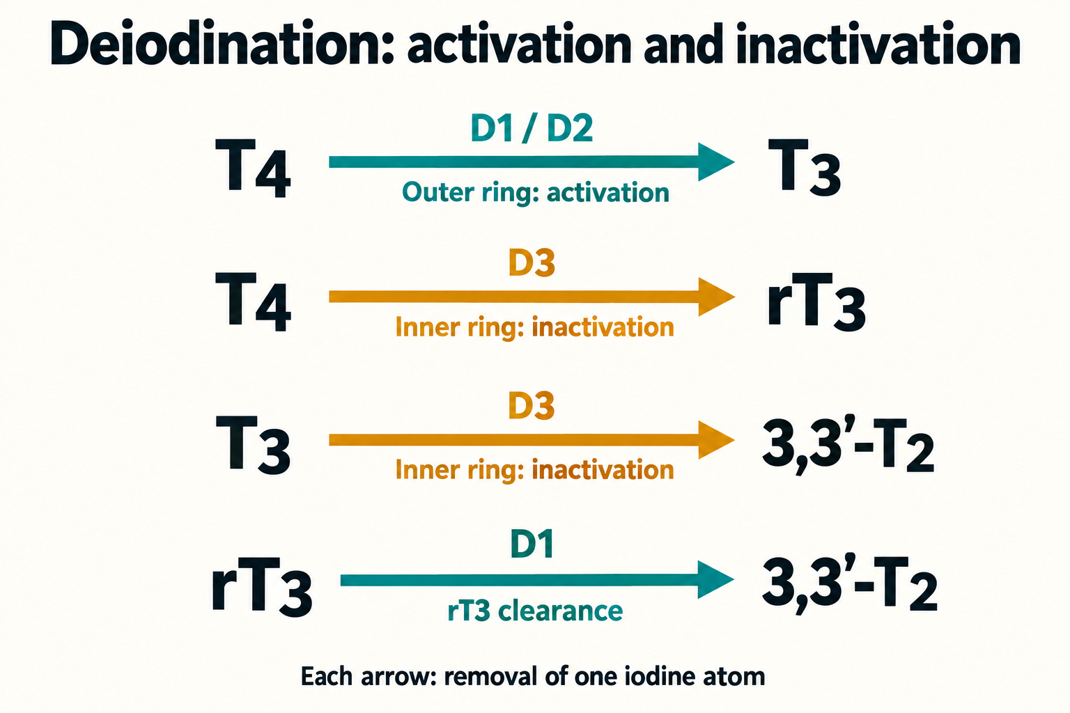
Which iodine atom is removed?
T4 is 3,5,3′,5′-tetraiodothyronine. Active T3 is 3,5,3′-triiodothyronine; reverse T3, rT3, is 3,3′,5′-triiodothyronine. The prime marks distinguish the outer phenolic ring from the inner tyrosyl ring. Losing an iodine at the outer ring activates T4, whereas inner-ring deiodination generally inactivates T4 or T3. Although T3 and rT3 have the same elemental composition, their iodine positions and receptor activity differ. The 3,3′-T2 discussed here is a particular diiodothyronine; it should not be confused with other T2 isomers studied for different biological effects. T4 has molecular formula C15H11I4NO4; T3 and rT3 both have C15H12I3NO4. Replacement of iodine by hydrogen explains the changed composition, whereas isomerism explains their different effects.
These are principal routes, not an exhaustive substrate list. D1 can carry out both outer- and inner-ring reactions, especially with certain conjugated substrates; D2 can also outer-ring deiodinate rT3. D3 prevents T4 activation and terminates T3 signalling. The reaction is reductive removal of iodine, not simple hydrolysis:
R denotes the remainder of the iodothyronine molecule. In cells, reducing equivalents arrive through enzyme-associated redox chemistry rather than as free electrons. A net donor model can be written as:
This balanced shorthand illustrates oxidation of two thiols to a disulfide. It does not establish that the same thiol is the exclusive physiological donor for all three enzymes (Bianco, 2002; Schweizer, 2014).
Selenocysteine is built into the enzyme
All three deiodinases are membrane-associated selenoproteins. Selenium is part of their catalytic selenocysteine residue, Sec, rather than a freely exchangeable metal cofactor like the iron in haem. Human cells encode 25 selenoproteins, including deiodinases, glutathione peroxidases and thioredoxin reductases. The shared element connects hormone metabolism with antioxidant systems without making their functions interchangeable.
Selenocysteine resembles cysteine but carries selenium in place of sulphur. Free Sec has a lower selenol pKa, commonly around 5.2, than the cysteine thiol, commonly around 8.3. A Henderson–Hasselbalch estimate gives the ionized fraction:
At pH 7.4, using pKa 5.2 gives approximately 0.994 for Sec; using 8.3 gives approximately 0.112 for cysteine. Protein microenvironments can shift these values, so this is an acid–base illustration, not a measurement of an enzyme's active site. Greater availability of the selenolate and its chemical properties help explain catalytic efficiency, but there is no universal 100–1000-fold turnover advantage. In recombinant human D2, changing catalytic Sec133 to cysteine increased the T4 Km from approximately 1.4 nM to 2.1 µM and lowered relative turnover about tenfold; substitution with alanine abolished activity. The catalytic position is Sec133, not Sec214 (Buettner, 2000).
Sec incorporation requires recoding of a UGA codon that would ordinarily terminate translation. A stem–loop SECIS element in the 3′ untranslated region recruits the necessary machinery. SECIS-binding protein 2, SECISBP2 or SBP2, interacts with the RNA and translation complex; eEFSec, the specialized eukaryotic elongation factor, delivers Sec-containing tRNA. Calling eEFSec simply bacterial SelB obscures this distinction. Sec is synthesized on a specialized tRNA initially charged with serine, through phosphorylation and selenium-dependent conversion involving PSTK, SEPSECS and selenophosphate generated by SEPHS2. Dietary selenomethionine is therefore not directly inserted into the catalytic Sec position.
Berry's 1991 experiments established the importance of the 3′ untranslated region for UGA recognition in D1. Rare pathogenic SECISBP2 variants subsequently demonstrated the human consequences of impaired selenoprotein synthesis: high T4, low T3 and high rT3 can accompany abnormalities affecting several tissues. Severity and residual synthesis vary; this is not proof that an ordinary low T3 result indicates a genetic defect or dietary selenium deficiency (Berry, 1991; Dumitrescu, 2005).
Catalytic chemistry and what structures actually show
Classical D1 assays support ping-pong kinetics, with a hormone-dependent oxidation step followed by reduction of the enzyme. D2 and D3 have different kinetic behaviour under common assay conditions; assigning one proven ping-pong scheme to all three is inaccurate. Dithiothreitol, DTT, is often used as an artificial reducing donor in vitro. Such assays help compare enzymes, but DTT concentration can change measured activity and apparent substrate constants.
A transient selenium–iodine species is part of proposed mechanisms, followed by oxidized selenium states and reduction. The 2014 mouse D3 catalytic-core structure suggested a peroxiredoxin-like mechanism involving resolving cysteine chemistry and proton transfer. Thioredoxin and glutaredoxin supported reduction in the accompanying biochemical experiments. Glutathione-linked systems may contribute to cellular reducing capacity, but the complete physiological donor system for each isoform remains less settled than a schematic cycle suggests. Oxidative stress can alter activity and donor availability; it should not be described as inevitable irreversible conversion of all deiodinases into a single inactive selenium acid.
Structural evidence must be distinguished from prediction. PDB 4TR3 and 4TR4 represent truncated mouse D3 catalytic domains with catalytic Sec replaced by cysteine, at 1.90 and 1.93 Å resolution, respectively. They are not intact human D1 structures. PDB 3L11 is an RNF168 RING domain, not D2. The D3 core shows a thioredoxin/peroxiredoxin-related fold, and biochemical work supports functional dimerization and an autoinhibitory loop. A crystal core, a full-length model and an experimentally determined membrane complex are different sources of evidence. Neither an AlphaFold model nor these deposits prove a fixed 90° hormone orientation or all asserted residue contacts in every human isoform (Schweizer, 2014).
D1, D2 and D3 occupy different compartments
| Feature | D1 | D2 | D3 |
|---|---|---|---|
| Gene and chromosomal region | DIO1, chromosome 1p32 | DIO2, chromosome 14q31 | DIO3, chromosome 14q32 |
| Typical localization | Plasma membrane; catalytic domain faces cytosol | Endoplasmic reticulum; catalytic domain faces cytosol | Plasma membrane and recycling/endosomal compartments |
| Principal reaction | T4 activation and efficient rT3 clearance; additional inner-ring substrates | T4-to-T3 activation | T4-to-rT3 and T3-to-3,3′-T2 inactivation |
| Common assay affinity scale | T4 Km in micromolar range; rT3 often a preferred substrate | T4 Km in low nanomolar range | T3 Km in low nanomolar range; substrate must be specified |
| Prominent expression | Liver, kidney and thyroid | Brain glia, pituitary, brown fat; muscle expression depends on species and context | Placenta, developing tissues, selected adult brain/skin compartments; inducible in disease |
| Response to PTU in usual assays | Sensitive | Relatively insensitive | Relatively insensitive |
| Turnover and regulation | Comparatively long-lived; T3-responsive expression | Short-lived; substrate-dependent ubiquitination and reversible rescue | Developmental, growth-factor and hypoxia-dependent expression |
Km is the substrate concentration giving half the maximal rate in an appropriate assay model; it is not a receptor dissociation constant or a whole-organ conversion efficiency. Comparing substrate affinity requires distinguishing D3 measurements for T3 from D1/D2 measurements for T4. Enzyme abundance, reducing donor, conjugation state and intracellular substrate delivery also matter. Blood free T4 is usually measured in picomolar units, so a nanomolar D2 Km should not be described as matching a nanomolar plasma free-T4 concentration.
D1 contributes to circulating T3 and rT3 clearance. Its T3-responsive gene expression and sensitivity to PTU help explain its greater contribution during hyperthyroidism. PTU also inhibits thyroid hormone synthesis through thyroid peroxidase; its clinical effect is not restricted to D1. Relative PTU insensitivity of D2 and D3 under usual conditions is more precise than absolute resistance to every concentration and circumstance.
D2-generated T3 can reach nuclear receptors efficiently and can also leave the cell. The idea that D1 exclusively supplies plasma while D2 exclusively supplies local tissues is therefore too rigid. Maia's 2005 intact-cell experiments and extrapolation to human tissue activities argued that D2 could supply a large part of extrathyroidal T3 in euthyroidism. That quantitative model depended on assumptions about tissue mass and enzyme activity; it is not direct measurement of each patient's organ contribution. D2's ER location supports access to nuclear hormone pools, but a sealed channel bypassing the cytosol has not been established.
D3 is necessary for developmental control of hormone exposure, including the placenta. Adult expression is not uniformly absent. Growth, hypoxia, injury and some tumours can increase its activity. Infantile haemangiomas with very high D3 activity can cause consumptive hypothyroidism, a distinct clinical state requiring specialist management. This demonstrates the importance of inactivation without making every adult low-T3 illness equivalent to a haemangioma (Huang, 2000; Gereben, 2008).
D2 ubiquitination is a reversible control point
D2 activity can change rapidly because substrate interaction accelerates its ubiquitination. WSB1 is a substrate-recognition component of an E3 complex involving Elongin B/C, Cullin5 and Rbx1; it should not be treated as an entire standalone ligase. Its recognition of a D2 instability region links Hedgehog signalling with local hormone activation in developing tissues. The ER ligase TEB4, also called MARCH6, provides another route, with tissue-dependent contributions.
Ubiquitination first inactivates D2 and can lead to ER-associated degradation by the proteasome. It is not synonymous with immediate irreversible destruction. USP33/VDU1 and USP20/VDU2 can remove ubiquitin and restore activity, as demonstrated in cell studies. Measured half-life varies with substrate, cell system and experimental conditions; reported short spans of tens of minutes should not be assigned to every tissue as a universal constant. ER stress can additionally affect D2 translation and processing (Curcio-Morelli, 2003; Dentice, 2005; Zavacki, 2009).
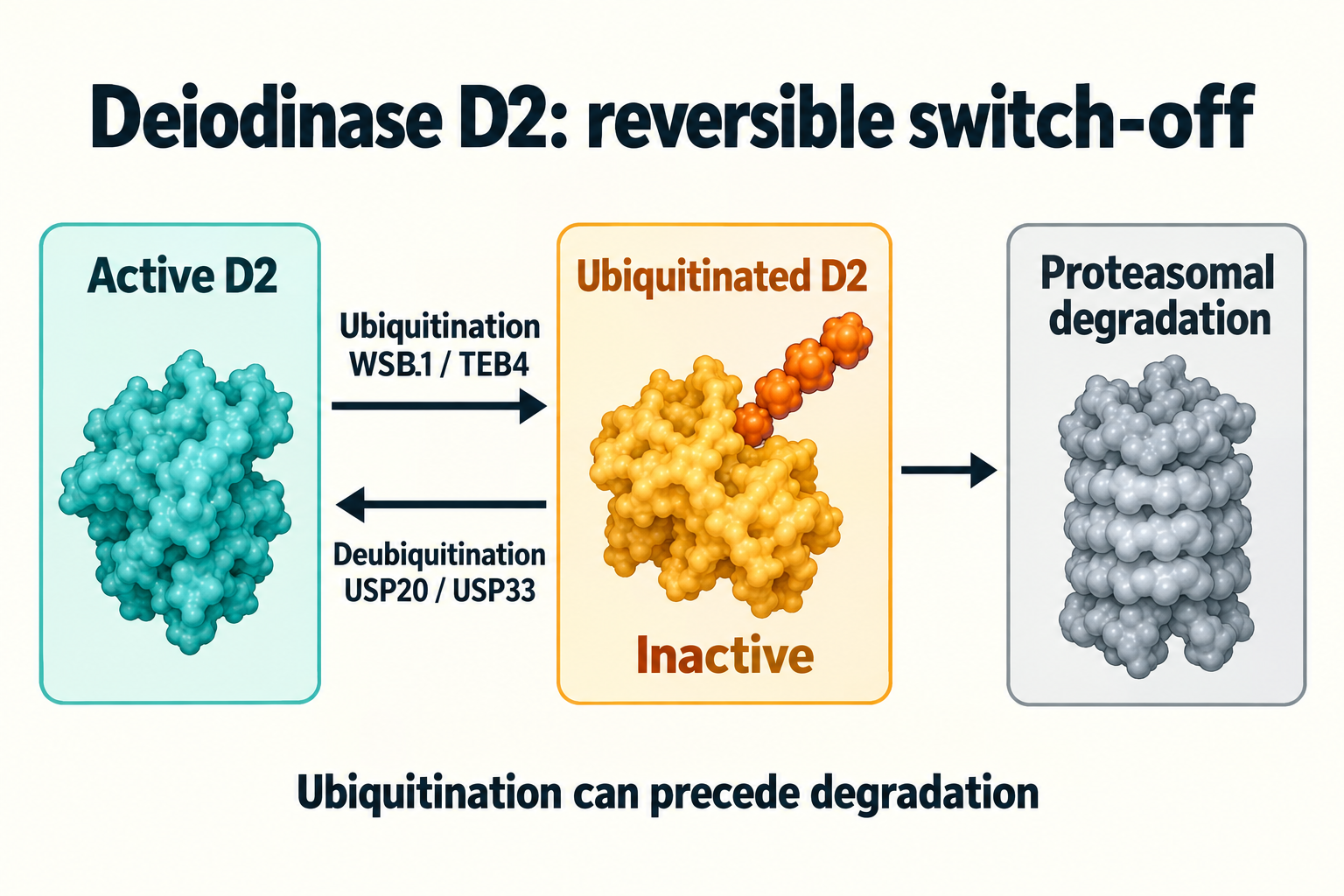
This feedback allows hormone availability to regulate activation locally. It does not mean a high serum T4 necessarily destroys all D2 or proves the need for T3 medication. Transcription, Sec incorporation, ubiquitin cycling, transport and receptor responses operate together.
Transport and nuclear responses
Most circulating T4 and T3 are bound to thyroxine-binding globulin, transthyretin or albumin. Their free fractions and binding differ. T4 is a relatively stable reservoir, with a typical euthyroid half-life around seven days; T3 turns over more rapidly, often roughly a day. These estimates vary with thyroid state and illness. Approximately four-fifths of circulating T3 normally comes from peripheral conversion, with the remainder secreted by the thyroid; these proportions are approximate and change with disease and treatment. T3 is a ligand, not itself a transcription factor.
Cellular transport is mediated by proteins including MCT8/SLC16A2, MCT10/SLC16A10 and OATP1C1/SLCO1C1. MCT8 transports T3 and T4, while MCT10 also transports aromatic amino acids. OATP1C1 has high affinity for T4 and rT3 and contributes to brain exposure in a species- and barrier-dependent manner. It is not a simple ATP-powered pump. Transporter biology rules out unrestricted lipid solubility as a sufficient model; it does not require an absolute assertion that any passive passage is thermodynamically impossible.
Loss-of-function SLC16A2 variants cause MCT8 deficiency, or Allan–Herndon–Dudley syndrome: profound neurodevelopmental impairment can coexist with high serum T3 and peripheral thyrotoxic features. Brain entry and metabolism are affected in different compartments; other tissues have alternative transporters. The disorder illustrates why a serum value does not fully describe every tissue, without proving universal “brain hypothyroidism” in adults with normal TSH. PDB 7W5N is a bacterial dehydrogenase structure and does not support MCT8 structural claims (Friesema, 2003, 2004). Human MCT8 cryo-EM with T3 or silychristin exists (Tan, 2025); it neither validates PDB 7W5N nor establishes clinical inhibitor use.
TRα1, TRβ1 and TRβ2 are nuclear thyroid hormone receptor isoforms with overlapping but different expression. TRα1 is important in heart, bone and nervous tissue; TRβ1 in liver and other organs; TRβ2 in central feedback and sensory structures. These are preferences rather than exclusive maps. T3 binding rearranges the ligand-binding domain and co-regulator interactions. T4 has lower affinity for classical nuclear receptors, but is not intrinsically incapable of binding or exerting effects; non-genomic actions are also investigated. A T3-bound receptor crystal cannot establish that T4 creates an insurmountable steric barrier.
TR frequently heterodimerizes with RXR on thyroid response elements. A common positive TRE contains two AGGTCA-like half-sites separated by four bases, a DR4 motif, but not every regulated gene follows this arrangement. In a classical positive-response model, unliganded TR recruits NCoR/SMRT and HDAC-containing complexes; T3 favours SRC-family coactivators and p300/CBP-associated chromatin regulation. PDB 1BSX supports TRβ–T3–coactivator interactions, 3GWS is a T3-bound receptor deposit, and 2NLL is a TR/RXR DNA-binding-domain heterodimer on response-element DNA. These are specific constructs. They do not prove continuous receptor binding at every site, total transcriptional silencing without hormone or an identical mechanism for negatively regulated TSHB/TRH genes (Darimont, 1998).
Non-thyroidal illness is a changing system
Non-thyroidal illness syndrome, NTIS, describes changes in thyroid tests during illness without necessarily indicating primary thyroid disease. “Euthyroid sick” is a historical label, not proof that every organ remains euthyroid. Low total or free T3 is common; T4 may remain normal early and fall in prolonged or severe disease. TSH is often normal or low, and a transient increase may occur during recovery. rT3 often rises through altered production and clearance, but may be normal or low. Fixed percentage falls, mandatory two-to-fourfold rT3 increases and two sharply separated stages are not universal diagnostic rules.
Changes involve central TRH/TSH drive, hormone binding and transport, D1/D2 activation, D3 inactivation, food intake, drugs and organ dysfunction. IL-6, TNF-α and IL-1β affect signalling through several pathways; individual tissues do not all suppress DIO2 in the same way. Hypothalamic D2 can increase in some illness models, helping alter central feedback. Therefore, the same plasma pattern need not imply the same brain, liver or muscle response (Boelen, 2011; Fliers, 2015).
Wajner's human-cell experiments showed that IL-6 could reduce T4 activation despite increased deiodinase expression, with redox mechanisms affecting intact-cell activity. Antioxidant rescue in that model does not establish clinical effectiveness of NAC or alpha-lipoic acid for NTIS. It also contradicts a simple account in which NF-κB completely silences every DIO1/DIO2 promoter. Transcriptional effects, substrate delivery and reducing cofactors must be distinguished (Wajner, 2011).
Peeters examined liver and muscle samples obtained shortly after death from critically ill patients and found reduced activation and increased D3-associated inactivation. This important human tissue evidence is observational and represents a selected, very ill population. It does not establish a tenfold fall in every patient's liver T3, a universal 80–90% D1 reduction or the cause of individual organ failure. Simonides demonstrated HIF-dependent DIO3 induction and local hormone inactivation in cell and rat hypoxic-ischemic models. Those findings support a mechanism; they do not establish all asserted fold changes in human sepsis or a specific D2-driven diaphragm-fibre atrophy syndrome (Peeters, 2003; Simonides, 2008).
T3 regulates processes relevant to cardiac SERCA2a, myosin expression, thermogenesis and energy use. Local reductions may affect repair and contractile responses, but animal–human differences and multifactorial injury limit causal attribution. The broader relationship between local thyroid activation and brown-fat thermogenesis provides one physiological example, rather than a treatment strategy for critical illness.
Whether particular NTIS changes are adaptive, maladaptive or both depends on duration, tissue and condition. Reducing energy expenditure during acute food deprivation can be adaptive; persistent central suppression may differ. It is too strong to claim that protective “hibernation” has been conclusively proved for every patient. Low hormone levels predict severity in some cohorts, but association with death does not show that raising the level improves survival.
Brent and Hershman's 1986 randomized study involved 23 intensive-care patients, not a large multicentre trial. T4 treatment raised circulating T4 but did not improve outcome; mortality was similar, about 75% versus 73%. This study did not establish a significant treatment-induced mortality increase. The ATA guideline recommends against routine T4 or T3 treatment for NTIS in hospitalized critically ill adults because useful clinical benefit has not been demonstrated and risks remain. Established hypothyroidism and suspected myxoedema coma are separate situations. Treating the underlying illness and assessing nutrition and medication effects should not be replaced by correcting a hormone number alone (Brent, 1986; Jonklaas, 2014).
Selenium, iron and zinc: different biological roles
Selenium supports synthesis of catalytic Sec and antioxidant selenoproteins. SELENOP transports selenium, with receptor-dependent uptake and tissue prioritization demonstrated particularly in experimental systems. Brain and reproductive tissues can preserve selenium differently from liver during deficiency. The thyroid is selenium-rich, but neither a universal serum threshold of 70–80 µg/L nor a fixed 30–50% fall in T3 production follows for every adult. Serum selenium and SELENOP also change with inflammation; a low concentration during illness is not automatically proof of inadequate dietary intake (Schomburg, 2012).
Thyroid hormone synthesis requires controlled hydrogen peroxide production by DUOX systems, especially DUOX2 with its maturation machinery. TPO uses hydrogen peroxide for iodide oxidation and coupling on thyroglobulin; TPO is not the principal generator of that peroxide. Glutathione peroxidases and other antioxidant systems help limit damage. Selenium deficiency may interact with iodine status and redox stress, but this mechanism does not prove that Hashimoto thyroiditis universally begins with selenium deficiency or resolves through supplementation (Moreno, 2002).
Iron is important for haem-containing TPO and oxygen transport. Thyroid hormones are iodinated amino-acid derivatives, not steroids, and iron is not a direct catalytic metal in D1/D2/D3. Deficiency can affect thyroid synthesis and overall physiology; ferritin rises with inflammation and requires interpretation alongside other iron measures. Hess's intervention in iron-deficient, goitrous children showed that iron correction improved the response to iodized salt. It did not establish that every levothyroxine-treated adult must reach ferritin 50–70 ng/mL to make T3. Non-anaemic deficiency should be evaluated in its clinical context, rather than assuming obligatory hypoxic D3 activation (Hess, 2002; Zimmermann, 2002).
Zinc stabilizes the cysteine-coordinated zinc fingers of nuclear receptor DNA-binding domains and participates in many cellular processes. It is not a replacement for catalytic Sec. Baltaci's 2004 thyroid hormone work concerned rats, not a human randomized trial demonstrating a universal 30% T3 decline or benefit from zinc picolinate. Clinical deficiency and laboratory associations require separate interpretation. High zinc intake can impair copper balance, while iron excess has its own risks; biochemical plausibility is not an indication for treating everyone (Baltaci, 2004).
NADPH helps recycle cellular reducing systems through glutathione reductase and thioredoxin reductases. The pentose phosphate pathway is one important NADPH source, alongside other metabolic routes. This supports redox chemistry but does not make dietary carbohydrate a unique direct deiodinase cofactor or establish a universal gram threshold. Nuclear receptor DNA-binding domains contain two Cys4 zinc fingers, each coordinating zinc through four cysteines; severe experimental disruption affects DNA binding, without proving complete receptor resistance from every mildly low zinc result. (Bianco, 2002; Schweizer, 2014; Zimmermann, 2002).
Nutrient trials and botanical proposals
| Evidence | What was investigated | What it does not establish |
|---|---|---|
| Gärtner, 2002 | Seventy autoimmune-thyroiditis patients randomized to sodium selenite, 200 µg/day, or placebo for three months; antibody concentrations were a major outcome | This was not a selenomethionine trial proving universal conversion correction or symptom relief |
| Larsen, 2024, CATALYST | 412 levothyroxine-treated adults with autoimmune thyroiditis randomized to selenium-enriched yeast, 200 µg/day, or placebo for 12 months; no additional quality-of-life benefit | Lower antibodies did not translate into improved LT4 dose requirements or hormone-ratio outcomes; trial dose is not a general prescription |
| Sharma, 2018 | Fifty adults with subclinical hypothyroidism randomized in a single-centre, eight-week pilot to ashwagandha root extract or placebo; serum thyroid indices changed | No direct measurement of D1/D2 activity or proof that lowering cortisol caused conversion improvement; short follow-up limits safety conclusions |
| Panda, 2005 | Guggulu studied in female mice with experimentally induced hypothyroidism | No demonstrated human “conversion” regimen or clinically validated guggulsterone dose |
Sharma, Basu and Singh’s 2018 pilot reports changes in serum T3/T4, without direct deiodinase activity measurements. A hormone change cannot establish its enzymatic or cortisol-mediated cause. Botanical effects on thyroid tests and reported thyrotoxicosis make unsupervised use inappropriate as a response to low T3. This distinction is developed in the article on ashwagandha and the HPA axis. (Sharma, 2018; van der Hooft, 2005).
Selenomethionine can enter the general methionine-containing protein pool and later supply selenium through metabolism. Inorganic selenite follows different metabolic handling. Neither form has a universal proven advantage for acute NTIS or guaranteed rapid conversion restoration. Correcting a documented deficit differs from adding selenium in a selenium-replete person. CATALYST demonstrates why an antibody change is insufficient evidence of clinical benefit.
A daily selenium supplement of 100–200 µg cannot be designated a universally safe “optimal window”. Total exposure includes food and all supplements. The US adult recommended allowance is 55 µg/day and the US upper level is 400 µg/day; EFSA's 2023 adult upper level is 255 µg/day, including pregnancy and lactation. These are jurisdiction-specific nutrition values, not therapeutic targets or guarantees for every individual. A 200-µg supplement plus diet can approach or exceed the European upper level. Excess selenium can cause hair/nail abnormalities, gastrointestinal symptoms and other toxicity; there is no universal sharp “acute toxicity starts at 800–1000” boundary. (NIH ODS; EFSA, 2023).
Fucus and kelp contain variable amounts of iodine and may carry contaminants. Iodine is substrate for thyroid synthesis, not a deiodinase activator. Excess can cause Wolff–Chaikoff-related inhibition in susceptible circumstances or iodine-induced hyperthyroidism. A stated iodine dose does not make a seaweed product reliable or appropriate for every patient. NAC, alpha-lipoic acid, methylfolate, methylcobalamin and SAM were proposed as a combined redox/epigenetic protocol, but human clinical evidence does not validate that stack for deiodinase dysfunction. Methyl donors do not simply erase presumed DIO1/DIO2 promoter hypermethylation.
Energy intake, hormones and daily rhythms
Food deprivation changes thyroid hormone economy through central signals and peripheral metabolism. Leptin links energy stores with hypothalamic TRH regulation, while glucocorticoids and some medications can suppress TSH or T4-to-T3 conversion. These effects depend on dose and setting. Insulin resistance, inflammation and AGE/RAGE-associated stress may interact with thyroid signalling, but there is no proven universal chain from visceral obesity to suppression of all peripheral deiodinases.
Spaulding's 1976 work compared fasting and 800-kcal diets with differing carbohydrate content. Fasting lowered T3 and raised rT3; a carbohydrate-free hypocaloric diet lowered T3 without a significant rT3 rise. Those observations do not establish absolute D1 dependence on a daily carbohydrate threshold, fixed NADPH depletion, a 150–300-kcal metabolic penalty or restoration within 24 hours after adding 50–75 g carbohydrate. Hormone concentrations were measured, not liver D1 activity in all participants. Energy deficit and macronutrient composition are different variables, and thyroid tests alone cannot diagnose a harmful ketogenic adaptation (Spaulding, 1976).
TSH has a circadian rhythm and pulsatile secretion, often with a nocturnal rise. Sleep timing, sampling time, illness and medication influence interpretation. Deiodinase rhythms have been studied in tissues and animal models, but a fixed 06:00–08:00 human muscle D2 peak and mandatory 20–30% T3 loss after sleeping less than six hours are not established clinical rules. Regular sleep is sensible for health; a specific bedroom temperature or 7.5–8.5-hour prescription is not a proven deiodinase treatment. Comparing samples obtained under similar conditions can be more informative than fitting them to an invented conversion timetable.
Tissue map and limits of inference
| Compartment | Important local processes | Interpretation boundary |
|---|---|---|
| Liver and kidney | D1-associated activation and metabolite clearance; inducible inactivation in some illnesses | No serum rT3 value uniquely measures hepatic D3 or renal D1 |
| Brain | Glial D2 activation, neuronal inactivation and transporter-mediated exchange | Rodent barrier pathways and local hormone fractions do not transfer unchanged to human adults |
| Pituitary/hypothalamus | Local hormone metabolism contributes to TSH/TRH feedback | Normal TSH is informative in primary thyroid assessment but does not describe every cellular hormone pool |
| Brown adipose tissue | D2 supports local T3 and thermogenic programmes alongside sympathetic cAMP signalling | Enzyme biology does not prove a safe hormone-based weight-loss intervention |
| Skeletal muscle | D2 expression/activity and transport vary with species, cell type and physiological condition | A fixed share of plasma T3 or uniform illness suppression cannot be assigned to all muscle |
| Heart | Circulating T3 entry and local inactivation during some injuries | Human failure and rodent D3 induction are related evidence, not identical causal experiments |
| Placenta/developing tissues | D3 limits excess exposure and shapes developmental timing | This barrier is physiological rather than inherently pathological |
Local hormone availability is influenced by production, uptake, export and destruction. A conceptual balance makes the distinction explicit:
Each J has concentration-per-time units in the same cellular compartment. This is a bookkeeping model, not a validated clinical calculator. Binding and compartment exchanges may need additional terms in a detailed model. No single serum ratio identifies all of these fluxes.
Persistent symptoms during levothyroxine treatment
Some patients with established hypothyroidism report symptoms despite appropriately interpreted TSH during levothyroxine treatment. Symptoms deserve assessment, but “resistance to T4 monotherapy” is not a specific diagnosis. Adherence, timing with meals, absorption, interacting medicines, dose stability, sleep, anaemia, mood disorders and other disease can contribute. Iron and calcium products can interfere with levothyroxine absorption; this must be distinguished from deiodinase catalysis.
DIO2 Thr92Ala, rs225014, is a common variant investigated for protein processing, cellular stress and treatment response. Its frequency depends on ancestry. Cell and animal findings do not establish a 20–35% reduction in every carrier's cerebral T3. Panicker's 2009 genetic analysis of 552 WATTS participants found an association with psychological scores and response to combination therapy, but the authors explicitly called for replication; circulating hormone levels were not affected. It was not proof that all Ala/Ala carriers benefit exclusively from T3. Other studies did not reproduce a clear genotype-based treatment effect, and a later 75-person crossover trial did not find Thr92Ala determined outcomes (Panicker, 2009; Appelhof, 2005; Shakir, 2021). In the Shakir trial, exploratory analysis of the most symptomatic third favoured T3-containing treatment on some symptom measures; this subgroup result did not establish genotype-based selection or consistent superiority for the whole group.
Levothyroxine remains standard replacement. The 2021 ATA/BTA/ETA consensus stated that 14 trials had not shown consistent benefit from combined LT4/LT3. The 2023 British consensus allows consideration of a specialist-supervised trial in selected patients with confirmed overt hypothyroidism, adequate LT4 treatment, persistent symptoms and other causes assessed. These positions do not endorse treating isolated low T3 or using a DIO2 genotype or rT3 ratio as an automatic prescription. The original's fixed 10:1–14:1 dosing rule and supplement-plus-liothyronine algorithm are not appropriate as universal advice (Jonklaas, 2021; Ahluwalia, 2023).
Autoimmunity, assays and environmental exposures
Autoimmune thyroiditis affects hormone production and inflammatory signalling. Anti-TPO positivity does not measure deiodinase activity. Antibodies to T3/T4 can interfere with some hormone assays, and heterophile antibodies or biotin can create misleading laboratory patterns. Discordance between symptoms and tests may warrant laboratory evaluation with an alternative method. It should not automatically be interpreted as antibody-mediated blockade of every MCT8 transporter or proven tissue hormone starvation.
Epigenetic regulation of DIO genes is studied in development, cancer and experimental systems. The claim that common Hashimoto disease permanently hypermethylates DIO1/DIO2 and requires methyl-donor therapy has not been clinically demonstrated. Retaining this research theme requires distinguishing a plausible molecular process from a validated treatment target.
Mercury, cadmium, PCBs, PBDEs, bisphenol A and phthalates have been investigated for effects on thyroid synthesis, binding proteins, transporters, deiodinases and receptors. Some compounds or metabolites affect enzyme activity in vitro or hormone economy in animals. Effects vary by chemical, concentration, life stage and model; they are not all selective D1/D2 inhibitors. TTR displacement is particularly relevant for certain hydroxylated metabolites, not a uniform action of every parent PCB/PBDE. Selenium–mercury chemistry is complex and does not justify a universal binding constant or complete enzyme paralysis after ordinary exposure. Associations in humans do not establish which molecular route dominates. Neither indiscriminate metal testing nor selenium-based “detoxification” follows from these mechanisms.
Interpreting laboratory tests without inventing a conversion index
| Test or observation | Useful contribution | Important limitation |
|---|---|---|
| TSH with free T4 | Initial evaluation of primary thyroid function; follow-up in appropriate replacement settings | Central disease, acute illness, pregnancy, medicines and assay problems require context |
| T3/free T3 | Selected evaluation of hyperthyroidism and illness-related patterns | A low result is not a direct assay of D1/D2, and free-T3 methods may be less reliable |
| rT3 | Specialized research or uncommon diagnostic contexts | Not routine diagnosis of hypothyroidism; can change through production and clearance |
| Thyroid antibodies | Supports assessment of autoimmune thyroid disease where indicated | Antibody concentration is not a conversion rate or severity score for every symptom |
| Selenium/SELENOP, iron and zinc assessment | Helps investigate a clinically plausible nutritional problem | Inflammation, binding and laboratory context influence values; no universal “optimal conversion” range |
| Medication and sample review | Identifies glucocorticoid, amiodarone, high-dose propranolol and assay/absorption effects | Essential medicines should not be stopped merely because they affect a thyroid test |
The popular calculation “free T3 in pg/mL divided by rT3 in ng/dL, multiplied by 100” is not the molar ratio its name implies. Because 1 ng/dL equals 10 pg/mL, and T3/rT3 have the same molecular mass, a true molar concentration ratio is:
The popular index is therefore 1000 times this ratio. More fundamentally, it compares free T3 with generally total rT3, different circulating pools. Cutoffs of 15 or 20 do not constitute validated diagnoses of tissue hypothyroidism or D3 dominance. Correcting units does not make the ratio clinically validated. ATA patient guidance states that rT3 does not help establish hypothyroidism in otherwise healthy, non-hospitalized people.
A normal TSH should not be dismissed categorically, and a low T3 during severe illness should not be equated with permanent primary hypothyroidism. Appropriate interpretation considers prior thyroid disease, free T4, medication, clinical findings and the course after recovery. Nutritional deficits should be investigated and corrected when supported, rather than targeting fixed selenium, ferritin, zinc, cortisol or HOMA-IR values to “unlock” conversion. The central scientific lesson is that deiodinases regulate hormone action within a larger transport, signalling and metabolic system; useful clinical decisions require evidence beyond a pathway diagram.
How this article was prepared
This is a narrative scientific review, with targeted checks of disputed mechanisms, structural deposits, bibliographic identifiers and clinical findings on 8 October 2026. Primary publications and accessible NCBI abstracts, publisher/JCI pages, RCSB records and ATA/BTA/ETA, EFSA and NIH guidance were used. Not every full text was available; checks are limited to the sections actually read. No systematic search, independent GRADE assessment, external peer review or clinical validation was performed. Cell mechanisms, animal experiments, observations and human trials are distinguished; study doses are not prescriptions.
FAQ
Can one rT3 result measure conversion?
No. Production and clearance both matter, while blood does not measure every tissue flux. Routine hypothyroidism diagnosis through rT3 is not established.
Does everyone with low T3 need selenium?
No. Deficiency differs from inflammation-associated changes; food and supplements contribute to excess risk. Selenium does not guarantee improved symptoms or conversion.
Is ubiquitinated D2 irreversibly switched off?
Not necessarily. Deubiquitinases can restore activity before degradation. This cellular regulation is not an independent indication for T3 medication.
Does Thr92Ala establish a need for liothyronine?
No. Clinical findings differ and genotype is not an automatic prescription. Selected supervised combination trials require an appropriate clinical context.
Sources
- Bianco AC, Salvatore D, Gereben B, Berry MJ, Larsen PR. Biochemistry, cellular and molecular biology, and physiological roles of the iodothyronine selenodeiodinases. Endocr Rev. 2002;23(1):38–89.
- Gereben B, Zavacki AM, Ribich S, Kim BW, Huang SA, Simonides WS, Zeöld A, Bianco AC. Cellular and molecular basis of deiodinase-regulated thyroid hormone signaling. Endocr Rev. 2008;29(7):898–938.
- Berry MJ, Banu L, Chen YY, Mandel SJ, Kieffer JD, Harney JW, Larsen PR. Recognition of UGA as a selenocysteine codon in type I deiodinase requires the 3' untranslated region. Nature. 1991;353(6341):273–276.
- Dumitrescu AM, Liao XH, Abdullah MS, Lado-Abeal J, Majed FA, Moeller LC, et al. Mutations in SECISBP2 result in abnormal thyroid hormone metabolism. Nat Genet. 2005;37(11):1247–1252.
- Boelen A, Kwakkel J, Fliers E. Beyond low plasma T3: local thyroid hormone metabolism during inflammation and infection. Endocr Rev. 2011;32(5):670–693.
- Fliers E, Bianco AC, Langouche L, Boelen A. Thyroid function in critically ill patients. Lancet Diabetes Endocrinol. 2015;3(10):816–825.
- Peeters RP, Wouters PJ, Kaptein E, van Toor H, Visser TJ, Van den Berghe G. Reduced activation and increased inactivation of thyroid hormone in tissues of critically ill patients. J Clin Endocrinol Metab. 2003;88(7):3202–3211.
- Friesema EC, Ganguly S, Abdalla A, Manning Fox JE, Halestrap AP, Visser TJ. Identification of monocarboxylate transporter 8 as a specific thyroid hormone transporter. J Biol Chem. 2003;278(41):40128–40135.
- Friesema EC, Grueters A, Biebermann H, Krude H, von Moers A, Reeser M, et al. Association between mutations in a thyroid hormone transporter and severe X-linked psychomotor retardation. Lancet. 2004;364(9443):1435–1437.
- Schomburg L. Selenium, selenoproteins and the thyroid gland: interactions in health and disease. Nat Rev Endocrinol. 2012;8(3):160–171.
- Duntas LH. Selenium and the thyroid: a close-knit connection. J Clin Endocrinol Metab. 2010;95(12):5180–5188.
- Gärtner R, Gasnier BC, Dietrich JW, Seling B, Rochhuber G. Selenium supplementation in patients with autoimmune thyroiditis decreases thyroid peroxidase antibodies concentrations. J Clin Endocrinol Metab. 2002;87(4):1687–1691.
- Zimmermann MB, Kohrle J. The impact of iron and selenium deficiencies on iodine and thyroid metabolism: biochemistry and relevance to human health. Thyroid. 2002;12(10):867–878.
- Hess SY, Zimmermann MB, Adou P, Torresani T, Hurrell RF. Treatment of iron deficiency in goitrous children improves the efficacy of iodized salt in Cote d'Ivoire. Am J Clin Nutr. 2002;75(4):743–748.
- Baltaci AK, Mogulkoc R, Kul A, Bediz CS, Ugur A. Opposite effects of zinc deficiency and supplementation on thyroid hormones in rats. Toxicology. 2004;195(1):69–75.
- Sharma AK, Basu I, Singh S. Efficacy and Safety of Ashwagandha Root Extract in Subclinical Hypothyroid Patients: A Double-Blind, Randomized Placebo-Controlled Trial. J Altern Complement Med. 2018;24:243–248.
- Panda S, Kar A. Guggulu (Commiphora mukul) potentially ameliorates hypothyroidism in female mice. Phytother Res. 2005;19:78–80.
- Brent GA, Hershman JM. Thyroxine therapy in patients with severe nonthyroidal illnesses and low serum thyroxine concentration. J Clin Endocrinol Metab. 1986;63(1):1–8.
- Curcio-Morelli C et al. Deubiquitination of type 2 iodothyronine deiodinase by von Hippel-Lindau protein-interacting deubiquitinating enzymes regulates thyroid hormone activation. J Clin Invest. 2003;112:189–196.
- Dentice M et al. The Hedgehog-inducible ubiquitin ligase subunit WSB-1 modulates thyroid hormone activation and PTHrP secretion in the developing growth plate. Nat Cell Biol. 2005;7:698–705.
- Huang SA, Tu HM, Harney JW, Venihaki M, Butte AJ, Kozakewich HP, et al. Severe hypothyroidism caused by type 3 iodothyronine deiodinase in infantile hemangiomas. N Engl J Med. 2000;343(3):185–189.
- Warner MH, Beckett GJ. Mechanisms behind the non-thyroidal illness syndrome. J Endocrinol. 2010;205(1):1–13.
- Wiersinga WM. Non-thyroidal illness. Pituitary. 2008;11(2):191–197.
- Wiersinga WM, Duntas L, Fadeyev V, Nygaard B, Vanderpump MP. 2012 ETA Guidelines: The Use of L-T4 + L-T3 in the Treatment of Hypothyroidism. Eur Thyroid J. 2012;1(2):55–71.
- Jonklaas J, Bianco AC, Bauer AJ, Burman KD, Cappola AR, Celi FS, et al. Guidelines for the treatment of hypothyroidism: prepared by the american thyroid association task force on thyroid hormone replacement. Thyroid. 2014;24(12):1670–1751.
- Kohrle J. Local activation and inactivation of thyroid hormones: the deiodinase family. Mol Cell Endocrinol. 1999;151(1-2):103–119.
- Kohrle J. The selenoenzyme family of deiodinase isozymes controls local thyroid hormone availability. Rev Endocr Metab Disord. 2000;1(1-2):49–58.
- Rayman MP. Multiple nutritional factors in the etiology of thyroid disease. Proc Nutr Soc. 2019;78(1):34–44.
- Arthur JR, Beckett GJ, Mitchell JH. The interactions between selenium and iodine deficiencies in man and animals. Nutr Res Rev. 1999;12(1):55–73.
- Bianco AC, Kim BW. Deiodinases: implications of the local control of thyroid hormone action. J Clin Invest. 2006;116(10):2571–2579.
- Visser TJ. Pathways of thyroid hormone metabolism. Acta Med Austriaca. 1996;23(1-2):10–16.
- Biondi B, Wartofsky L. Combination treatment with L-T4 and L-T3 for hypothyroidism: are we there yet? Thyroid. 2012;22(3):227–229.
- Schweizer U et al. Crystal structure of mammalian selenocysteine-dependent iodothyronine deiodinase suggests a peroxiredoxin-like catalytic mechanism. PNAS. 2014;111:10526–10531.
- Buettner C, Harney JW, Larsen PR. The role of selenocysteine 133 in catalysis by the human type 2 iodothyronine deiodinase. Endocrinology. 2000;141:4606–4612.
- Maia AL et al. Type 2 iodothyronine deiodinase is the major source of plasma T3 in euthyroid humans. J Clin Invest. 2005;115:2524–2533.
- Wajner SM et al. IL-6 promotes nonthyroidal illness syndrome by blocking thyroxine activation while promoting thyroid hormone inactivation in human cells. J Clin Invest. 2011;121:1834–1845.
- Simonides WS et al. Hypoxia-inducible factor induces local thyroid hormone inactivation during hypoxic-ischemic disease in rats. J Clin Invest. 2008;118:975–983.
- Zavacki AM et al. The E3 ubiquitin ligase TEB4 mediates degradation of type 2 iodothyronine deiodinase. Mol Cell Biol. 2009;29:5339–5347.
- Panicker V et al. Common variation in the DIO2 gene predicts baseline psychological well-being and response to combination thyroxine plus triiodothyronine therapy in hypothyroid patients. J Clin Endocrinol Metab. 2009;94:1623–1629.
- Appelhof BC et al. Polymorphisms in type 2 deiodinase are not associated with well-being, neurocognitive functioning, and preference for combined thyroxine/3,5,3′-triiodothyronine therapy. J Clin Endocrinol Metab. 2005;90:6296–6299.
- Shakir MKM et al. Comparative Effectiveness of Levothyroxine, Desiccated Thyroid Extract, and Levothyroxine+Liothyronine in Hypothyroidism. J Clin Endocrinol Metab. 2021;106:e4400–e4413.
- Larsen CB et al. Selenium supplementation and placebo are equally effective in improving quality of life in patients with hypothyroidism. Eur Thyroid J. 2024;13:e230175.
- Jonklaas J et al. Evidence-Based Use of Levothyroxine/Liothyronine Combinations in Treating Hypothyroidism: A Consensus Document. Eur Thyroid J. 2021;10:10–38.
- Ahluwalia R et al. Use of liothyronine (T3) in hypothyroidism: Joint British Thyroid Association/Society for endocrinology consensus statement. Clin Endocrinol. 2023;99:206–216.
- Moreno JC et al. Inactivating mutations in the gene for thyroid oxidase 2 (THOX2) and congenital hypothyroidism. N Engl J Med. 2002;347:95–102.
- Darimont BD et al. Structure and specificity of nuclear receptor–coactivator interactions. Genes Dev. 1998;12:3343–3356.
- Spaulding SW et al. Effect of caloric restriction and dietary composition of serum T3 and reverse T3 in man. J Clin Endocrinol Metab. 1976;42:197–200.
- EFSA NDA Panel. Scientific opinion on the tolerable upper intake level for selenium. EFSA J. 2023;21:7704.
- NIH Office of Dietary Supplements. Selenium: Health Professional Fact Sheet. Nutrition reference values and excess-intake risks; accessed 8 October 2026.
- American Thyroid Association. Thyroid Function Tests. Patient guidance on TSH, FT4, T3, rT3 and assay context; accessed 8 October 2026.
- van der Hooft CS et al. Thyrotoxicosis following the use of ashwagandha. Ned Tijdschr Geneeskd. 2005;149:2637–2638. Case report.
- RCSB PDB 4TR3. Mouse D3 catalytic core, Sec-to-Cys mutant, 1.90 Å. Structural construct metadata; accessed 8 October 2026.
- RCSB PDB 4TR4. Mouse D3 catalytic core, Sec-to-Cys mutant, 1.93 Å. Structural construct metadata; accessed 8 October 2026.
- RCSB PDB 3L11. RNF168 RING domain: not deiodinase D2. Structural construct metadata; accessed 8 October 2026.
- RCSB PDB 7W5N. Bacterial sorbose dehydrogenase: not MCT8. Structural construct metadata; accessed 8 October 2026.
- RCSB PDB 1BSX. Thyroid hormone receptor beta, T3 and GRIP1 coactivator peptide. Structural construct metadata; accessed 8 October 2026.
- RCSB PDB 3GWS. T3-bound thyroid hormone receptor deposit. Structural construct metadata; accessed 8 October 2026.
- RCSB PDB 2NLL. TR/RXR DNA-binding domains on thyroid response-element DNA. Structural construct metadata; accessed 8 October 2026.
- Tan J et al. Structural insights into thyroid hormone transporter MCT8. Nat Commun. 2025;16:2958.