Spermidine: polyamine metabolism, EP300, eIF5A hypusination, and the limits of cardioprotection
Spermidine enzymology, autophagic flux and eIF5A–TFEB, heart and brain models, the negative SmartAge trial, cohort associations, oncology duality, and safety.
Spermidine belongs to the polyamines that cells synthesize, take up, consume in translation, convert into other polyamines, and eliminate at the same time. Its concentration is therefore not a simple indicator of “youth”: the same high pool may reflect active growth, increased external supply, or reduced catabolism. Interest in spermidine concerns two distinct mechanisms. It may alter the acetylation of proteins involved in autophagy, and it serves as the indispensable donor of the aminobutyl moiety for eIF5A hypusination. These cellular functions do not establish that a dietary supplement prevents heart failure or dementia in humans. Assessing that possibility requires tracing reactions and compartments, measuring autophagic flux, and considering controlled studies.
Polyamines as a chemical and metabolic system
Putrescine (H₂N–(CH₂)₄–NH₂), spermidine (H₂N–(CH₂)₃–NH–(CH₂)₄–NH₂), and spermine (H₂N–(CH₂)₃–NH–(CH₂)₄–NH–(CH₂)₃–NH₂) differ in the number of amino groups and in chain length. In physiological aqueous media, these groups are substantially protonated. Their spatially distributed positive charge promotes interactions with RNA, DNA, acidic phospholipids, and proteins, but it does not imply a fixed “+3 charge” in every microenvironment: protonation depends on pH and the local surroundings. A simple stereochemical contrast between “natural L-spermidine” and “synthetic D-spermidine” does not apply to this structure.
Stabilizing nucleic acids is one function of polyamines, but specific binding depends on ionic strength and competing cations. The geometry of methylene groups cannot establish a universal distance to phosphates, a precise effect on chromatin compaction, or protection of DNA in a living cell. It is especially important to distinguish the free intracellular fraction from polyamine bound to macromolecules: mass-spectrometric analysis of a whole extract measures their sum, whereas an enzyme can access only part of the pool. In tissues, dietary delivery, microbial synthesis, cellular transport, and renal elimination also contribute.
From ornithine and SAM to spermidine
Arginase forms ornithine from arginine in the context of urea metabolism. Cytosolic ornithine decarboxylase ODC1 (EC 4.1.1.17) removes the carboxyl group from ornithine, yielding putrescine and CO₂. The reaction depends on pyridoxal 5′-phosphate (PLP); the active enzyme is a dimer, and both subunits contribute to its catalytic center. Calling ODC1 “rate-limiting” is useful only for a particular cellular state. Flux through the entire network is also constrained by ornithine availability, the second donor-substrate branch, transport, and catabolism. Perturbing ODC1 is not equivalent to selectively lowering spermidine alone.
L-ornithine → putrescine + CO₂ (ODC1, PLP).
ODC1 turns over unusually rapidly. An increase in the polyamine pool promotes programmed ribosomal frameshifting on OAZ1 mRNA, formation of antizyme, and its binding to ODC1. Antizyme dissociates the active dimer and promotes proteasomal degradation of ODC1 without requiring polyubiquitination. AZIN1, a structural ODC homologue without decarboxylase activity, binds antizyme and modifies this feedback. Exact ODC half-life values depend on cell line and conditions, so a single interval cannot be generalized to every human tissue. This regulation illustrates why polyamine concentrations may change compensatorily after supplementation.
In parallel, AMD1, or AdoMetDC (EC 4.1.1.50), decarboxylates S-adenosylmethionine (SAM). The AMD1 precursor undergoes autocatalytic cleavage to form a covalently bound pyruvoyl group required for the reaction. The resulting decarboxylated SAM (dcSAM) donates an aminopropyl group to polyamine synthases; putrescine can stimulate dcSAM formation, coupling the two branches. Km and Vmax values for an isolated enzyme do not determine the strength of this coupling in vivo without substrate concentrations, compartment, pH, and competing SAM reactions. SAM also participates in methylation, so flux through AMD1 alters the availability of network metabolites, but spermidine concentration alone cannot diagnose a methylation deficiency.
SAM → dcSAM + CO₂ (AMD1, pyruvoyl group).
Spermidine synthase SRM/SPDS (EC 2.5.1.16) transfers an aminopropyl group from dcSAM to putrescine; spermine synthase SMS (EC 2.5.1.22) uses another dcSAM molecule to convert spermidine into spermine. Both reactions release 5′-methylthioadenosine (MTA):
Putrescine + dcSAM → spermidine + MTA (SRM); spermidine + dcSAM → spermine + MTA (SMS).
MTA should not be described as uniformly beneficial or toxic. At particular exposures it can inhibit certain enzymes; MTAP routes it into methionine and purine salvage. MTAP activity differs between normal and tumor cells, which matters when interpreting polyamine metabolism in oncology. The presence of spermidine in food does not mean that all intracellular spermidine passed through ODC1, AMD1, and SRM: exogenous uptake is a separate source.
The reverse pathway: acetylation, oxidation, and excretion
SAT1/SSAT uses acetyl-CoA to N¹-acetylate spermine and spermidine. The change in charge facilitates further oxidation of the acetylated forms and may promote export; the specific transporter and direction depend on the cellular system, so SLC3A2/SLC7A5 cannot be called universal exporters of acetylpolyamines in all tissues without separate evidence. Peroxisomal, FAD-dependent PAOX/APAO converts N¹-acetylspermine back to spermidine and N¹-acetylspermidine to putrescine. Cytosolic SMOX directly converts unmodified spermine into spermidine. These oxidative reactions are coupled to formation of aldehydes and H₂O₂; biological consequences depend on compartment, local antioxidant capacity, and flux magnitude, not merely on the presence of peroxide.
| Conversion | Enzyme and compartment | Products relevant to interpretation |
|---|---|---|
| Spermine/spermidine + acetyl-CoA → N¹-acetyl form + CoA | SAT1, predominantly cytosolic | Substrate for back-conversion or export |
| N¹-acetylspermine → spermidine; N¹-acetylspermidine → putrescine | PAOX, peroxisome, FAD | 3-acetamidopropanal and H₂O₂ |
| Spermine → spermidine | SMOX, cytosol, FAD | 3-aminopropanal and H₂O₂ |
The biosynthesis and back-conversion diagram represents a network, not a closed shuttle with mass conservation: some metabolites are exported and consumed, MTA enters its own salvage pathway, and acetyl-CoA and dcSAM have other cellular roles. 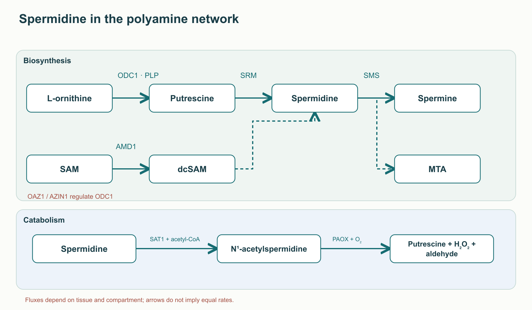
Age and measurement of the tissue pool
Polyamine composition changes with age in experimental systems, but the statement that “spermidine falls by 40–70% in all human tissues” is not supported by a single representative sample. Results depend on species, age, organ, diet, microbiota, and quantitative assay. Plasma concentration need not mirror the free pool in a cardiomyocyte or neuron; a high value in a long-lived person does not prove that the polyamine caused longevity. Senescence is characterized not by one polyamine measure but by a combination of durable cell-cycle arrest, DNA damage, metabolic change, and a secretory phenotype; p16, p21, and γH2AX are not absolutely specific tests on their own.
The polyamine network may affect proteostasis, oxidative stress, and proliferation. This provides a biological rationale for studying aging, but without tissue-specific evidence it does not allow an age-related change to be attributed solely to ODC1/AMD1 methylation or increased OAZ1 or SAT1. It is especially important not to treat every form of autophagy as “rejuvenation”: autophagosome formation may increase even when lysosomal degradation is blocked and clearance remains incomplete.
EP300 and measurable autophagic flux
Macroautophagy begins with formation of the phagophore, proceeds through ATG12–ATG5 conjugation and lipidation of ATG8/LC3 with phosphatidylethanolamine, and then continues with fusion of the mature autophagosome with a lysosome and hydrolysis of its cargo. A snapshot showing many LC3-positive puncta cannot distinguish accelerated autophagosome formation from stalled degradation. Inferring increased flux requires dynamic measurements, for example comparing markers with lysosomal inhibition and assessing p62/SQSTM1 with appropriate controls. This distinction is fundamental to any claim of “cellular cleansing.”
EP300/KAT3B transfers an acetyl group from acetyl-CoA to the ε-amino group of a protein lysine: acetyl-CoA + protein–Lys–NH₂ → CoA + protein–Lys–NH–COCH₃. EP300 acts in the nucleus and on non-histone proteins; its function is not a single autophagy “off switch.” The nucleocytosolic acetyl-CoA level depends, among other factors, on citrate delivery and ATP-citrate lyase ACLY, but changing acetyl-donor availability affects many reactions. mTORC1 regulates the ULK1 complex and other processes; acetylation is another regulatory circuit. These circuits cannot be declared fully independent in every experiment.
In the primary study by Pietrocola and colleagues, knockdown of EP300 and NAA20 among 43 acetyltransferases examined was accompanied by signs of autophagy and mTORC1 suppression in a selected cellular system. The EP300 inhibitor C646 also had an effect in enucleated cytoplasts, supporting the existence of a cytoplasmic mechanism. In a cell-free assay, recombinant EP300 acetylated histone H3 less efficiently in the presence of spermidine; raising acetyl-CoA from 10 to 100 μmol/L attenuated the inhibition. This is consistent with competition for the acetyl donor, but it is not structural evidence that spermidine occupies specific atomic contacts. The data do not support a universal Ki of 5–10 μmol/L, a list of Asp/Tyr/Trp contacts, ITC/SPR results, or equivalence between this mechanism and any level of oral exposure. 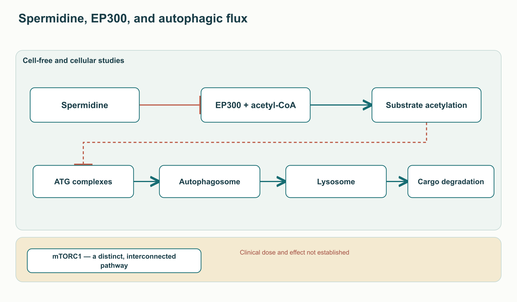
The original claim of a strictly mTORC1-independent action requires an especially explicit correction: the abstract of the same experiment describes signs of autophagic flux together with mTORC1 inhibition. Accordingly, the primary study does not support contrasting spermidine with rapamycin as a substance that is guaranteed to preserve basal mTORC1 activity and avoid associated risks. Dietary spermidine is not pharmacologically equivalent to rapamycin; their safety cannot be compared without direct human data.
ATG proteins, acetylation, and compartment
ATG7 acts as an E1-like enzyme in two conjugation systems, activating ATG12 and ATG8/LC3; the ATG12–ATG5–ATG16L1 complex provides an E3-like function during LC3 conjugation to phosphatidylethanolamine. ATG4 processes the LC3 precursor and also participates in its subsequent delipidation. Beclin 1 with VPS34/PIK3C3 participates in PI3P formation at nucleation membranes. Acetylation of individual components alters their interactions and localization in different model systems. However, the precise set of residues Lys347/352 in ATG7, Lys130 in ATG5, Lys49/51 in LC3B, and Lys437 in Beclin 1 cannot all be attributed to direct EP300 action in the spermidine experiment without an independent site map and functional mutations. Spermidine may lower overall acetylation, but that does not mean it instantly and completely “releases the brake” on every listed protein.
Nuclear EP300 regulates transcription through histone and non-histone substrates. Reduced acetylation of selected histone marks cannot automatically be interpreted as globally beneficial chromatin compaction: consequences depend on locus, cell type, and repair state. ATM primarily responds to double-strand DNA breaks, whereas ATR responds to replication stress and single-stranded DNA regions; BRCA1 and RAD51 act in a particular homologous-recombination context. A linear scheme of “inhibit EP300 → activate ATM/ATR → repair DNA without error” has not been established as a universal response to spermidine intake. γH2AX and 53BP1 are markers of a damage response, not stand-alone quantitative proof of genome integrity.
Telomeres consist of TTAGGG repeats and are protected by the shelterin complex, which includes TRF1, TRF2, TIN2, RAP1, TPP1, and POT1. Guanine-rich sequences are vulnerable to oxidation, and disruption of shelterin can trigger a damage response. This explains the possible interest in polyamine–DNA interactions, but there is no basis for describing spermidine as a proven direct stabilizer of every shelterin protein or for promising telomere-length preservation after dietary intake. Telomere length also depends on the cellular composition of the sample and cell-division history.
From spermidine to eIF5A hypusination and lysosomes
Spermidine’s effect on translation is fundamentally distinct from cell-free EP300 inhibition. Deoxyhypusine synthase DHPS uses spermidine as a donor of the 4-aminobutyl moiety and transfers it to a conserved lysine on eIF5A. The reaction involves NAD⁺; 1,3-diaminopropane is a by-product. Iron-containing deoxyhypusine hydroxylase DOHH then hydroxylates deoxyhypusine using O₂, completing hypusine formation. Schematically: spermidine + eIF5A–Lys → 1,3-diaminopropane + eIF5A–deoxyhypusine → eIF5A–hypusine. NAD⁺, O₂, and their products should be accounted for when writing the full enzymatic balance; the arrows here show sequence, not a closed stoichiometric equation.
Despite its historical name, “initiation factor,” hypusinated eIF5A is important for translation elongation and termination. It helps the ribosome traverse sequences at which peptide-bond formation is difficult, especially some polyproline stretches. This does not mean that every Pro-Pro motif inevitably stalls protein synthesis without eIF5A, or that all TFEB translation depends on a single such sequence. Reduced eIF5A hypusination affects a range of transcripts and cellular functions, so supplementation is not a selective “TFEB switch.”
The study by Zhang and colleagues demonstrated the spermidine–hypusinated eIF5A–TFEB axis in aging B cells and model systems. Aged B cells had a reduced autophagic response; interventions affecting this pathway restored some B-cell memory measures ex vivo. This is meaningful mechanistic evidence in the specified immune compartment, but it is not a trial of oral supplementation on human cognitive outcomes, myocardial infarction incidence, or lifespan. A B-cell-specific finding cannot be generalized to every neuron, cardiomyocyte, and tumor cell without testing.
TFEB coordinates expression of lysosomal, autophagy, and cellular-metabolism genes through CLEAR elements. The network includes V-ATPase subunits, lysosomal hydrolases, membrane proteins, and other regulators. Yet TFEB synthesis and its nuclear entry are distinct control steps: nuclear localization depends, among other factors, on mTORC1 phosphorylation and calcineurin signaling in the relevant context. Even an increase in lysosome number does not guarantee completed degradation if acidification, membrane fusion, or enzyme function is impaired. Thus, EP300 and eIF5A–TFEB should be viewed as two potentially interacting branches, not a fully closed mechanism that “prevents a bottleneck” in autophagy in every cell.
The heart: from cardiomyocyte autophagy to diastolic mechanics
The aging heart cannot be reduced to a single autophagy measure. A postmitotic cardiomyocyte must renew both mitochondria and the contractile apparatus; interstitial collagen adds extrasarcomeric stiffness, while the endothelium regulates vascular load and NO signaling. Heart failure with preserved ejection fraction (HFpEF) is a clinically heterogeneous syndrome involving impaired filling, elevated pressures, and systemic comorbid factors. Neither an aged-mouse model nor a Dahl salt-sensitive rat reproduces the full human syndrome. This is essential when interpreting striking experimental cardioprotection.
What the Eisenberg study actually showed
In the study by Eisenberg and colleagues, oral spermidine supplied in drinking water increased mouse lifespan, including when the intervention began in late life; older animals also had less cardiac hypertrophy and better-preserved diastolic function. The heart showed increased autophagy and mitophagy, improved mitochondrial respiration, increased titin phosphorylation, and fewer signs of subclinical inflammation. In Dahl rats on a high-salt diet, spermidine lowered blood pressure, attenuated hypertrophy, and delayed development of heart failure. These are a set of distinct but related animal outcomes, not evidence of fewer myocardial infarctions or treatment of HFpEF in patients. In the human part of the study, the association between dietary spermidine and lower blood pressure and cardiovascular morbidity was observational (Eisenberg et al., 2016).
The causal role of cellular degradation was tested in mice lacking Atg5 in cardiomyocytes: much of the protection associated with spermidine supplementation disappeared. ATG5 is required for ATG12–ATG5 conjugation and LC3 lipidation; its loss itself impairs basal proteostasis. The result therefore supports a requirement for a functional cardiomyocyte autophagy apparatus in this model, but does not prove that EP300, TFEB, or a single titin site fully mediates all observed effects. To distinguish autophagosome formation from completed lysosomal degradation, the investigators used leupeptin blockade of proteolysis, a tandem fluorescent LC3 reporter, and mitochondrial Mito-Keima. Each method has limitations: an LC3-II/LC3-I ratio at one time point does not by itself measure flux (Eisenberg et al., 2016).
Exact percentages of change in median or maximum lifespan and the size of a blood-pressure reduction depend on the specific curve, sex, strain, intervention start, and dose. Directly converting “18 mouse months = 55–60 human years” is not a biological calibration, and a value from one group is not a universal effect size.
Titin, NO, and two components of stiffness
Titin extends from the Z-disc to the M-line across each half-sarcomere. Its I-band segments, including Ig-like domains, the cardiomyocyte-specific N2B region, and the proline/glutamate/valine/lysine-rich PEVK segment, form an elastic system. The N2B and N2BA isoforms differ in length and passive stiffness; isoform composition, phosphorylation, and oxidation modify force during stretch. In several conditions, phosphorylation of N2B by cGMP-dependent protein kinase G lowers passive tension. Eisenberg and colleagues measured isoform composition and overall N2B phosphorylation, including a signal for Ser4080, after spermidine supplementation in mice (Eisenberg et al., 2016). This links the intervention with mechanics but does not establish the chain “spermidine directly activates eNOS → only PKG phosphorylates Ser4080 and Ser4099 → tension falls by >40% in every patient.” Phosphorylation of Ser4099, the specific magnitude of the mechanical effect, and the separate role of each enzyme require the appropriate primary experiment.
The physiological endothelium/cardiomyocyte axis is defined by the reactions L-arginine + O₂ + NADPH → L-citrulline + NO + NADP⁺ (NOS3/eNOS, with heme and pterin cofactors), NO → soluble guanylyl cyclase (sGC), GTP → cGMP, and cGMP → PKG. eNOS uncoupling during tetrahydrobiopterin deficiency or excess oxidative stress can shift the enzyme toward superoxide production; ADMA competes with L-arginine. This biochemistry explains why endothelial dysfunction can affect titin, but it does not prove that spermidine intake normalizes ADMA, eNOS acetylation, or human flow-mediated dilation (FMD). The path from supplementation to a human vascular outcome remains a testable hypothesis.
Ventricular passive stiffness comprises the cardiomyocyte titin component and the extracellular matrix. Under TGF-β signaling through serine/threonine kinase receptors and phosphorylated SMAD2/3, fibroblasts may differentiate into α-SMA-positive myofibroblasts and increase deposition of collagen I/III. Lysosomal degradation of components in this cascade and a reduction in fibrosis are biologically possible, but the specific claim that “spermidine rapidly destroys SMAD2/3 and reverses fibrosis” has not been established as a necessary mechanism by the cited cardiac study. Even histological reduction in fibrosis in a model would not equal regression in patients. Pulse-wave velocity (PWV) reflects arterial stiffness, whereas FMD measures an endothelium-dependent response; neither should be presented as a demonstrated human effect of spermidine without a corresponding intervention study.
Mitochondria, inflammation, and atherosclerotic plaque
In cardiomyocytes, continuous oxidative phosphorylation requires renewal of respiratory complexes, cristae, and damaged organelles. With substantial outer-membrane depolarization, PINK1 may accumulate on mitochondria; ubiquitin phosphorylation and Parkin/PARK2 enhance ubiquitination of membrane proteins, while OPTN and NDP52 receptors link this signal to LC3-positive membranes. Parkin-independent mitophagy pathways also exist, and the specific mechanism depends on the cell and injury. Mito-Keima in the Eisenberg experiments supports altered delivery of mitochondrial material to an acidic compartment, but does not prove that spermidine necessarily triggers the precise PINK1–Parkin–VDAC1/MFN1/2 sequence in all mice or humans (Eisenberg et al., 2016).
Damaged mitochondria may increase reactive oxygen species and release danger signals that contribute to activation of the NLRP3 inflammasome, caspase-1, and maturation of IL-1β/IL-18. The connection between mitophagy and inflammation is plausible; reduced inflammatory markers measured in animals do not justify describing NLRP3 as completely switched off. An ApoE-deficient mouse model and atherosclerotic-plaque morphology require a separately identified primary experiment. Even confirmed morphology would not equal a clinical outcome such as myocardial infarction or arterial elasticity in humans. Cardiac findings therefore form a research map, not a ready-to-use cardiology protocol.
Neurons, active zones, and the limits of cognitive inference
Neurons are long-lived and polarized, and depend on axonal transport and local disposal of damaged proteins. Their autophagy is therefore not a simple central “garbage disposal”: autophagosomes also form in distal processes and then move toward regions with greater lysosomal competence. Amyloid peptides Aβ40/Aβ42 arise through sequential cleavage of APP by β- and γ-secretases, while tau regulates microtubules and, when abnormally phosphorylated, may form pathological aggregates. Lysosomal flux contributes to proteostasis in both systems, but this does not establish that oral spermidine in humans selectively removes Aβ42 before plaque formation or tau bearing AT8/PHF1 epitopes in CA1/CA3. The processes of formation, intracellular degradation, extracellular plaque deposition, and clinical memory cannot be collapsed into one inevitable chain. These effects should be measured separately rather than inferred from a general ATG mechanism (Schroeder et al., 2021).
Microglia respond to injury and can remove synapses; their cytokines TNF-α and IL-1β and pathways such as iNOS/NOS2 have context-dependent roles. The old “M1/M2” dichotomy describes microglial states in the human brain too crudely. Preclinical observations of reduced inflammatory signals with spermidine do not demonstrate global microglial suppression, cessation of synaptic pruning, or dementia prevention. Cognitive improvement in aged animals in the study by Schroeder and colleagues should be distinguished from specific molecular mediators and from the negative large human trial (Schroeder et al., 2021; Schwarz et al., 2022).
Bruchpilot: a strong model and a weak automatic translation
The presynaptic active zone organizes the proximity of calcium channels and vesicles ready for release. In Drosophila, the structural protein Bruchpilot (BRP) forms the characteristic T-bar architecture. Gupta and colleagues studied age-related memory in flies and found that restoration of the polyamine pool, including with spermidine, improved behavioral performance when autophagy was functional. They linked age-related accumulation of BRP at the active zone to these phenotypes (Gupta et al., 2013). This was not an experiment in the human hippocampus, and the names of the measured tests cannot be replaced by a promise of “full restoration of human spatial memory.”
CAST/ELKS/ERC, Bassoon, and Piccolo organize the active zone in mammals, but evolutionary functional analogy does not prove that dietary spermidine directly clears human CAST or preserves Synaptotagmin-1 and the SNARE complex (syntaxin-1, SNAP-25, VAMP2). Their inclusion is useful for presynaptic anatomy and hypothesis generation, but every step from “BRP in flies” to “CAST in humans” to “clinical memory” requires its own experimental bridge. Where Bruchpilot findings are established in flies, the article names that model; when discussing human neurodegeneration, it does not substitute the model for clinical evidence.
Three distinct human trials
| Study and population | Intervention and outcome | What the result permits us to conclude |
|---|---|---|
| Wirth et al., 3-month pilot RCT, 30 older adults with subjective memory decline | Wheat-germ extract providing 1.2 mg spermidine/day; mnemonic discrimination | A signal in a small exploratory sample that requires replication; not evidence of dementia prevention (Wirth et al., 2018). |
| Schwarz et al., SmartAge, 100 randomized participants with subjective complaints, 12 months | 0.9 mg/day versus placebo; primary Mnemonic Similarity Task | Negative primary result: between-group difference −0.03; 95% CI −0.11 to 0.05; p=0.47. Eighty-nine participants completed the intervention; secondary outcomes in the ITT analysis also showed no convincing effect (Schwarz et al., 2022). |
| Pekar et al., preliminary 3-month multicenter trial in six nursing homes, 85 people aged 60–96 years | Different levels of spermidine intake; CERAD/MMSE tests | In the higher-intake group and the mild-dementia subgroup, MMSE increased by 2.23 points, p=0.026; phonemic fluency changed by +1.99 with p=0.47. The small size and subgroup nature do not establish an effective dose threshold (Pekar et al., 2021). |
The final SmartAge report was published by Schwarz and colleagues in 2022; verbal memory and inflammation were exploratory signals, not confirmed primary effects. The authors discussed the low supplemental dose (about 10% of average daily intake in developed countries), no increase in blood polyamines, preserved cognitive function in subjective cognitive decline (SCD), and a short period for prevention. Each is a hypothesis explaining the null result; the trial did not prove that a precisely higher dose would work. Hippocampal MRI atrophy and hair cortisol should not be described as reliably improved without the corresponding analysis in the primary report (Schwarz et al., 2022).
Pekar and colleagues studied people who already had mild or moderate dementia, not the same SCD population. The food product, baseline function, scale, and measures differed; therefore, the contrast “0.9 mg does not work, 3.3 mg works” is not a dose–response curve from a single trial. When counseling a patient, this preliminary study cannot be placed above the negative prespecified SmartAge endpoint to recommend supplementation as treatment for Alzheimer’s disease.
What observational epidemiology tells us
In the population-based Bruneck Study, 829 people aged 45–84 years completed repeated dietary assessments in 1995, 2000, 2005, and 2010 (2,540 assessments in total); 341 deaths were recorded through 2015. Per standard-deviation increase in estimated spermidine intake, the all-cause mortality HR adjusted for age, sex, and energy intake was 0.74 (95% CI 0.66–0.83); after broader adjustment for lifestyle, risk factors, and dietary features, it was 0.76 (0.67–0.86). In SAPHIR, the direction of the mortality association was replicated: HR per SD was 0.71 (0.53–0.95) in the reported model. The difference in risk between the top and bottom tertiles was statistically compared with an age difference of 5.7 years (95% CI 3.6–8.1); this is a way of describing the model, not a measured slowing of each participant’s biological age (Kiechl et al., 2018).
| Evidence level | What was measured | Main limitation |
|---|---|---|
| Repeated food-frequency questionnaires in Bruneck and SAPHIR | Estimated dietary intake and subsequent mortality | FFQs and food-composition tables have measurement error; plant-rich dietary patterns, income, and health may remain residual confounders. |
| Wirth pilot RCT and preliminary Pekar trial | Short-term memory tests in different populations | Unstable effect estimates, multiple tests, and different baseline states. |
| Larger SmartAge RCT | Prespecified memory test over 12 months | No effect was detected at this dose and with this design; dementia prevention and lifespan were not tested. |
| Aged heart and brain models | Physiology, autophagic flux, and animal behavior | Cross-species translation and dose exposure remain open questions. |
Exact tertile boundaries, HRs for cause-specific mortality, and NHANES estimates should not be used without checking the corresponding primary tables and outcomes; the published Bruneck abstract reports primarily the analysis per standard deviation. SAPHIR is an independent replication of mortality, not the PWV and myocardial hypertrophy outcomes asserted in the source draft. Any HR is an association with diet, not an estimate of assigning spermidine in a tablet. Likewise, LC3-II/LC3-I and p62/SQSTM1 in a single PBMC sample do not establish systemic flux; flux requires dynamic degradation blockade or an appropriate compartment-specific reporter.
Dietary spermidine, transport, and exposure
Polyamines come not only from supplements: cellular synthesis, gut microorganisms, food, cellular uptake, and catabolism interact. Wheat germ, legumes, some mushrooms, fermented soy products, and aged cheeses may provide spermidine, but concentrations vary substantially with cultivar, fermentation, dry versus wet weight, and analytical method. A value from one food-composition table cannot be applied to every serving or treated as a universal range of 250–350 mg/kg for wheat germ. It is essential to distinguish mg of extract from mg of analytically confirmed spermidine. Fermentation also changes putrescine and spermine profiles; vitamin E and phospholipids in the food matrix may be accompanying components, but “absorption synergy” has not been demonstrated in a comparative trial (Madeo et al., 2020).
In the gut, polyamines bind anionic surface components, undergo cellular uptake, and are partly metabolized in the epithelium; the microbiota both synthesizes and consumes them. The identity and contribution of specific transporters in human enterocytes remain under study. Thus, claims of obligatory absorption predominantly through OCT1/2/3 (SLC22A), as well as universal sequestration of 85–90% of a dose in erythrocytes/leukocytes, should not be presented as established human pharmacokinetics. Plasma, whole-blood, and cellular-fraction analyses yield different results. Polyamines bind negatively charged macromolecules and distribute among compartments; a single serum concentration is not equivalent to exposure in the heart or brain. Passage of supplemented spermidine across the blood–brain barrier and saturation of the pathway in humans require direct tracer data; an animal observation cannot be replaced with the claim that a “free fraction readily crosses the BBB.”
In cells, SAT1 uses acetyl-CoA for N¹-acetylation of polyamines; the acetylated form may be exported or converted by PAOX, producing a shorter polyamine, 3-acetamidopropanal, and H₂O₂. Direct oxidation of spermine by SMOX yields spermidine, 3-aminopropanal, and H₂O₂. Aldehydes and peroxide are potentially toxic, so greater oral exposure need not monotonically increase the beneficial intracellular pool. Repeated measurement of free and acetylated polyamines in the relevant matrix is more informative than mechanically converting dietary dose into EP300 activity.
The proposed range of 1–6 mg/day, morning fasting intake, and a 16/8 window do not come from a comparative clinical trial. A general therapeutic dose for cardioprotection or neuroprotection, a required time of day, synergy with fasting, and a threshold for EP300 activation have not been established in humans. Doses in individual RCTs describe study conditions, not a clinical protocol. Even 0.9 mg/day in SmartAge had a null prespecified cognitive outcome; higher dietary intake in the Bruneck cohort was not a randomized drug dose (Schwarz et al., 2022; Kiechl et al., 2018).
Why mechanistic “stacks” are not yet therapy
In cell models, resveratrol and spermidine affected acetylation and autophagy through different pathways; this does not establish a durable “double epigenetic lock” in humans or additive lifespan extension (Morselli et al., 2011). Urolithin A is studied in the context of mitochondrial quality; PINK1/Parkin are not its only targets, and a combined RCT with spermidine would be needed before claiming synergy. NMN/NR replenish the NAD⁺ pool through their own metabolic pathways, but the NAD⁺ dependence of sirtuins and DHPS does not mean that adding a precursor enhances spermidine’s effects. Moreover, NAD⁺ is a substrate for many enzymes, not direct “fuel for the Krebs cycle”; NADH/NAD⁺ participate in redox reactions. Possible combinations remain research questions, each requiring separate doses, outcomes, and safety assessment.
Oncological duality and immune context
Polyamines are needed for normal proliferation, differentiation, translation, and repair, but tumors use the same resources. The MYC oncogenic program can increase ODC1 expression and polyamine flux. ODC1 converts ornithine to putrescine; the irreversible ODC1 inhibitor eflornithine/DFMO is studied and used in certain oncologic contexts, often together with restriction of exogenous polyamine uptake. The rationale is clear: a tumor may bypass synthesis blockade by importing polyamines. This mechanism is a strong reason for caution with concentrated supplements during active cancer, but it does not prove the universal rule that any dietary dose accelerates every tumor (Casero et al., 2018).
Another side of the issue involves immune cells. In human CD8⁺ T cells from older donors ex vivo, Alsaleh and colleagues observed a reduced polyamine pool and functional autophagic response; added spermidine restored some flux measures and the effector molecules IFN-γ and perforin through eIF5A/TFEB. The study also compared T-cell autophagy with immune responses after vaccination, but did not show reduced cancer incidence, restoration of tumor-specific exhausted CD8⁺ cells after oral supplementation, or survival benefit in cancer patients (Alsaleh et al., 2020). Translational findings on B-cell hypusination by Zhang are likewise not a cancer-protection trial (Zhang et al., 2019).
Thus, “cancer protection in healthy people” and “an absolute contraindication in every active tumor” are not two clinically established extremes. Observational dietary associations do not justify calling a pill cancer prevention; the metabolic dependence of some tumors supports avoiding self-directed concentrated supplementation during cancer treatment and discussing any intervention with the treating team. Product-specific official contraindications should not be replaced by an improvised universal list. Potential effects on a tumor depend on its genotype, transport and catabolic enzymes, local immune environment, and therapy.
Safety: what has and has not been measured
Over 12 months, the randomized SmartAge trial at 0.9 mg/day found no imbalance in adverse events between spermidine and placebo; this conclusion applies to that older population, dose, product, and follow-up period (Schwarz et al., 2022). Small studies can inform tolerability but cannot exclude rare events, risks in pregnancy, interactions with anticancer therapy, or effects over many years. A “human equivalent” calculated by simple body-weight scaling from a purported rodent NOAEL >50 mg/kg is not valid: an established toxicity endpoint, allometric scaling, bioavailability, and an applicable regulatory approach are required. The original conclusion that “>250 mg/day is safe for 70 kg” is unsupported.
Wheat-germ extract requires separate checking of its composition: for celiac disease or wheat allergy, the gluten and allergen data for the specific product matter. A pure spermidine salt is not equivalent to an extract in substance mass or impurities; a claim of “>99% pharmaceutical purity” applies only to the tested batch. Pregnancy and lactation remain populations without reliable data. A routine panel of ALT, eGFR, ApoB, NT-proBNP, PWV, and echocardiographic E/e′ every 3–6 months has not been validated as monitoring for this supplement by any of the reviewed trials; studies selected outcomes to address their own questions. When heart disease or cognitive decline is suspected, clinical assessment should be guided by the patient’s condition, not a spermidine protocol.
How to interpret the overall evidence
The molecular plausibility of spermidine is high because it is not an external switch for one receptor but part of a metabolic network: synthesis from putrescine through dcSAM, breakdown, and intercellular uptake sustain the pool; EP300/acetylation and DHPS–DOHH/eIF5A–TFEB provide distinct points of proteostasis regulation. The strongest functional evidence concerns aged-heart models and several cell types with genetic and flux-based tests. The weaker step is the assumption that an ordinary dietary or supplemental dose reproduces the required concentration and compartment in a human organ. In observational cohorts, a promising association between diet and mortality does not eliminate residual confounding. In clinical memory studies, pilot signals coexist with a null result on a larger prespecified endpoint. The cited studies contain no human cardiac RCT with myocardial infarction, HFpEF, or survival as an outcome and a confirmed spermidine mechanism.
A research program should therefore prespecify the product and pure dose, exposure matrix, age and baseline state, suitable tissue or indirect flux biomarker, clinically meaningful endpoint, and adequate duration. A modest increase in polyamine-containing foods may be discussed as part of an overall dietary pattern and according to tolerability, but it is not a specific prescription for “anti-aging treatment.” Practical promises of preventing myocardial infarction, dementia, or cancer remain unsupported.
How this review was prepared
This is a narrative review of spermidine, polyamine metabolism, and autophagy. In the editorial review dated 4 October 2026, selected mechanistic and clinical claims and identified bibliographic conflicts were checked directly; the final selection includes 16 publications. No systematic search using a prespecified protocol, meta-analysis, or formal risk-of-bias assessment for each study was conducted. The selection is not exhaustive, and information from unavailable full texts is not considered independently verified.
Enzyme, cell, and animal experiments are distinguished from population associations and human trials. An association between dietary intake and an outcome does not prove an effect of supplementation. Cognitive trials are assessed in light of the population studied, regimen, and primary endpoint, including negative results. Interest in autophagy and cardioprotection does not make polyamines a proven treatment; any possible effect depends on biological context.
FAQ
Has spermidine been shown to treat heart failure with preserved ejection fraction?
No. Improved diastolic function and autophagy have been shown in mice and rats; the reviewed evidence contains no clinical trial with this outcome.
Why was a small memory study positive while SmartAge was negative?
Sample size, dose, population, and duration differed; SmartAge did not meet its prespecified endpoint with 0.9 mg/day for 12 months. This does not establish that an effective dose threshold exists.
Can spermidine be used to prevent cancer?
Cohort associations with dietary intake and cellular immune mechanisms do not prove cancer prevention by a supplement. During active cancer treatment, concentrated supplements should be discussed with the treating team.
Sources
- Pegg AE. Functions of Polyamines in Mammals
- Eisenberg T et al. Induction of autophagy by spermidine promotes longevity
- Pietrocola F et al. Spermidine induces autophagy by inhibiting the acetyltransferase EP300
- Eisenberg T et al. Cardioprotection and lifespan extension by the natural polyamine spermidine
- Zhang H et al. Polyamines Control eIF5A Hypusination, TFEB Translation, and Autophagy to Reverse B Cell Senescence
- Mariño G et al. Regulation of autophagy by cytosolic acetyl-coenzyme A
- Morselli E et al. Spermidine and resveratrol induce autophagy by distinct pathways converging on the acetylproteome
- Kiechl S et al. Higher spermidine intake is linked to lower mortality: a prospective population-based study
- Gupta VK et al. Restoring polyamines protects from age-induced memory impairment in an autophagy-dependent manner
- Schroeder S et al. Dietary spermidine improves cognitive function
- Wirth M et al. The effect of spermidine on memory performance in older adults at risk for dementia: a randomized controlled trial
- Schwarz C et al. Effects of Spermidine Supplementation on Cognition and Biomarkers in Older Adults With Subjective Cognitive Decline: A Randomized Clinical Trial
- Pekar T et al. The positive effect of spermidine in older adults suffering from dementia: First results of a 3-month trial
- Alsaleh G et al. Autophagy in T cells from aged donors is maintained by spermidine and correlates with function and vaccine responses
- Casero RA Jr et al. Polyamine metabolism and cancer: treatments, challenges and opportunities
- Madeo F et al. Nutritional Aspects of Spermidine
Related materials
- S-adenosylmethionine: methylation, neurochemistry and clinical limits
- L-Arginine in the Human Body: Nitric Oxide, Endothelium, Blood Pressure, Immunity, and the Limits of Supplementation
- Fermented Foods: Microbiota, Digestion, and Safety
- Vitamin B3: NAD⁺, Nicotinic Acid, Nicotinamide, and Why a Better Lipid Panel Does Not Necessarily Mean Better Cardiovascular Protection