
Silymarin and silibinin: membranes, rRNA and clinical evidence
Extract composition, stereochemistry, oral and IV exposure, membrane experiments, OATP1B3, rat Pol I/rRNA, redox and fibrosis pathways. Primary liver trials, negative endpoints, interactions and formulation limits.
Silymarin is a mixture extracted from milk-thistle fruits, whereas silibinin, also called silybin, is principally a pair of flavonolignan diastereomers. Those names do not designate interchangeable substances. A conventional oral extract, a silybin–phosphatidylcholine complex and intravenous silibinin succinate differ in composition, absorption, metabolism and exposure. A beneficial effect of one preparation in a cell experiment cannot establish the effectiveness of every product bearing the milk-thistle name.
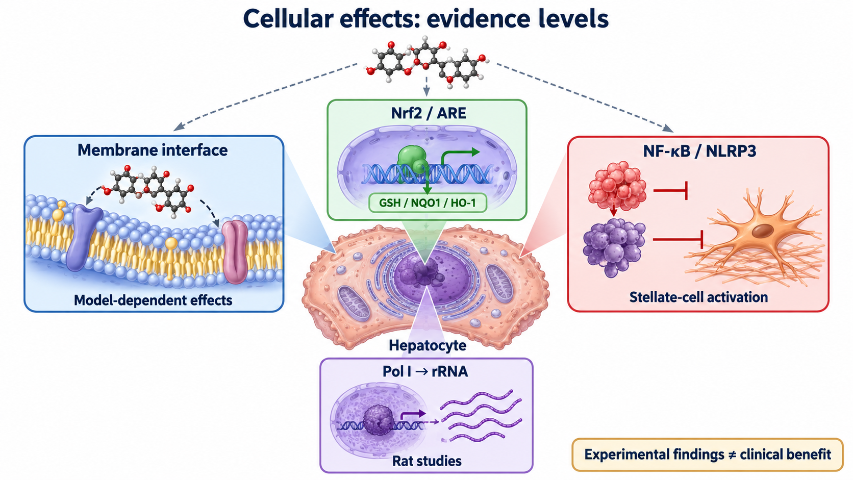
Several mechanisms make these compounds scientifically interesting: interaction with membrane lipids and uptake transporters, experimental stimulation of ribosomal RNA synthesis, redox regulation, and modification of inflammatory and fibrogenic responses. Their evidential strength differs. In particular, descriptions of membrane stabilization or regeneration must accommodate experiments with opposite membrane effects and clinical trials with negative primary outcomes. This article follows the route from chemistry to cell compartments to human studies, keeping laboratory activity separate from validated clinical benefit.
1. What is in the preparation?
A plant extract, a constituent and a derivative
The fruits of Silybum marianum (L.) Gaertn., family Asteraceae, are technically achenes. Their extracts contain flavonolignans formed from a flavonoid and a phenylpropanoid. Oxidative coupling of taxifolin with coniferyl alcohol, followed by cyclization, explains the combined scaffold; plant peroxidase activity participates in this biosynthesis (Lv et al., 2017). Different coupling orientations produce regioisomers, while stereochemistry produces diastereomers. Neither botanical origin nor membership in the same chemical family establishes identical pharmacology.
Silymarin commonly contains silybin A and B, isosilybin A and B, silychristin and related constituents, silydianin, and the flavonoid taxifolin. Taxifolin lacks the lignan-derived portion and should not be counted as a flavonolignan. Minor constituents and oxidation products also occur. Commercial analytical profiles vary with plant material, extraction, purification, storage and the assay used. A claim such as “80% silymarin” requires an analytical definition: percentage of an extract, measured flavonolignans, and a calculation using a reference compound are not necessarily the same quantity.
Silybin often supplies a large fraction of the measured flavonolignans, with approximately half frequently reported, but the proportions of silychristin, silydianin and isosilybins are not fixed universal constants. Nor does a standardized total percentage guarantee an identical A:B ratio, minor-constituent profile or dissolution behaviour. Kroll et al. (2007) discussed the consequences of inconsistent nomenclature; Davis-Searles et al. (2005) separated constituents and demonstrated different effects in prostate-cancer cell assays. Those experiments establish the importance of identifying the material, not a general superiority ranking for liver treatment.
| Term | Chemical or pharmaceutical meaning | What it does not establish |
|---|---|---|
| Silymarin | An extract containing several flavonolignans and other constituents | A single molecule or invariant mixture |
| Silibinin / silybin | Usually the silybin A and B pair | The whole extract, or a fixed ratio in every preparation |
| Silybin A and B | Diastereomers sharing connectivity | Identical metabolism and biological responses |
| Isosilybin A and B | Regioisomers relative to silybin, also a diastereomeric pair | Merely different names for silybin A and B |
| Silybin–phosphatidylcholine complex | A formulation designed to alter delivery | Proven equivalence to an intravenous drug or universal clinical superiority |
| Intravenous silibinin succinate | A water-soluble derivative formulated for infusion | An oral supplement with the same exposure |
Stereochemistry needs an explicit numbering convention
Silybin has the molecular formula C₂₅H₂₂O₁₀ and a molar mass of about 482.44 g/mol. Its structure includes a dihydroflavonol portion, a benzodioxane-containing phenylpropanoid portion, a methoxy group and five hydroxyl groups. Only some hydroxyls are phenolic; the secondary alcohol and hydroxymethyl alcohol should not be assigned the same radical-donating chemistry as a phenolic group.
With one commonly used silybin numbering system, the major A form is assigned (2R,3R,10R,11R), and B is assigned (2R,3R,10S,11S). Alternative numbering labels the benzodioxane stereocentres differently. A stereochemical specification without its numbering scheme is therefore ambiguous. Four independent stereocentres allow up to 2⁴ = 16 theoretical stereoisomers; this counting statement does not mean that a routine extract contains all sixteen. The major natural pair shares the flavonoid-centre configuration and differs at the lignan-derived centres. Isosilybins have different connectivity, so identical R/S letters cannot by themselves describe the same structure across the two families.
NMR, electronic circular dichroism and crystallographic work support the stereochemical assignments. Studies of cis–trans isomerization further show that other configurations can be generated under particular chemical conditions (Novotná et al., 2014). The major natural A and B forms are diastereomers, not an enantiomeric pair. Differences in solubility, conformation, conjugation or activity are possible, but a universal assertion that A is the aqueous antioxidant and B is the more potent transporter or nuclear-receptor ligand would require measurements in specified systems. An exact solvent-independent “half-chair versus axial” map is not needed to explain their experimentally observed differences.
Measuring a reproducible substance
A useful analytical description combines chromatographic identity and amount with purity, degradation products and batch information. RP-HPLC or LC–MS can distinguish components that a total-extract label combines. Dissolution, formulation mass and the mass of actual silibinin should also be reported. For example, 200 mg of a phospholipid complex is not automatically 200 mg of silybin. The same issue arises when an infusion vial gives both derivative mass and the equivalent mass of silibinin.
2. Absorption, conjugation and delivery
Low oral exposure is a formulation-dependent problem
Silybin is poorly soluble in water, and absorption is limited by dissolution, permeability and extensive presystemic metabolism. Reported solubility and octanol–water partition coefficients depend on temperature, pH, crystal form and measurement method. A precise solubility below 0.04 mg/mL or a particular log P range should not be applied as a universal specification for the entire extract. Likewise, assigning a multicomponent botanical preparation one biopharmaceutics-classification number hides differences among components and formulations.
After oral administration, absorbed material enters intestinal and hepatic metabolism. Glucuronides and sulfates are prominent circulating species, and free unconjugated flavonolignans form a different exposure measure from “total” analyte measured after hydrolysis. Wen et al. (2007) measured these fractions separately and reported short half-lives, approximately 1–3 hours for free and 3–8 hours for conjugated flavonolignans in their study. This does not supply a universal half-life for every product, disease state or infusion regimen.
Two general phase-II reactions illustrate the distinction:
UDP-GlcA is the activated glucuronyl donor; PAPS is the activated sulfate donor, and PAP is its desulfated product. UGT and SULT denote enzyme families, rather than proof that one isoform makes every positional metabolite. Conjugation at different hydroxyls can be stereoselective. The main conjugates cannot simply be declared inactive: their tissue distribution, deconjugation and biological activity require separate study.
Biliary excretion of conjugates, intestinal hydrolysis and reabsorption can contribute to enterohepatic cycling. Canalicular efflux systems, including MRP2/ABCC2 and BCRP/ABCG2, are relevant to disposition, but a transporter observed in an experimental model should not become an exact whole-dose mass balance in humans. Unabsorbed material, microbial metabolism, biliary return and urinary elimination are distinct routes. Claims that a fixed 80–85% of every swallowed dose is secreted in bile confuse administered, absorbed and recovered quantities. Absolute oral bioavailability also cannot be inferred from a low plasma concentration without an appropriate intravenous comparator and analyte definition.
Schrieber et al. (2008, 2011) found that exposure varied between liver-disease populations and healthy comparators or between disease groups. Thus, hepatic dysfunction can change the pharmacokinetics of the botanical constituents; “liver-directed” does not imply that an impaired liver automatically clears them normally. Hawke et al. (2010) also observed nonlinear pharmacokinetics during ascending oral dosing in chronic hepatitis C. Increasing dose or bioavailability may therefore change interaction risk as well as putative therapeutic exposure.
Phospholipid complexes are not interchangeable with liposomes
Complexing silybin with phosphatidylcholine can change its dispersion and absorption. Associations between polar groups and the amphiphilic lipid environment can help explain delivery, but a static drawing of a phospholipid completely wrapping one molecule does not establish its structure in gastrointestinal fluids. Hydrogen bonds are polar interactions. A liposome, in contrast, is a lipid vesicle whose aqueous compartment and bilayer can carry different classes of cargo; not every liposomal drug sits exclusively in the aqueous core.
Barzaghi et al. (1990) compared a silybin–phosphatidylcholine complex with silymarin in nine healthy volunteers using doses equivalent to 360 mg silybin. Their work supports formulation-dependent improvement in exposure. It does not prove a universal 4.6–10-fold AUC increase, a fixed peak-concentration multiplier, or an unrestricted passage through mucus by pinocytosis and chylomicron transport. AUC, Cmax and Tmax must be compared for the same analyte, equivalent dose and sampling conditions. Improved absorption is not itself a clinical-outcome trial.
Micronization, amorphous dispersions, self-emulsifying systems, solid lipid nanoparticles, polymeric carriers such as PLGA, and albumin-based nanoparticles address different delivery barriers. These approaches range from physicochemical studies to animal experiments and selected human pharmacokinetic studies. They should not all be described as established clinical replacements. A simple dissolution model explains why increased surface area can help:
Here M is dissolved mass, D is a diffusion coefficient, A is exposed particle area, h is an effective diffusion-layer thickness, Cs is the saturation concentration and C is bulk-medium concentration. This Noyes–Whitney-type expression is a simplified model. Smaller particles can increase A, but aggregation, wetting, crystal state, intestinal conditions and metabolism determine whether an improvement in dissolution produces more systemic exposure. A particle-size claim alone does not guarantee a twofold clinical benefit.
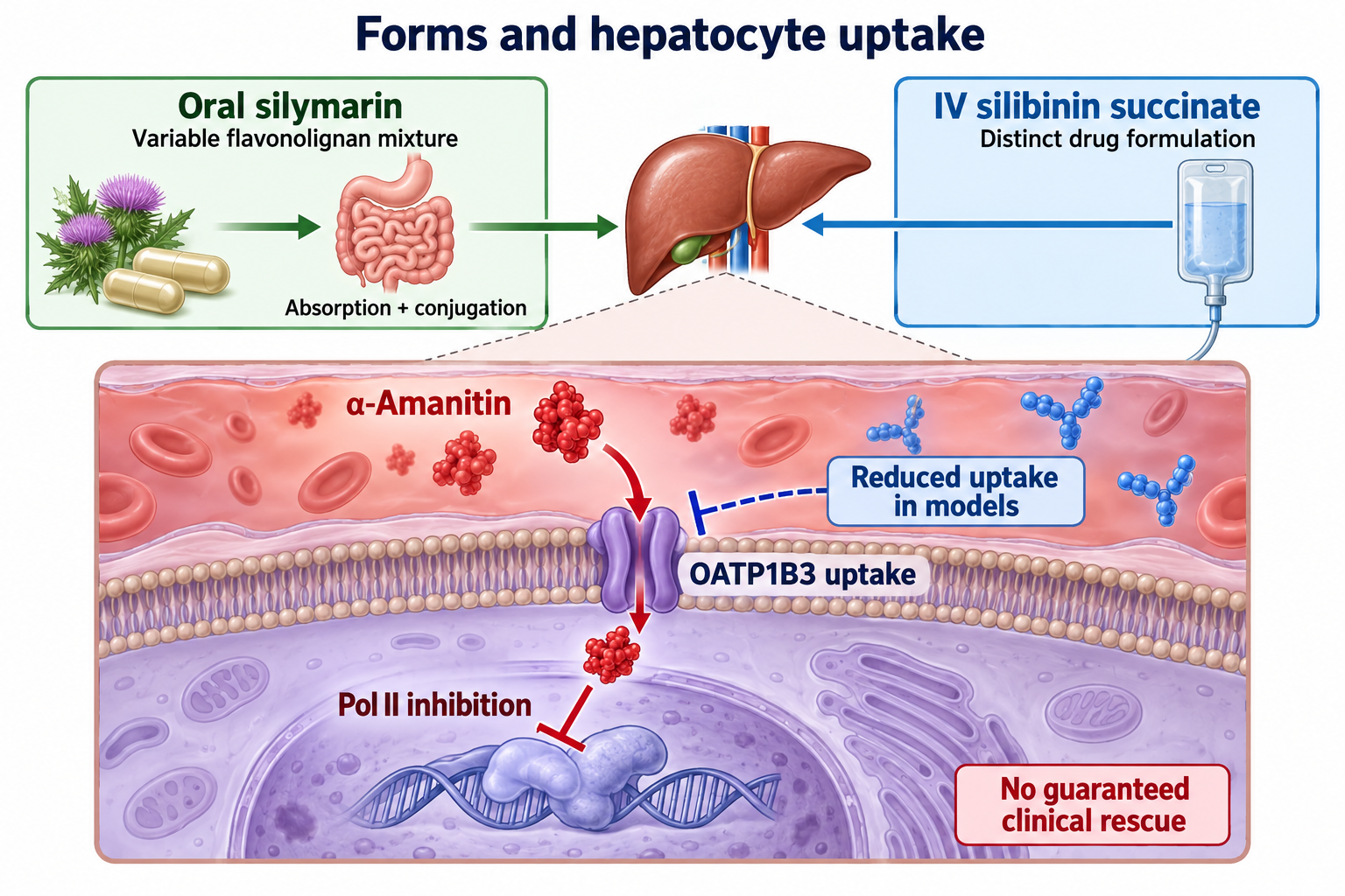
3. Membrane interaction and toxin uptake are different mechanisms
What a membrane experiment measures
The hepatocyte has a sinusoidal surface facing blood and a canalicular surface forming bile canaliculi. These surfaces contain different transport systems. Membrane lipids, cortical actin, adhesion complexes and tight junctions contribute to cellular integrity; the plasma membrane is not merely a passive bag. Oxidants, bile acids, solvents and other toxins can disturb lipid organization, proteins and ion gradients. Their effects vary with concentration, compartment and exposure time.
Flavonolignans can partition into lipid–water interfaces and interact with polar head groups or acyl-chain environments. Fluorescence probes such as DPH report local behaviour, but anisotropy is influenced by probe distribution and rotational motion as well as the surrounding membrane. A change in fluorescence cannot establish that all membranes have become less permeable, more ordered or resistant to every toxin.
Parasassi et al. (1984) examined rabbit liver microsomal membranes and observed modest probe changes consistent with incorporation near the hydrophobic–hydrophilic interface and altered acyl-chain packing. These were intracellular membrane preparations, not direct measurements of a functioning human sinusoidal barrier. Exact insertion depths between specified carbon atoms, a universal reduction of lipid flip-flop or a mechanical exclusion of all oxygen radicals are stronger propositions than these observations support.
Most importantly, whole silymarin and purified silibinin can behave differently. Basiglio et al. (2009) tested isolated membranes and rat hepatocytes under detergent or osmotic stress. At the investigated high concentration, the extract protected against disruption, whereas silibinin could increase Triton-associated or osmotic lysis and was less protective against taurochenodeoxycholate. This does not establish harm from customary oral exposure, but it directly challenges the assumption that isolated silibinin always reproduces the extract's membrane stabilization.
LDH or ALT released into culture medium is a useful indicator of damage under a specified condition. Less enzyme leakage can support a cytoprotective finding; it is not proof of restored bile secretion, rebuilt human liver architecture or survival benefit. Changes in fluidity must also be described consistently: viscosity and fluidity are related, so “reducing excessive viscosity” and “preventing excessive fluidization” cannot be treated as the same unconditional action.
OATPs are uptake carriers, not ATP-driven toxin pumps
OATP1B1 and OATP1B3, encoded by SLCO1B1 and SLCO1B3, are prominent human hepatic sinusoidal uptake transporters. Their substrate ranges include endogenous compounds and drugs. OATP2B1 has a different expression pattern and substrate profile. Their physiological localization does not mean that OATP1B3 is absent from every other tissue or tumour context. OATPs are distinct from canalicular ABC efflux transporters and are not ATP-hydrolysing pumps.
Inhibitory potency depends on the tested transporter, probe substrate, exposure time and preparation. Köck et al. (2013), for example, reported IC50 values for silymarin of 1.3, 2.2 and 0.3 µM at OATP1B1, OATP1B3 and OATP2B1, respectively. The corresponding values differed for silybin A, silybin B and silychristin. Such comparisons demonstrate component- and assay-specific inhibition, rather than one fixed affinity attached to the word silibinin.
IC50 is the concentration giving half-maximal inhibition under an assay's conditions; it is not automatically a binding dissociation constant or an in-vivo effective concentration. Ki also depends on the inhibition model. An experimental inhibitor need not completely stop transport at its plasma concentration, particularly when unbound concentration, protein binding, metabolites and substrate competition differ. A modelled interaction cannot establish a resolved binding pose involving particular OATP residues or a universally arrested transport conformation.
Amatoxins: the supported transporter distinction
Amatoxins and phallotoxins are different toxin families. Amatoxins are bicyclic octapeptides, whereas phallotoxins such as phalloidin are bicyclic heptapeptides. Phalloidin binds filamentous actin and is important in experimental cytoskeletal injury, but its poor gastrointestinal absorption means that it should not be presented as the principal cause of systemic human death-cap poisoning. Amatoxins, including α-amanitin, can cause severe liver injury after ingestion. Mushroom toxin content is variable, so a numerical lethal-dose estimate is not a reliable way to judge an individual ingestion.
Letschert et al. (2006) used transporter-expressing cells and primary human hepatocytes. Among the tested OATPs, OATP1B3 transported the labelled amanitin derivative; the measured Km was about 3.7 ± 0.6 µM. Silibinin dihemisuccinate and other compounds inhibited uptake. The experimental conditions and labelled substrate matter: this work supports a hepatic uptake mechanism, not the claim that OATP1B1 was demonstrated to carry amanitin in the same panel or that a single transporter explains every aspect of poisoning.
Once α-amanitin reaches its intracellular target, RNA polymerase II inhibition disrupts messenger-RNA transcription and subsequent protein production. The major mechanistic sequence is therefore uptake followed by intracellular toxicity. Uptake inhibition is different from displacing toxin already bound to Pol II, chemically neutralizing it, or restoring cells already lost to necrosis. Renal elimination and enterohepatic recirculation contribute to toxin disposition, while renal injury itself can complicate poisoning. Blocking an uptake route does not guarantee that all circulating toxin will be harmlessly and rapidly removed.
Model choice is important. Primary hepatocytes and some engineered cell systems express relevant uptake transporters better than commonly used hepatoma lines. Experiments comparing formulations found that HepG2-based systems can be insufficiently sensitive when the required transporters are poorly expressed (2020 primary-hepatocyte model study). A negative result in an unsuitable model and a positive result in a high-exposure model both need this qualification.
Cytoskeleton, calcium and blebbing
Cortical actin and actomyosin support the canalicular pole, while junctional proteins help separate bile from the blood-facing environment. Oxidative or toxic injury can alter actin organization, membrane attachment and intracellular calcium. Calpain activation may cleave cytoskeletal or adhesion-associated proteins; changes in phosphorylation and energy supply can disturb contractility. Membrane blebs are protrusions, not automatically extracellular vesicles. They occur in several settings, including apoptosis and reversible stress, and their appearance alone does not identify a specific death mechanism.
Silymarin-associated reductions in experimentally induced leakage or blebbing can fit a broader cytoprotective response. Nevertheless, a demonstrated reduction in cell injury does not prove direct strengthening of ezrin–radixin–moesin attachment, selective PLC inhibition, prevention of all IP3-dependent calcium release or a specific restoration of myosin-light-chain phosphorylation. Those are separable molecular propositions. The relationship between membrane organization, calcium, proteases and junctions is useful biological context, but clinical prevention of cholestasis requires measurements beyond an illustrative pathway.
4. Pol I, ribosomal RNA and regeneration
The historical biochemical observation
Machicao and Sonnenbichler (1977) studied isolated rat liver nuclei, nucleoli and RNA-polymerase preparations. Silybin increased RNA synthesis, particularly ribosomal RNA, and increased the activity assigned to polymerase A, corresponding to Pol I, while polymerase B/Pol II was not stimulated in that system. Their kinetic analysis described an increased rate parameter with unchanged Km. This is a historically important experimental finding, but it is not a structural demonstration of a binding pocket on human Pol I.
A generic Michaelis–Menten relationship helps interpret the kinetic wording:
Here v is reaction rate, S is a specified substrate concentration, Vmax is the limiting rate in that model and Km is the substrate concentration associated with half of Vmax. A higher Vmax at unchanged Km supports altered catalytic performance under the measured conditions. It cannot independently identify an allosteric residue, distinguish every possible change in enzyme availability or establish a clinical regeneration rate. RNA transcription is also a multicomponent process, so this equation is an interpretation aid rather than a complete model of a living nucleolus.
Sonnenbichler and Zetl (1984) studied rat liver in vivo after intraperitoneal administration. Nuclear RNA synthesis increased, with a maximal increase of about 60% reported eight hours after administration, and newly formed ribosomal RNA was incorporated into mature ribosomes. Species, route and timing belong to that observation. It is not evidence that an ordinary oral supplement doubles human ribosome production or accelerates liver-mass recovery by a fixed percentage.
The machinery that produces ribosomes
In mammalian cells, Pol I transcribes the precursor containing the future 18S, 5.8S and 28S rRNAs. The human primary transcript is commonly designated 47S; early processing produces the 45S precursor and later intermediates. Terminology in older literature may emphasize 45S. Pol II transcribes protein-coding mRNAs and various noncoding RNAs, while Pol III supplies tRNAs and 5S rRNA. The 5S transcript is not a segment cut out of the Pol-I precursor.
UBF, the SL1/TIF-IB promoter-selectivity complex and RRN3/TIF-IA participate in Pol-I transcription. UBF can shape rDNA architecture; SL1 contains TBP and Pol-I-associated TAFs; RRN3 helps recruit initiation-competent Pol I. Regulatory features differ among species and experimental descriptions. A schematic of this apparatus provides context for the historical finding, but it does not show that silibinin directly phosphorylates UBF through CK2, recruits SL1 or converts all inactive rDNA repeats into euchromatin. These particular drug actions have not been established by the cited classical experiments.
The general processing and assembly sequence is:
Small nucleolar RNPs guide modifications such as methylation and pseudouridylation; fibrillarin and dyskerin are examples of relevant enzymes. Cleavage and trimming remove external and internal transcribed spacers. The 18S rRNA enters the small 40S subunit; 28S, 5.8S and separately synthesized 5S rRNA enter the large 60S subunit together with ribosomal proteins. Export and final maturation generate translationally competent particles. Sedimentation coefficients are not arithmetically additive: a joined eukaryotic ribosome is designated 80S, not 100S.
More RNA is not automatically complete organ recovery
Hepatocytes need translation to replace damaged proteins and produce albumin, coagulation factors, metabolic enzymes and transporters. Ribosome biogenesis is therefore a plausible part of recovery. However, recovery also requires viable cells, energy, amino acids, appropriate growth signals, vascular and biliary integrity, and control of ongoing injury. More labelled uridine incorporated into RNA does not demonstrate restoration of BSEP, MDR3/ABCB4 or MRP2 function, normal portal pressure or survival in a patient.
Experimental partial-hepatectomy studies investigated DNA synthesis and regenerative responses. Sonnenbichler et al. (1986) reported stimulation in partially hepatectomized rat liver with nonresponse in the tested malignant-cell models. The result does not justify a universal 25–40% reduction in human liver-regeneration time. Animal surgical regeneration, toxic injury and chronic fibrotic disease are biologically different settings.
The tumour-safety claim has limits
A lack of stimulation in selected hepatoma or other malignant-cell lines is narrower than proof that every pre-existing tumour clone cannot be stimulated. Ribosome biogenesis, Myc, PI3K/Akt/mTOR and cell-cycle pathways are relevant to both regeneration and cancer, but tumour cells are not all locked at an identical maximal Pol-I rate. A kinetic maximum in one assay is not an oncological safety mechanism for every genotype and dose.
Other studies have reported cell-cycle arrest, changes in cyclins or CDK inhibitors, and altered EGFR, STAT3 or Wnt signalling in cancer models. These observations support investigation of antiproliferative activity, not a guaranteed anticancer benefit in humans. Nor does failure to stimulate Pol II in an isolated preparation establish absence of mutagenicity, teratogenicity or long-term tumour risk. Such questions require their own toxicological and clinical evidence.
5. Redox chemistry and endogenous defence
Direct radical reactions
Silybin's hydroxyl-bearing aromatic structure can support radical-scavenging reactions in chemical and biological systems. A phenolic group can donate a hydrogen atom, generating a phenoxyl radical whose stability depends on the local structure and environment:
Electron transfer followed by proton transfer is another possible route:
Ar denotes an aromatic group, and the superscript dot denotes an unpaired electron. These are conceptual reactions, not a complete quantitative account of every metabolite or radical species. Hydroxyl, superoxide, lipid alkoxyl and lipid peroxyl radicals, and nonradical oxidants such as hydrogen peroxide or hypochlorous acid, have different kinetics and reactivities. Hydrogen peroxide is an oxidant but not itself a free radical. An in-vitro IC50 against one assay substrate cannot be used as an interchangeable reaction-rate constant against another.
Lipid peroxidation proceeds through initiation, propagation and termination. Intercepting lipid-derived radicals can reduce chain propagation in a suitable environment. Measures such as MDA, 4-HNE, conjugated dienes or dye-based ROS signals provide experimental information but differ in specificity. A decrease in one signal does not identify the affected molecular step or prove clinically important regeneration. The free, protein-bound and conjugated concentrations achieved at the membrane also matter; a large test-tube concentration may not match routine oral exposure.
Keap1–Nrf2–ARE is a regulated response, not a universal switch
Keap1 serves as a substrate adaptor in a Cul3-containing ubiquitin-ligase system that promotes Nrf2 turnover. Oxidative or electrophilic perturbations can alter this regulation, allowing Nrf2 to accumulate and act in the nucleus with small Maf proteins at antioxidant-response elements. The pathway integrates stress sensing, transcription and subsequent protein synthesis. It does not directly neutralize a radical merely because its name appears on a diagram.
Nrf2-associated targets include glutamate–cysteine ligase subunits GCLC/GCLM, NQO1 and HMOX1/HO-1, with consequences for glutathione availability and redox handling. GPX4 reduces phospholipid hydroperoxides using glutathione; SOD isoforms and catalase participate in distinct oxidant-handling steps. They should not all be described as equivalent conjugating “phase-II enzymes.” Direct drug modification of Keap1 Cys151, Cys273 or Cys288 is a more specific claim than observing altered Nrf2-related proteins. It requires site-specific evidence rather than inference from pathway activation.
Preclinical silibinin formulations have produced Nrf2/ARE-associated changes, including a 2022 study of silibinin–albumin nanoparticles in acute liver-injury models. This supports a model-specific defence response, not proof that an oral extract universally raises hepatic GSH by 30–60% or prevents human ferroptosis. The formulation, injury model and available exposure need to accompany the result (Ding et al., 2022).
The GPX4 reaction illustrates a supported biochemical function:
PLOOH is a phospholipid hydroperoxide, PLOH its reduced alcohol, GSH reduced glutathione and GSSG the disulfide. The distinction between this membrane-lipid reaction, glutathione synthesis and other antioxidant processes is developed in GPX4, ferroptosis and lipid peroxides. Preservation of GPX4-associated markers is not automatically proof that ferroptosis was the dominant death pathway in a particular patient.
HO-1 can produce biliverdin, carbon monoxide and iron during haem breakdown. Biliverdin/bilirubin and carbon monoxide can have signalling or protective effects, but released iron also requires controlled handling. Inducing HO-1 is not intrinsically protective in every iron-loaded system. Persistent Nrf2 activation may also benefit some malignant cells; biological context matters for a response often summarized as antioxidant.
Iron binding and the Fenton reaction
Labile pools of transition metals can promote oxidative chemistry. Ferritin and transferrin normally contribute to controlled iron handling; they do not eliminate all reactive intracellular iron under pathological conditions. Two reactions explain a potential redox cycle:
The first is the Fenton reaction; the second can regenerate ferrous iron under suitable conditions. Metal binding by oxygen-containing groups can alter accessibility and redox behaviour. Coordination depends on pH, ligand concentration, competing ligands and metal oxidation state. A depicted 1:1 or 1:2 complex does not prove irreversible inactivation of all hepatic iron or replacement of a clinical iron-removal treatment.
Hutchinson et al. (2010) studied ten fully treated patients homozygous for the HFE C282Y variant in a crossover meal experiment. Silybin taken with the meal reduced the postprandial serum-iron response. This suggests an effect on absorption in that small setting; it does not demonstrate removal of accumulated body iron, prevention of fibrosis or a mortality benefit. Iron deficiency and iron overload are different conditions: “sideropenic overload” confuses them. Studies of adjunctive silymarin in transfusional iron overload also involve standard chelation and cannot be read as evidence for substituting the extract for it.
6. Inflammation and fibrosis involve several cell populations
NF-κB regulation
Kupffer cells are resident liver macrophages, while infiltrating monocytes, neutrophils, hepatocytes and stellate cells can contribute to inflammatory injury. LPS and damage-associated signals can activate innate immune pathways. In a canonical TLR-associated route, MyD88/IRAK/TRAF-related signalling leads to IKK activation, IκB phosphorylation and degradation, and nuclear activity of NF-κB subunits such as p50/p65. This is a pathway description, not proof that every inflammatory stimulus follows only one branch.
Manna et al. (1999) found that silymarin reduced TNF-induced NF-κB-related responses in cell models, including effects on IκB phosphorylation/degradation and reporter transcription. Such work supports an anti-inflammatory mechanism for investigation. It does not establish a universal IKK IC50 in Kupffer cells, specific occupancy of a kinase pocket or reduction of all TNF, IL-1β and IL-6 production in patients. Reduced phosphorylation of p65 or expression of a downstream gene can arise through several upstream mechanisms.
Suppressing an inflammatory readout is not equivalent to beneficially suppressing all hepatic innate immunity. Macrophages also clear debris, defend against pathogens and participate in resolution. The relevant question is whether a preparation improves an injury response at an achievable exposure without unacceptable effects, not whether all immune activation disappears.
NLRP3: priming and assembly
The commonly used two-signal model distinguishes priming, which increases components such as NLRP3 and pro-IL-1β, from activation and assembly with ASC and procaspase-1. Potassium efflux, mitochondrial disturbance and other stress-associated events can contribute, depending on the stimulus. Cleaved caspase-1 can process IL-1β and IL-18 and contribute to gasdermin-D-dependent membrane permeabilization. Not every hepatocyte injury is pyroptosis, and a decrease in inflammatory cytokines does not prove that gasdermin pore formation was directly inhibited.
Zhang et al. (2017) investigated high-fat-diet-fed mice and cultured hepatocytes. Their findings linked silybin-related inhibition of inflammasome assembly to NAD⁺/SIRT2-associated regulation and microtubule acetylation, alongside reduced endoplasmic-reticulum stress. This is a mechanistically informative animal/cell study. It should not be replaced by an unsupported assertion that silibinin directly binds the NLRP3 pyrin domain and prevents its contact with ASC in human Kupffer cells. Associations with ROS, mitochondrial DNA or membrane potential likewise do not identify a unique drug-binding target.
Eicosanoids
Release of arachidonic acid from phospholipids supplies cyclooxygenase and lipoxygenase pathways. 5-LOX participates in leukotriene formation, while COX enzymes support prostanoid synthesis. Experimental silymarin/silybin work has reported effects on these inflammatory pathways, including 5-LOX-associated responses and COX-2 expression. Enzyme inhibition, transcriptional suppression and altered substrate availability are different mechanisms (Gupta et al., 2000).
A universal reduction of hepatic LTB4 by more than 70% cannot be assumed from a selected model. COX-1 is not exclusively protective and COX-2 not exclusively harmful; prostanoid functions depend on tissue and timing. Most importantly, a cell-based anti-inflammatory profile does not establish NSAID-like clinical efficacy with guaranteed freedom from gastrointestinal bleeding or ulceration.
Stellate cells and extracellular matrix
Quiescent stellate cells store retinoids in lipid droplets. During ongoing injury, activated stellate cells can acquire a myofibroblast-like phenotype, increase α-smooth-muscle actin and produce extracellular matrix, including collagens I and III. TGF-β receptor signalling through Smad2/3 and Smad4 is a major fibrogenic pathway, while PDGF-associated pathways contribute to proliferation and migration. Matrix turnover depends on MMPs, TIMPs, cell state and the persistence of injury. Fibrosis is dynamic and can regress; not every liver injury inevitably produces cirrhosis.
Trappoliere et al. (2009) studied cultured human hepatic stellate cells and found effects on inflammatory and fibrogenic responses, including PDGF-related proliferation/migration and TGF-β-associated collagen synthesis. These findings support biological plausibility. A reduced Smad signal does not itself prove direct inhibition of the ALK5 catalytic site. Likewise, altered TIMP/MMP expression is not equivalent to demonstrated dissolution of an established human scar. Human fibrosis endpoints require clinical assessment, ideally with prespecified histological or validated outcome measures.
7. Antiviral findings: formulation and assay matter
Hepatitis C research provides a clear example of why route matters. Ahmed-Belkacem et al. (2010) reported inhibition of HCV RNA-dependent RNA polymerase NS5B by silibinin-related compounds in biochemical and experimental systems. Other studies examined viral entry, membrane fusion, cell-to-cell spread and lipid-associated steps (Wagoner et al., 2010). Different results across preparations and replicon or infection models mean that one simple, universally established NS5B allosteric pocket should not be asserted.
HCV entry involves several host factors, including CD81, SR-BI and tight-junction-associated proteins. Inhibiting infection or a membrane-fusion assay does not prove direct competitive binding to every one of those receptors. A concentration effective against isolated NS5B cannot be translated directly into an oral dose because solubility, protein binding, conjugation and intracellular delivery intervene.
Ferenci et al. (2008) investigated intravenous silibinin in patients who had not responded to peginterferon/ribavirin. Viral RNA fell during infusion in this historical setting. This differs from demonstrating sustained virological cure, improved long-term survival or success with an ordinary oral extract. Transplant case reports and small series also differ from randomized proof of prevention in every recipient. The historical observation remains relevant to mechanism but should not be presented as a general substitute for a validated antiviral regimen.
Fried et al. (2012) randomized 154 patients with chronic HCV infection and prior unsuccessful interferon therapy to oral silymarin 420 mg or 700 mg three times daily, or placebo, for 24 weeks. The study found no meaningful advantage in its ALT-based primary outcome and no convincing HCV-RNA reduction. The doses were per administration, not the whole daily amount. This negative oral trial does not negate every intravenous observation, but it decisively limits claims of interchangeable antiviral efficacy.
8. Human liver disease: what the trials actually support
Amatoxin poisoning is an emergency setting
Intravenous silibinin succinate has a specific toxicological role in some clinical settings and is pharmacologically distinct from taking milk-thistle capsules. A product-specific summary of characteristics describes a daily amount equivalent to 20 mg silibinin/kg, distributed over four two-hour infusions with attention to fluid balance. Other specialist protocols and reports have used a loading dose followed by continuous infusion. These are descriptions of supervised drug use, not a universal regimen for self-treatment or for every liver disease.
Enjalbert et al. (2002) analysed historical reports retrospectively. Such evidence can support the use of an intervention in a rare dangerous poisoning but is vulnerable to differences in timing, co-treatment, illness severity and case reporting. Comparing mortality across historical groups cannot isolate the drug effect as a randomized comparison would. A claim that early infusion invariably lowers mortality below 5%, eliminates transplantation or rescues every lethal ingestion exceeds that evidence.
Toxin-related gastrointestinal symptoms can be delayed and may be followed by a deceptive improvement before liver failure develops. Management therefore requires urgent toxicology assessment and monitoring of hepatic and renal injury, coagulation and clinical state. Fluid and electrolyte replacement, selected decontamination strategies, other adjuncts and assessment for transplantation are separate parts of care. “Forced diuresis” is not an automatic harmless method of toxin elimination; fluid balance and renal function require individualized decisions. N-acetylcysteine in this context is an adjunctive clinical question, not proof that oral silymarin and NAC are interchangeable antidotes.
MASLD and MASH: plausible mechanisms, mixed clinical results
Metabolic dysfunction-associated steatotic liver disease and steatohepatitis involve lipid accumulation, metabolic stress, inflammation and, in some patients, progressive fibrosis. Older trials used NAFLD/NASH terminology and diagnostic criteria. Their populations should not be retrospectively assumed to meet every newer definition without reassessment. ALT, steatosis, insulin-resistance indices, fibrosis stage and clinical outcomes measure different aspects of the disease.
Loguercio et al. (2012) studied a silybin–phosphatidylcholine–vitamin E combination for 12 months. Of 179 treated participants with NAFLD, 36 were also HCV-positive; 41 withdrew, and 138 were analysed per protocol. Improvements in enzymes, HOMA and some histological measures were reported. Attrition and baseline histological imbalance complicate interpretation. Because the intervention combined several ingredients, this trial cannot identify an isolated silybin effect or establish a general phase-III claim for all MASH. Combination-product results also should not be transferred to a different formulation merely because it contains a milk-thistle ingredient.
A relevant fasting index is:
The denominator applies to the stated glucose units; another convention uses mg/dL and a different denominator. Changes in HOMA-IR indicate changes in a surrogate estimate and are influenced by sampling conditions and assay characteristics. They are not proof of fibrosis reversal. These distinctions are developed in HOMA-IR, TyG and QUICKI.
Chan et al. (2017) randomized 99 people with biopsy-proven NASH to silymarin 700 mg three times daily or placebo for 48 weeks. The primary outcome, at least a 30% reduction in NAS, occurred in 32.7% versus 26.0%, respectively, without a significant between-group difference (P = 0.467). Histological fibrosis improvement was more frequent in the active group, 22.4% versus 6.0% (P = 0.023), as a secondary finding. This signal deserves investigation but does not turn the negative primary outcome into an established disease-modifying indication.
Navarro et al. (2019) randomized 78 participants to 420 mg or 700 mg silymarin three times daily or placebo. The prespecified histological response rates were 19%, 15% and 12%, respectively (P = 0.79), without evidence of primary benefit. Central review found that many participants did not meet the histological entry criteria, an important limitation. The two monotherapy trials differ from the combination study in formulation and design, so neither a blanket benefit nor a blanket equivalence conclusion is appropriate.
Alcohol-related disease and cirrhosis
Ethanol metabolism through alcohol dehydrogenase and aldehyde dehydrogenase generates acetaldehyde and changes the NADH/NAD⁺ balance; CYP2E1 can contribute to oxidative stress. Experimental preservation of glutathione or reduction of lipid-peroxidation markers is relevant to this injury biology. It does not show that silibinin reliably reoxidizes excess NADH, neutralizes continued alcohol exposure or treats severe alcohol-related hepatitis.
Ferenci et al. (1989) randomized 170 patients with cirrhosis to silymarin or placebo. Reported four-year survival was 58 ± 9% versus 39 ± 9%, with the ± values given as standard errors and P = 0.036. Subgroup findings and the historical setting limit broad extrapolation. This is not a universal survival improvement applicable to contemporary patients with all etiologies or stages.
Parés et al. (1998) randomized 200 people with alcohol-related cirrhosis to silymarin or placebo and found no survival or clinical-course benefit. Their result must accompany the earlier positive study. Changes in liver enzymes are not direct measures of portal pressure, hepatic clearance capacity or long-term synthetic function. Evidence from heterogeneous studies cannot guarantee a reduction in transplantation or death from cirrhosis.
Drug-induced injury and oncology
Paracetamol/acetaminophen toxicity involves formation of NAPQI, glutathione depletion and mitochondrial injury at toxic exposure. Silymarin-related protection in animal or cell models does not establish an effective replacement for time-sensitive overdose treatment. Route, timing, toxin dose and formulation can strongly determine experimental success. A pretreated animal is not equivalent to a patient presenting after an overdose.
Antituberculosis drug-associated injury is another heterogeneous setting. Luangchosiri et al. (2015) reported a preventive signal in a small trial with 55 participants. Other preventive and treatment trials have not consistently shown benefit, and preventing injury is a different question from treating injury already present. Definitions of DILI, follow-up duration and co-treatment differ. These studies do not justify adding silymarin to every antituberculosis regimen with a guaranteed threefold risk reduction or assured preservation of antimicrobial efficacy (Marjani et al., 2016; treatment trial, 2019).
Ladas et al. (2010) enrolled 50 children with acute lymphoblastic leukaemia and hepatotoxicity in a pilot randomized study. There was no significant difference in ALT, AST or bilirubin at day 28. At day 56, AST was lower with milk thistle (P = 0.05), while ALT showed a nonsignificant trend (P = 0.07). Chemotherapy dose reductions were numerically less frequent but not significantly different. These are exploratory pilot findings, not proof of better cancer outcomes or a reason to change chemotherapy without specialist review.
| Study / setting | Preparation and design | Main interpretation |
|---|---|---|
| Enjalbert 2002, amatoxin poisoning | Historical retrospective analysis of treatments | Supports clinical investigation/use in a rare emergency; cannot establish a randomized mortality effect |
| Ferenci 1989, cirrhosis | Randomized oral silymarin study, 170 participants | A survival signal in one historical trial |
| Parés 1998, alcohol-related cirrhosis | Randomized study, 200 participants | No survival or course benefit |
| Loguercio 2012, NAFLD | Combination with phosphatidylcholine and vitamin E; 179 treated, 138 per-protocol | Improvements in selected measures; combination attribution and attrition limit conclusions |
| Fried 2012, chronic HCV | Oral 420 or 700 mg three times daily, 154 participants | Negative primary ALT result; no convincing antiviral benefit |
| Chan 2017, NASH | Oral silymarin, 99 participants | Negative primary NAS outcome; secondary fibrosis signal |
| Navarro 2019, NASH | Oral silymarin, 78 participants | No primary histological benefit; entry-criteria limitation |
| Ladas 2010, childhood ALL | Pilot oral preparation, 50 participants | Selected delayed enzyme findings, not established chemotherapy-outcome benefit |
9. Safety, interactions and dose interpretation
Tolerability is not absence of all risk
Many oral studies reported reasonable tolerability, with gastrointestinal symptoms, pruritus or other minor reactions occurring in some participants. Allergy is possible, including sensitivity associated with Asteraceae plants. Product identity, contamination and additional ingredients matter. Evidence sufficient to support a universal statement of absent teratogenic or mutagenic potential is not supplied by a high animal LD50 or by a small tolerability trial. Pregnancy, lactation, children and severe hepatic dysfunction require population-specific data rather than extrapolation from healthy adults.
Higher-exposure formulations and experimental high-dose regimens cannot inherit the safety conclusions of customary oral use without assessment. Free and conjugated concentrations, unbound fraction and portal exposure may differ. Transient bilirubin or liver-enzyme changes in high-dose studies also show why “hepatoprotective” should not mean incapable of altering hepatic laboratory tests.
Enzyme and transporter findings need human context
Silybin constituents can inhibit CYP2C9-mediated warfarin metabolism in vitro (Brantley et al., 2010). CYP3A-related and UGT-associated inhibition has also been investigated. The relevant concentration, substrate and form matter; an IC50 from a microsomal assay is not a guaranteed clinical interaction. Conversely, a low average exposure in one formulation does not exclude interactions with every higher-bioavailability product or in every susceptible patient.
UGT1A1 participates in bilirubin and SN-38 glucuronidation. SN-38 is the active metabolite of irinotecan, so any change in its disposition must be distinguished from changes in parent irinotecan. Van Erp et al. (2005) studied a particular oral milk-thistle regimen in a small cancer-patient pharmacokinetic study and did not find a meaningful effect on irinotecan disposition. This human result qualifies the extrapolation from enzyme inhibition; it does not certify all preparations as interaction-free.
P-glycoprotein/ABCB1-related observations have similarly varied by system and substrate. Gurley et al. (2006) did not find a meaningful digoxin pharmacokinetic change with the tested milk-thistle preparation in healthy volunteers. It is therefore inaccurate to present increased digoxin bioavailability as an established universal effect. Hepatic OATP inhibition is another potential interaction mechanism, particularly as exposure rises. OATP-mediated reuptake of bilirubin conjugates and UGT-mediated conjugation are distinct processes and can produce different bilirubin-fraction patterns.
For a narrow-therapeutic-index drug, such as warfarin, a potential interaction is clinically relevant even when the extent is uncertain. Assessment should identify the exact product, route, dose, other medicines and monitoring context. This is more informative than asserting either that all milk-thistle products inhibit every enzyme or that customary dosing makes all interactions impossible.
Trial doses are not a general protocol
The literature contains extract amounts of several hundred milligrams per day, high-dose oral trial regimens, phospholipid complexes and weight-based intravenous drug amounts. These figures describe specific interventions. They cannot be combined into one standard prevention regimen or converted across preparations using a universal factor. A total extract amount, assayed flavonolignan mass, silibinin-equivalent dose and derivative mass are different quantities.
| Quantity or endpoint | Useful meaning | Important limitation |
|---|---|---|
| Product composition / HPLC profile | Identity and measured constituent amounts | “Standardized” totals do not make all extracts identical |
| Free versus total plasma analyte | Different exposure definitions | Hydrolysis-derived total is not unconjugated concentration |
| AUC / Cmax / Tmax | Formulation-specific pharmacokinetics | More exposure is not proof of clinical benefit |
| ALT / AST | Injury-associated biochemical measures | Lower enzymes do not automatically show restored synthesis or survival |
| Albumin / INR and clinical state | Components of synthetic function and severity assessment | Affected by illness and other factors; not interchangeable with ALT |
| NAS / fibrosis / liver stiffness | Different activity or structural measures | Secondary changes or stiffness are not automatically a mortality effect |
| HCV RNA during infusion | Antiviral activity at that route and exposure | Short-term decline is not sustained cure or evidence for oral extracts |
| Mortality / transplantation | Patient-important outcomes | Historical comparisons and rare-event series require careful causal interpretation |
The strongest conclusion is that chemical identity, formulation, experimental context and endpoint must travel together. Membrane interaction, RNA synthesis and redox signalling provide mechanisms worth studying. They do not erase negative randomized results or supply a universal hepatoprotection prescription.
How this article was prepared
This narrative scientific synthesis was prepared on 8 October 2026. Targeted primary-publication checks addressed compound identity, membrane experiments, OATP transport, classical rat RNA studies, pharmacokinetics, human liver trials and drug interactions. Accessible journal or PubMed abstracts were used to verify specific study populations, interventions and principal results; selected accessible full texts or product-specific documents supplied additional context. Access differed among sources, and an abstract check is not a complete audit of every analysis, supplement or batch specification.
The work was not a systematic literature search or a GRADE assessment. No independent external peer review or clinical validation of the article was performed. General biochemical context is separated from directly tested actions, and animal, cell, observational and randomized evidence are identified. Reported doses describe their study or specialist-drug context and do not define a universal treatment protocol.
Molecular motifs in the illustrations are conceptual and do not represent experimentally determined structural binding maps.
FAQ
Are silymarin and silibinin the same?
No. The extract contains several constituents; silibinin usually describes silybin A/B. Derivatives and delivery forms also differ.
Does more bioavailability prove better treatment?
No. Higher exposure is a pharmacokinetic result and can also change interaction risk. Clinical endpoints require their own trials.
Does Pol I stimulation establish human liver regeneration?
The classical evidence is experimental and rat-specific. More rRNA does not by itself establish organ function or survival.
Can oral capsules replace IV treatment for amatoxin poisoning?
No. The intravenous derivative is a distinct specialist drug. Poisoning requires urgent toxicological assessment; capsules are not an equivalent antidote.
Sources
- Kroll DJ, Shaw HS, Oberlies NH. Milk thistle nomenclature: why it matters in cancer research and pharmacokinetic studies. 2007.
- Davis-Searles PR et al. Milk thistle and prostate cancer: differential effects of pure flavonolignans from Silybum marianum on antiproliferative end points in human prostate carcinoma cells. Cancer Research. 2005;65:4448–4457.
- Lv Y et al. Spatial organization of silybin biosynthesis in milk thistle. The Plant Journal. 2017.
- Novotná M et al. Cis–trans isomerization of silybins A and B. Beilstein Journal of Organic Chemistry. 2014.
- Parasassi T, Martellucci S, Conti F, Messina B. Drug-membrane interactions: silymarin, silibyn and microsomal membranes. Cell Biochemistry and Function. 1984;2:85–88.
- Basiglio CL et al. Differential effects of silymarin and its active component silibinin on plasma membrane stability and hepatocellular lysis. Chemico-Biological Interactions. 2009;179:297–303.
- Köck K et al. Interaction of silymarin flavonolignans with organic anion-transporting polypeptides. Drug Metabolism and Disposition. 2013.
- Letschert K, Faulstich H, Keller D, Keppler D. Molecular characterization and inhibition of amanitin uptake into human hepatocytes. Toxicological Sciences. 2006;91:140–149.
- Assessment of α-amanitin toxicity and effects of silibinin and penicillin in different in vitro models. 2020.
- Machicao F, Sonnenbichler J. Mechanism of the stimulation of RNA synthesis in rat liver nuclei by silybin. Hoppe-Seyler’s Zeitschrift für Physiologische Chemie. 1977;358.
- Sonnenbichler J, Zetl I. Mechanism of action of silibinin. V. Effect of silibinin on the synthesis of ribosomal RNA, mRNA and tRNA in rat liver in vivo. Hoppe-Seyler’s Zeitschrift für Physiologische Chemie. 1984;365:555–566.
- Sonnenbichler J et al. Stimulatory effect of Silibinin on the DNA synthesis in partially hepatectomized rat livers: non-response in hepatoma and other malign cell lines. Biochemical Pharmacology. 1986;35:538–541.
- Wen Z et al. Pharmacokinetics and metabolic profile of free, conjugated, and total silymarin flavonolignans in human plasma after oral administration of milk thistle extract. Drug Metabolism and Disposition. 2007.
- Schrieber SJ et al. The pharmacokinetics of silymarin is altered in patients with hepatitis C virus and nonalcoholic fatty liver disease and correlates with plasma caspase-3/7 activity. Drug Metabolism and Disposition. 2008;36:1909–1916.
- Schrieber SJ et al. Differences in the disposition of silymarin between patients with nonalcoholic fatty liver disease and chronic hepatitis C. Drug Metabolism and Disposition. 2011.
- Hawke RL et al. Silymarin ascending multiple oral dosing phase I study in noncirrhotic patients with chronic hepatitis C. Journal of Clinical Pharmacology. 2010.
- Barzaghi N, Crema F, Gatti G, Pifferi G, Perucca E. Pharmacokinetic studies on IdB 1016, a silybin-phosphatidylcholine complex, in healthy human subjects. European Journal of Drug Metabolism and Pharmacokinetics. 1990;15:333–338.
- Trappoliere M et al. Silybin, a component of silymarin, exerts anti-inflammatory and anti-fibrogenic effects on human hepatic stellate cells. Journal of Hepatology. 2009;50:1102–1111.
- Manna SK, Mukhopadhyay A, Van NT, Aggarwal BB. Silymarin suppresses TNF-induced activation of NF-κB, c-Jun N-terminal kinase, and apoptosis. Journal of Immunology. 1999;163:6800–6809.
- Zhang B et al. Silybin inhibits NLRP3 inflammasome assembly through the NAD+/SIRT2 pathway in mice with nonalcoholic fatty liver disease. 2017.
- Ding Y et al. Preclinical validation of silibinin/albumin nanoparticles as an applicable system against acute liver injury. Acta Biomaterialia. 2022;146:385–395.
- Hutchinson C et al. The iron-chelating potential of silybin in patients with hereditary haemochromatosis. European Journal of Clinical Nutrition. 2010.
- Combined therapy of silymarin and desferrioxamine in patients with beta-thalassemia major. 2009.
- Enjalbert F et al. Treatment of amatoxin poisoning: 20-year retrospective analysis. Journal of Toxicology: Clinical Toxicology. 2002;40:715–757.
- Legalon SIL, Meda GmbH. Summary of product characteristics. July 2015; product-specific infusion and fluid-balance information.
- Loguercio C et al. Silybin combined with phosphatidylcholine and vitamin E in patients with nonalcoholic fatty liver disease: a randomized controlled trial. Free Radical Biology and Medicine. 2012;52:1658–1665.
- Chan WK et al. A randomized trial of silymarin for the treatment of nonalcoholic steatohepatitis. Clinical Gastroenterology and Hepatology. 2017;15:1940–1949.e8.
- Navarro VJ et al. Silymarin in non-cirrhotics with non-alcoholic steatohepatitis: a randomized, double-blind, placebo controlled trial. PLOS ONE. 2019.
- Fried MW et al. Effect of silymarin (milk thistle) on liver disease in patients with chronic hepatitis C unsuccessfully treated with interferon therapy: a randomized controlled trial. JAMA. 2012;308:274–282.
- Ferenci P et al. Randomized controlled trial of silymarin treatment in patients with cirrhosis of the liver. Journal of Hepatology. 1989;9:105–113.
- Parés A et al. Effects of silymarin in alcoholic patients with cirrhosis of the liver: results of a controlled, double-blind, randomized and multicenter trial. Journal of Hepatology. 1998;28:615–621.
- Luangchosiri C et al. A double-blinded randomized controlled trial of silymarin for the prevention of antituberculosis drug-induced liver injury. BMC Complementary and Alternative Medicine. 2015.
- Marjani M et al. Evaluation of Hepatoprotective Effect of Silymarin Among Under Treatment Tuberculosis Patients: A Randomized Clinical Trial. Iranian Journal of Pharmaceutical Research. 2016;15:247–252.
- Evaluation of Silymarin for management of anti-tuberculosis drug induced liver injury: a randomized clinical trial. 2019.
- Ladas EJ et al. A randomized, controlled, double-blind, pilot study of milk thistle for the treatment of hepatotoxicity in childhood acute lymphoblastic leukemia. Cancer. 2010;116:506–513.
- Ferenci P et al. Silibinin is a potent antiviral agent in patients with chronic hepatitis C not responding to pegylated interferon/ribavirin therapy. Gastroenterology. 2008;135:1561–1567.
- Ahmed-Belkacem A et al. Silibinin and related compounds are direct inhibitors of hepatitis C virus RNA-dependent RNA polymerase. Gastroenterology. 2010;138:1112–1122.
- Wagoner J et al. Multiple effects of silymarin on the hepatitis C virus lifecycle. Hepatology. 2010;51:1912–1921.
- Brantley SJ et al. Two flavonolignans from milk thistle inhibit CYP2C9-mediated warfarin metabolism at clinically achievable concentrations. Drug Metabolism and Disposition. 2010.
- Gurley BJ et al. Effect of milk thistle and black cohosh supplementation on digoxin pharmacokinetics in humans. Drug Metabolism and Disposition. 2006;34:69–74.
- van Erp NP et al. Effect of milk thistle (Silybum marianum) on the pharmacokinetics of irinotecan. Clinical Cancer Research. 2005.
- Gupta OP et al. Anti-inflammatory and anti-arthritic activities of silymarin acting through inhibition of 5-lipoxygenase. Phytomedicine. 2000;7:21–24.