
Quercetin and luteolin: mast-cell signaling pathways and the limits of clinical evidence
How mast-cell receptors and calcium signals work, what quercetin and luteolin studies show, and why cell mechanisms do not establish direct CRAC blockade or MCAS treatment.
Mast cells are tissue immune cells that recognize allergens, some medicines, complement signals, tissue injury, and neuropeptides. After activation, they rapidly release histamine and other granule contents, then synthesize lipid mediators and cytokines. Calcium signals, including entry through Orai CRAC channels, participate in these responses. In cell and animal models, quercetin and luteolin changed calcium signaling and the release of selected mediators. This does not show that they directly block Orai1 or treat mast cell activation syndrome (MCAS): the clinical evidence is limited to small studies and does not replace diagnostic criteria or clinician-directed care.
1. The mast cell is a tissue effector with several activation modes
Mast cells arise from hematopoietic progenitors but complete their maturation in tissues under the influence of the local environment. Stem cell factor (SCF), acting through c-Kit (CD117), supports their survival and development. Their granule contents, receptors, and enzymes vary with the organ, inflammation, age, and experimental model. Textbooks often distinguish mucosal and connective-tissue phenotypes, for example by predominance of tryptase or a combination of tryptase and chymase. In people, this is not an invariant binary classification; mediator profiles can also vary within a tissue.
The high-affinity receptor FcεRI binds IgE. When an allergen cross-links adjacent receptors carrying IgE, one of the best-studied activation routes begins. But not every mast-cell response starts with IgE. Human MRGPRX2 and its mouse relative Mrgprb2 recognize some medicines, cationic compounds, and neuropeptides; C3a and C5a receptors respond to complement signals. These pathways can converge at intracellular nodes, but they are not interchangeable and are not expressed identically across all cell populations. A response to one stimulus in culture therefore does not describe responses to every possible trigger.
Granules contain preformed substances: histamine; proteases, including tryptase and, depending on phenotype, chymase; heparin-containing proteoglycans; and some protein mediators. After activation, membrane phospholipids supply arachidonic acid. Cyclooxygenase and lipoxygenase pathways produce, among other products, prostaglandin D₂ and cysteinyl leukotrienes. Cytokines and chemokines may be synthesized later, although timing and composition depend on stimulus and tissue. A single laboratory measure, such as histamine release, does not capture all these branches at once.
The branches also differ in timing. Granule mediators can appear in the extracellular environment soon after stimulation. Producing prostanoids and leukotrienes requires processing membrane lipids, while some cytokine responses depend on new transcription and protein synthesis. No single mediator is a specific signature of one receptor: its measured concentration depends on cell type, culture conditions, stimulus strength, sampling time, and assay. Thus, results for β-hexosaminidase, histamine, PGD₂, calcium flux, and a cytokine cannot be collapsed into an unconditional statement that “degranulation was suppressed.”
Mast cells interact with epithelium, blood vessels, nerve endings, and other immune cells. In tissue models, their mediators can change vascular permeability, itch, bronchial reactivity, and sensory signaling. These mechanisms make symptoms biologically plausible, but mast cells near a nerve or sensitivity to one trigger do not establish MCAS in an individual.
2. From receptor to mediator release: calcium and CRAC
In FcεRI-dependent signaling, Src-family tyrosine kinases, including Lyn and Fyn, participate in phosphorylation of ITAM motifs in the receptor complex. Syk associates with the γ chains, followed by formation of a signaling platform with the adaptors LAT and SLP-76. This cascade activates phospholipase Cγ (PLCγ). PLCγ cleaves membrane PIP₂ into inositol 1,4,5-trisphosphate (IP₃) and diacylglycerol (DAG): IP₃ binds its receptors on the endoplasmic reticulum (ER), while DAG remains in the membrane and regulates several downstream enzyme pathways.
IP₃-receptor-mediated Ca²⁺ release from the ER lowers calcium in the ER lumen. STIM1 senses this depletion, changes conformation, and interacts with Orai in the plasma membrane. The opened channel allows extracellular Ca²⁺ to enter. This store-operated calcium entry (SOCE) includes the CRAC current. Structural studies have shown the architecture of the Orai pore and its activation by STIM proteins. Orai/CRAC-like currents have been measured in primary human lung mast cells; experimental channel inhibitors reduced calcium entry and some mediator responses in that system. This supports a role for CRAC in FcεRI-dependent secretion, but does not make it the only calcium route or a universal off-switch for degranulation.
Calcium signaling helps move secretory granules and fuse them with the membrane, affects lipid-mediator synthesis, and regulates transcriptional responses. Secretion also depends on protein phosphorylation, the cytoskeleton, SNARE machinery, granule contents, and stimulus strength and duration. MRGPRX2 initiates its own receptor cascade, which can also raise intracellular Ca²⁺. The fact that different stimuli change calcium concentration does not by itself show that an intervention acts directly on Orai: it may act upstream, affect ER calcium release, membrane potential, or other targets.
This is where claims need care. Studies of quercetin and luteolin have measured calcium responses, mediators, or phosphorylation of selected proteins in cells and models. Those observations are not electrophysiological evidence of direct flavonoid binding to the Orai1 pore, do not identify a binding site or selectivity for Orai over other channels, and do not establish therapeutic action in people. CRAC is a plausible part of the cellular context, but quercetin and luteolin should not be described as direct CRAC blockers.
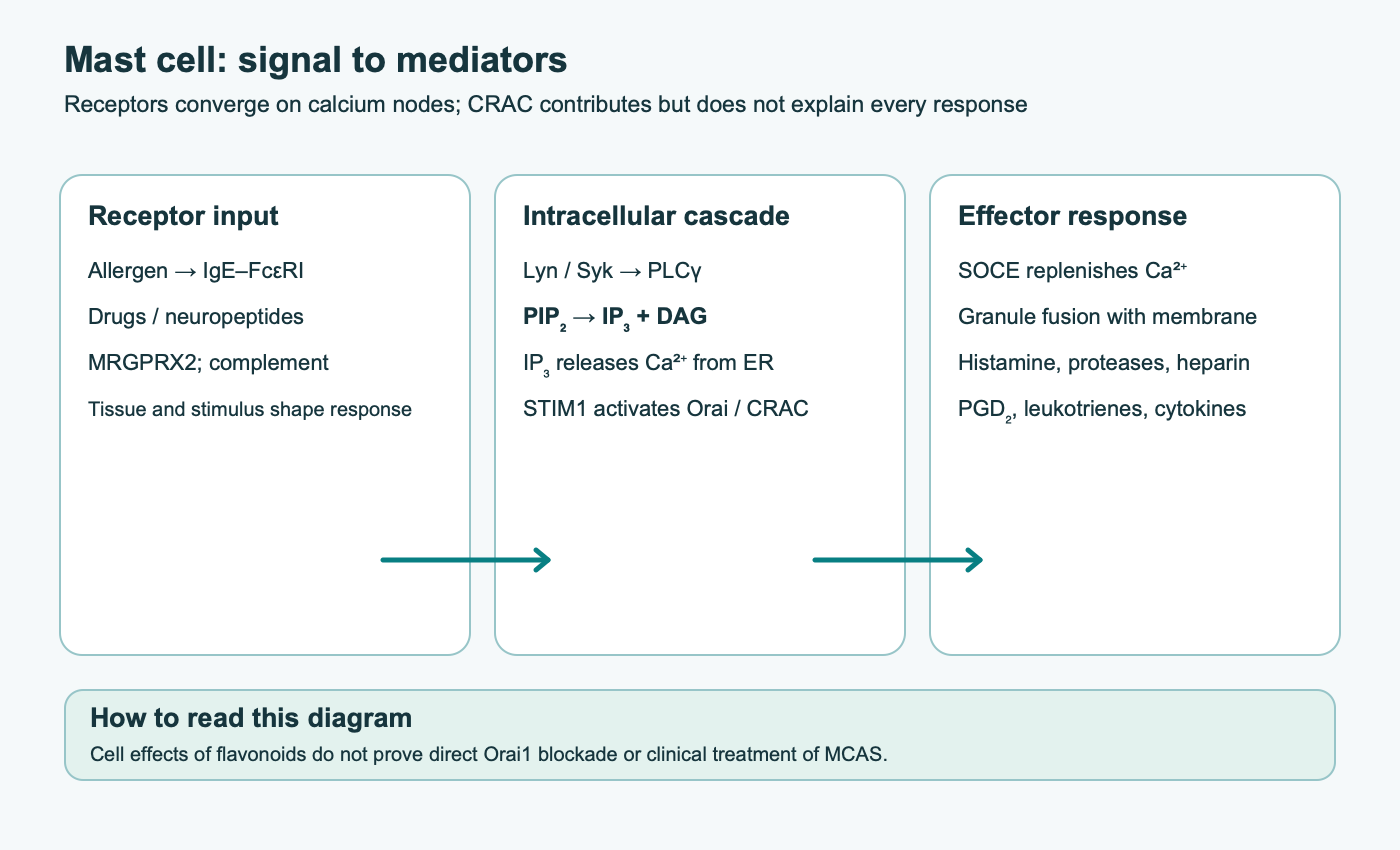
Experiments may measure intracellular Ca²⁺ concentration, electrical current across the membrane, or subsequent secretion. Under controlled conditions, only voltage-clamp/patch-clamp measurement directly characterizes an ion current; a change in calcium signaling after adding a polyphenol does not localize its target to the Orai pore. Orai subunit composition, STIM-protein abundance, and the contribution of other channels also depend on mast-cell type and stimulus.
3. What cell-model studies show about quercetin and luteolin
Quercetin is a flavonol and luteolin a flavone. Quercetin has molecular formula C₁₅H₁₀O₇ and molar mass about 302.24 g/mol; luteolin is C₁₅H₁₀O₆ and about 286.24 g/mol. Both have hydroxyl groups on aromatic rings, including a catechol group. Quercetin also has a hydroxyl at C3, which distinguishes flavonols from flavones. This structure affects redox properties and interactions with biological targets, but does not by itself identify which protein a molecule binds in living tissue.
Shared structural features include a C2=C3 double bond, a carbonyl group at C4, and a pair of catechol hydroxyls on ring B. Quercetin has an additional 3-hydroxyl group. These features influence planarity, electron donation, polarity, and conjugation. This chemical description cannot rank clinical potency: comparing activity requires the same cell model, stimulus, concentration, incubation time, endpoint, and solvent control. IC₅₀ values from different laboratories or mediators cannot be treated as a universal measure of a compound’s “strength.”
In experiments with cultured human mast cells, compounds were added directly to the cells. For example, Weng and colleagues compared quercetin and a water-soluble form with cromolyn in LAD2 cells and primary mast cells from umbilical cord blood. Some protocols used concentrations around 100 μM and about 30 minutes of pre-incubation; they measured histamine, prostaglandin D₂, leukotrienes, selected cytokines, intracellular calcium, and NF-κB translocation. Effects depended on the cell model and timing: pre-incubation and adding the compound together with the trigger produced different results. These laboratory conditions cannot be directly converted into an oral dose: the free concentration reaching tissue after food or a supplement is unknown and usually differs from the concentration in a culture dish.
Luteolin studies have also reported changes in activation of cell lines and some human cell cultures. A 2022 study examined FcεRI- and MRGPRX2-dependent activation and linked the effect to changes in calcium signaling and PLCγ phosphorylation. In a mouse model, luteolin reduced some signs of response, including edema and serum mediator measures. Mouse skin, a cultured cell line, and clinical MCAS are three different evidence levels; a result at one level cannot be transferred to another as established treatment.
A study has also examined quercetin in an MRGPRX2-dependent pseudoallergic response using LAD2 cells, HEK293 cells expressing MRGPRX2, and a mouse model. The authors described changes in calcium mobilization, degranulation, and the PLCγ–IP₃-receptor pathway. Molecular contact modeling and reduced response in a model suggest a mechanism, but do not replace direct biophysical confirmation of receptor binding or establish clinical efficacy.
Much of the literature uses “membrane stabilization” as a convenient description for reductions in selected cell responses. This does not mean a flavonoid physically strengthens the membrane, decreases its fluidity, or uniformly suppresses fusion of all granules. Such claims require direct measurements of membrane properties and exocytosis, not only less histamine in supernatant. Likewise, findings on individual phosphokinases, NF-κB, cyclooxygenase, or oxidative stress in cells do not prove comprehensive blockade of all these targets at concentrations achievable in the body.
The source material lists additional proposed mechanisms: changes in membrane fluidity; inhibition of phosphodiesterases, TRP and potassium channels; inhibition of phospholipase A₂, COX/5-LOX, IKK, NADPH oxidase, and synthesis of PAF and VEGF. Some of these proteins and pathways are biologically linked to inflammation or calcium regulation. However, the sources underlying this review do not test them as one unified mechanism of quercetin or luteolin in human mast cells at attainable tissue concentrations. These claims should remain hypotheses unless a target and its causal contribution to secretion are directly tested.
| Claim | What would need to be measured | What the available evidence supports |
|---|---|---|
| Direct Orai1/CRAC blockade | ICRAC current by patch clamp during controlled STIM–Orai activation, selectivity, and target confirmation | Cell studies report calcium and mediator outcomes; a direct flavonoid target mechanism is not established here |
| Syk/Lyn/PLCγ inhibition | Phosphorylation and enzyme activity with appropriate controls, including tissue-achievable concentrations | A downstream change is compatible with several sites of action and does not prove direct kinase inhibition |
| Reduced membrane “fluidity” | Physical measures of membrane order/fluidity and direct exocytosis analysis | Lower levels of selected secreted markers do not confirm physical bilayer stabilization |
| PDE, TRP, or KCa blockade | Separate functional channel/enzyme tests in mast cells and causal linkage to secretion | These mechanisms cannot be assigned to specific compounds based on general antioxidant or anti-inflammatory activity |
| Suppression of COX/5-LOX, PLA₂, ROS, PAF, or VEGF | Measurement of the relevant product and pathway validation in a defined model | Histamine alone does not describe lipid phases, transcription, or vascular responses |
The table does not mean these hypotheses are biologically impossible; it distinguishes plausibility from an established mechanism. In particular, reduced PLCγ phosphorylation in an experiment does not show whether a compound bound PLCγ directly, changed an upstream kinase, or affected cell state nonspecifically.
4. Oral exposure and metabolites
Food contains quercetin mainly in sugar-linked forms; composition varies by food and processing. Gut enzymes and microbiota hydrolyze glycosides to varying degrees. After absorption, the parent molecule is rapidly conjugated, including by glucuronidation, sulfation, and methylation. Plasma therefore contains a mixture of metabolites rather than the sustained high free-aglycone concentration used in many cell experiments. Exposure depends on food matrix, formulation, meals, microbiota, metabolism, and assay method.
Phospholipid complexes and other formulations can change measured pharmacokinetics. Human studies of quercetin phytosome found changes in systemic exposure to total quercetin and its conjugates in healthy volunteers. This describes a specific formulation and study; it does not show that free quercetin reaches mast cells at the concentration used in a dish or that improved absorption reduces MCAS symptoms. Products labeled “liposomal,” “micronized,” or “phytosome” are not interchangeable without comparative data.
Quercetin and luteolin from ordinary foods cannot be equated with concentrated supplements: dose, composition, and exposure differ. Human pharmacokinetic data for luteolin are limited; extrapolating quercetin parameters to luteolin is inappropriate.
After quercetin intake, detectable forms depend on food source and product. Glycosides may be hydrolyzed in the gut, and absorbed aglycone can be quickly conjugated. Thus, total plasma “quercetin” is not the same as free active molecule at a mast cell. Conjugates may have their own biology, but that cannot automatically be equated with the effect of aglycone added to culture medium. Human pharmacokinetic data for luteolin are even more limited, and its metabolic profile differs.
A phytosome, liposome, nanoparticle, and ordinary powder are not interchangeable delivery systems. A study of one commercial phospholipid formulation may show a change in AUC or peak concentration relative to a particular comparator, but does not establish an equivalent dose for another product, target-tissue penetration, or clinical benefit. A claim of crossing the blood–brain barrier requires direct data on brain exposure; a higher plasma concentration does not establish it.
Proposals to combine quercetin with vitamin C, zinc, bromelain, DAO, or a low-histamine diet should not be presented as one mast-cell-stabilization protocol. These interventions have different proposed targets, products, and risks; synergy must be shown in controlled comparisons of the specific combinations. The presence of a vitamin or enzyme in a blend, or laboratory antioxidant activity, does not guarantee better outcomes, lower histamine, or prevention of an episode. Elimination diets may restrict intake and need individualized justification and follow-up assessment of tolerance. A change in bioavailability is an intermediate pharmacokinetic measure, not proof of allergy prevention or a safe long-term dose. General differences between dietary polyphenols, metabolites, and supplement formulations are discussed in polyphenols and human evidence.
5. Clinical evidence: allergic symptoms are not MCAS
Human quercetin data include small pilot skin studies and a limited supplement trial for pollinosis. In work by Weng and colleagues, ten volunteers with nickel sensitivity had repeated skin patch tests before and after a short course of water-soluble quercetin; a separate protocol assessed skin response to UVB. This was a small open within-subject experiment without a randomized parallel control. It can generate a hypothesis about specific short-term skin outcomes, but does not prove long-term prevention, efficacy across allergic diseases, or MCAS treatment.
A randomized, placebo-controlled study in 66 adults with pollinosis symptoms evaluated a specific quercetin phytosome taken daily for four weeks. The authors reported improvement in some subjective symptoms and quality-of-life measures. This is a more direct clinical outcome than a cell assay, but it applies to one formulation, a defined population, a short period, and rhinoconjunctivitis symptoms. The trial did not study diagnosed MCAS, compare treatments for mastocytosis, or establish a universal quercetin dose.
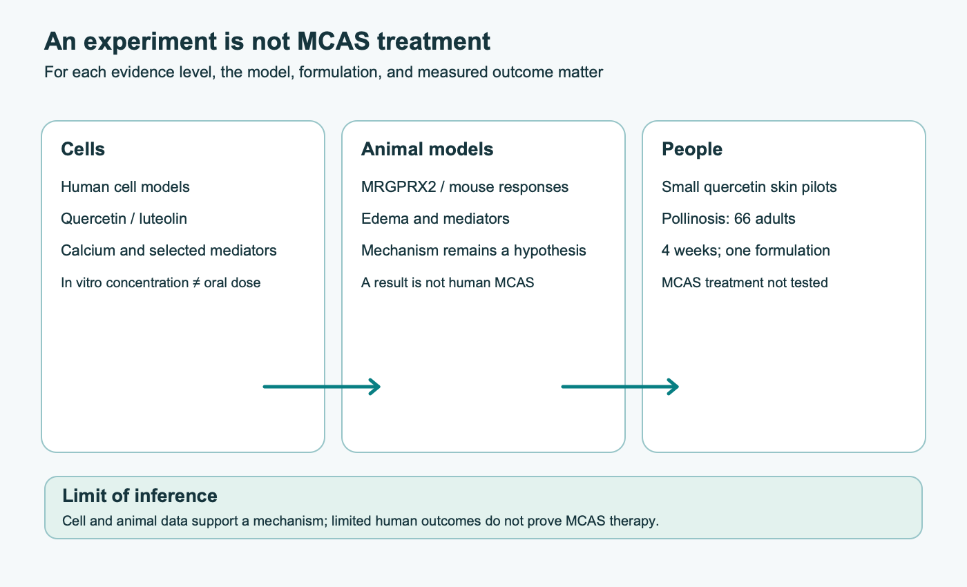
In the reviewed evidence base, luteolin research is mainly in cells and animals; it does not establish a clinical protocol for MCAS. Neither comparative in-vitro concentrations nor reduced edema in mice demonstrate that luteolin is superior to cromolyn, ketotifen, or antihistamines for treating people. Such comparisons require clinical trials with comparable populations, doses, outcomes, and controls.
In irritable bowel syndrome, associations have been reported between mucosal mast cells, nerve structures, and symptoms, and a separate trial studied ketotifen. This is not a quercetin or luteolin study and does not allow one drug’s effect to be transferred to another. Similarly, publications on luteolin in neuroinflammation or cognitive symptoms do not turn a laboratory hypothesis about brain mast cells into an established treatment for post-COVID syndrome or ME/CFS.
In clinical practice, discussion of a supplement should be tied to an established indication and a specific outcome. Findings in seasonal rhinoconjunctivitis do not answer questions about asthma, atopic dermatitis, systemic mastocytosis, or MCAS. For bowel symptoms, studies of mast cells near nerves and a ketotifen trial are separate evidence lines, not trials of quercetin or luteolin. There is no confirmed clinical outcome here for luteolin in post-COVID cognitive complaints or ME/CFS; proposed brain availability does not replace studies of exposure and outcomes.
Comparisons with cromolyn, H₁ antihistamines, ketotifen, or omalizumab also require caution. These medicines act at different points and are prescribed for clinical indications. A reduced cell response to a flavonoid cannot be compared with a clinical drug effect without a direct study with an appropriate design, comparable patients, and a prespecified outcome. An antihistamine blocks histamine action at its receptor but does not necessarily stop release of other mediators; a cell-dish observation does not show that a flavonoid acts “more broadly” or can replace prescribed treatment.
6. MCAS, mastocytosis, and “histaminosis”: diagnostic boundaries
MCAS is a clinical syndrome of recurrent episodes of mast-cell activation, not a synonym for chronic urticaria, allergy, histamine intolerance, irritable bowel syndrome, or a collection of nonspecific symptoms. Consensus criteria consider characteristic episodic symptoms involving multiple systems, an objective mediator change during an episode relative to the individual baseline, and clinical response to therapy directed at mast-cell mediators. For tryptase, the rule is an increase of at least 20% plus 2 ng/mL over baseline at an appropriate time after an episode; interpretation depends on proper sampling time and clinical context. The criteria are for diagnosis, not self-checking from one test.
Systemic mastocytosis is a clonal disease with separate diagnostic criteria; it is not the same as MCAS. Histamine intolerance also has a different proposed mechanism and unresolved measurement and diagnostic issues. More on this distinction is available in histamine intolerance and DAO. Elevated histamine in a single sample, nonspecific improvement after a diet, or a subjective response to a supplement does not establish any of these conditions.
A lower mediator level in a cell dish does not confirm MCAS treatment. The sources included in this review do not provide sufficient randomized clinical evidence that quercetin or luteolin treats diagnosed MCAS, prevents anaphylaxis, or replaces standard evaluation. Episodes involving breathing difficulty, tongue or throat swelling, fainting, or low blood pressure require emergency medical care; supplements do not replace a prescribed anaphylaxis action plan.
7. Safety and appropriate use of evidence
An experiment with a purified compound is not a ready-made dosing plan. Quercetin products include different salts, glycosides, complexes, and extracts; “aglycone equivalent” labeling and active-content amounts may vary. A formulation studied for pharmacokinetics or pollinosis is not equivalent to every retail product. Clinical data on luteolin products and long-term use are even more limited.
Supplement tolerability varies; gastrointestinal symptoms and reactions to excipients may occur. High doses and prolonged use warrant particular care because chronic safety data for specific groups and interaction data are incomplete. During pregnancy or breastfeeding, with chronic illness, before planned surgery, or with regular medicines, discuss supplements with a clinician or pharmacist. This is especially relevant for medicines with a narrow therapeutic range and when taking several products. Do not stop or reduce prescribed treatment because of a laboratory hypothesis about flavonoids.
Clinical decisions depend on diagnosis, goal, measurable outcome, specific product, duration, and adverse effects. If a clinician considers a nutritional supplement as an adjunct, assessment should include not only “histamine levels” but also frequency and severity of defined episodes, functional symptoms, and safety. Combining quercetin, luteolin, bromelain, vitamins, zinc, and diamine oxidase on one’s own is not an evidence-based mast-cell-stabilization protocol.
8. Methods and limits of this review
This narrative review covers basic mast-cell biology, calcium signaling, experimental studies of quercetin and luteolin, and limited clinical trials. On October 9, 2026, the primary records behind the source text’s strongest claims were checked individually: quercetin and human mast cells/skin pilots by Weng and colleagues; cell and mouse studies of quercetin through MRGPRX2; a 2022 luteolin study; a quercetin-phytosome study in pollinosis; phytosome pharmacokinetics; functional Orai/CRAC data in human lung mast cells; and the publication of consensus MCAS diagnostic criteria.
This targeted check corrected an incorrect PMID for the main Weng paper and an incorrect PMID for the phytosome pharmacokinetic study in the source bibliography. It was not a comprehensive independent audit of all 56 source records, a systematic search, a meta-analysis, or a formal risk-of-bias assessment. Structural models, cell experiments, animal studies, skin pilots, and a randomized pollinosis study are treated as different evidence types. The clinical studies available here did not test quercetin or luteolin in patients with confirmed MCAS.
9. Conclusion
Mast-cell activation involves several receptors and signaling routes. SOCE through STIM–Orai/CRAC is one functionally important part of calcium responses, but cell data on quercetin and luteolin do not establish direct Orai1 blockade or a therapeutic dose. Quercetin has limited evidence for pollinosis symptoms and small pilot skin observations; luteolin has an even less defined clinical evidence base. Neither establishes MCAS treatment. Diagnostic and treatment decisions should rely on clinical criteria, objective data, and proven care.
FAQ
What do mast cells do?
They are tissue immune cells that respond to several classes of signals and release granule mediators or synthesize mediators after activation. Their receptors and mediator profiles vary by tissue and model.
Are quercetin and luteolin direct CRAC/Orai1 blockers?
That mechanism has not been established in humans. Cell studies measured calcium responses and other signaling steps; they do not prove direct binding to Orai1 or a clinical effect.
Is there evidence that they treat MCAS?
The included clinical data do not establish quercetin or luteolin as treatment for diagnosed MCAS. Cell and mouse models are not treatment trials in patients.
Has quercetin been studied in people?
To a limited extent: small pilot skin studies and one small study of a specific formulation for pollinosis. Those outcomes do not answer the MCAS question.
Are there clinical trials of luteolin for MCAS?
The reviewed evidence is mainly from cell and animal studies; it does not establish a clinical MCAS treatment protocol.
Can cell-study concentrations be used as a dosing schedule?
No. A culture-medium concentration cannot be converted directly into an oral dose; absorption, metabolism, and tissue exposure differ.
How does MCAS differ from histamine intolerance and mastocytosis?
They are distinct conditions with separate diagnostic approaches. MCAS requires suitable episodic symptoms, objective evidence of mediator activation, and clinical response to directed therapy; one nonspecific test is not enough.
Sources
- Galli, S. J., & Tsai, M. (2012). IgE and mast cells in allergic disease. Nature Medicine, 18(5), 693–704.
- Frossi, B., Mion, F., Sibilano, R., Danelli, L., & Pucillo, C. E. (2017). Is it time for a new classification of mast cells? What do we know about mast cell heterogeneity? Immunological Reviews, 282(1), 35–46.
- McNeil, B. D., Pundir, P., Meeker, S., Han, L., Undem, B. J., Kulka, M., & Dong, X. (2015). Identification of a mast-cell-specific receptor crucial for pseudo-allergic drug reactions. Nature, 519(7542), 237–241.
- Siraganian, R. P. (2003). Mast cell signal transduction from the high-affinity IgE receptor. Current Opinion in Immunology, 15(6), 639–646.
- Ashmole, I., Duffy, S. M., Leyland, M. L., Morrison, V. S., Begg, M., & Bradding, P. (2012). CRACM/Orai ion channel expression and function in human lung mast cells. Journal of Allergy and Clinical Immunology, 129(6), 1628–1635.e2.
- Feske, S., Gwack, Y., Prakriya, M., Srikanth, S., Puppel, S. H., Tanasa, B., et al. (2006). A mutation in Orai1 causes immune deficiency by abrogating CRAC channel function. Nature, 441(7090), 179–185.
- Roos, J., DiGregorio, P. J., Yeromin, A. V., Ohlsen, K., Lioudyno, M., Zhang, S., et al. (2005). STIM1, an essential and conserved component of store-operated Ca2+ channel function. Journal of Cell Biology, 169(3), 435–445.
- Hou, X., Pedi, L., Diver, M. M., & Long, S. B. (2012). Crystal structure of the calcium release-activated calcium channel Orai. Science, 338(6112), 1308–1313.
- Hou, X., Burstein, S. R., & Long, S. B. (2018). Structures reveal opening of the store-operated calcium channel Orai. Nature, 562(7728), 539–544.
- Kimata, M., Shichijo, M., Miura, T., Serizawa, I., Inagaki, N., & Nagai, H. (2000). Effects of luteolin, quercetin and baicalein on immunoglobulin E-mediated mediator release from human cultured mast cells. Clinical & Experimental Allergy, 30(4), 501–508.
- Weng, Z., Zhang, B., Asadi, S., Sismanopoulos, N., Butcher, A., Fu, X., et al. (2012). Quercetin is more effective than cromolyn in blocking human mast cell cytokine release and inhibits contact dermatitis and photosensitivity in humans. PLOS ONE, 7(3), e33805.
- Asadi, S., Zhang, B., Weng, Z., Angelidou, A., Kempuraj, D., Skevaki, C., et al. (2010). Luteolin and tetramethoxyluteolin inhibit mast cell activation. Journal of Allergy and Clinical Immunology, 126(6), 1290–1295.
- Kempuraj et al. (2005). Flavonols inhibit proinflammatory mediator release, intracellular calcium ion levels and protein kinase C theta phosphorylation in human mast cells. British Journal of Pharmacology, 145(7), 934–944.
- Ding, Y., Che, D., Li, C., Cao, J., Wang, J., Ma, P., Zhao, T., An, H., & Zhang, T. (2019). Quercetin inhibits Mrgprx2-induced pseudo-allergic reaction via PLCγ-IP3R related Ca2+ fluctuations. International Immunopharmacology, 66, 185–197.
- Hao, Y., Che, D., Yu, Y., Liu, L., Mi, S., Zhang, Y., et al. (2022). Luteolin inhibits FcεRI- and MRGPRX2-mediated mast cell activation by regulating calcium signaling pathways. Phytotherapy Research, 36(5), 2197–2206.
- Yamada, S., Shirai, M., Inaba, Y., & Takara, T. (2022). Effects of repeated oral intake of a quercetin-containing supplement on allergic reaction: a randomized, placebo-controlled, double-blind parallel-group study. European Review for Medical and Pharmacological Sciences, 26, 4331–4345.
- Riva, A., Ronchi, M., Petrangolini, G., Bosisio, S., & Allegrini, P. (2019). Improved oral absorption of quercetin from Quercetin Phytosome, a new delivery system based on food grade lecithin. European Journal of Drug Metabolism and Pharmacokinetics, 44(2), 169–177.
- Manach, C., Scalbert, A., Morand, C., Rémésy, C., & Jiménez, L. (2004). Polyphenols: food sources and bioavailability. American Journal of Clinical Nutrition, 79(5), 727–747.
- Walle, T. (2004). Absorption and metabolism of flavonoids. Free Radical Biology and Medicine, 36(7), 829–837.
- Valent, P., Akin, C., Bonadonna, P., Hartmann, K., Broesby-Olsen, S., Brockow, K., et al. (2019). Proposed diagnostic criteria and classification of basophil and mast cell disorders. Journal of Allergy and Clinical Immunology, 144(4), 938–944.
- Maintz, L., & Novak, N. (2007). Histamine and histamine intolerance. American Journal of Clinical Nutrition, 85(5), 1185–1196.
- Barbara, G., Stanghellini, V., De Giorgio, R., Cremon, C., Cottrell, G. S., Santini, D., et al. (2004). Activated mast cells in proximity to colonic nerves correlate with abdominal pain in irritable bowel syndrome. Gastroenterology, 126(3), 693–702.
- Klooker, T. K., Braak, B., Koopman, K. E., Welting, O., Wouters, M. M., van der Heide, S., et al. (2010). The mast cell stabiliser ketotifen decreases visceral hypersensitivity and improves intestinal symptoms in patients with irritable bowel syndrome. Gut, 59(9), 1213–1221.
- Akin, C. (2017). Mast cell activation syndromes. Journal of Allergy and Clinical Immunology, 140(2), 349–355.
- Weinstock, L. B., Pace, L. A., Rezaie, A., Afrin, L. B., & Molderings, G. J. (2021). Mast cell activation syndrome: a primer for the gastroenterologist. Digestive Diseases and Sciences, 66(4), 965–982.