Postbiotics, peptidoglycan, and NOD2: mechanisms and clinical evidence — NutriFit
Postbiotics and the Bacterial Cell Wall: MDP, NOD2, and the Limits of Immune Effects
A professional review of bacterial wall composition, MDP access to NOD2, signaling and genetic pathways, and the limits of clinical evidence for specific inactivated preparations.
An inactivated bacterium remains a collection of chemical signals: its peptidoglycan, teichoic acids, and lipoproteins can interact with host cells without microbial replication. But recognition of a cell-wall fragment by NOD2 is only one level of explanation; it does not make every powder of dead bacteria beneficial or turn purified muramyl dipeptide into a proven postbiotic. Professional assessment must connect product composition, access of its components to cellular compartments, established human outcomes, and safety in the population concerned.
1. What exactly is called a postbiotic?
The 2021 ISAPP expert consensus defines a postbiotic as a preparation of inanimate microorganisms and/or their components that confers a health benefit on the host. Metabolites may be present alongside cellular structures, but purified metabolites alone do not meet the definition. The source microorganism, inactivation method, composition and standardization of the final product, and beneficial effect all need to be characterized. “Inactivated,” “lysate,” and “postbiotic” are therefore not complete synonyms: a lysate without demonstrated benefit remains an investigational lysate. Purified, chemically synthesized MDP is a valuable molecular probe or drug compound, but it is not automatically a postbiotic preparation under the consensus definition.
Inactivation prevents replication when viability is reliably controlled, but it does not by itself guarantee heat stability, unchanged epitopes, absence of biogenic amines or pyrogenicity, or complete safety in severe immunodeficiency. Heat, high pressure, and enzymatic lysis produce different proportions of intact cells, wall debris, DNA, and soluble substances. A dried product is assessed for residual viability, particle count, strain identity, and activity of the specific batch. “10⁹ inactivated cells” cannot be equated with 10⁹ CFU of a live probiotic or with milligrams of purified MDP.
Object
What it contains
What the name itself establishes
Live probiotic
Viable, identified strain
Class membership only; benefit is tested for the strain and indication
Inactivated preparation
Whole nonviable cells, fragments, and sometimes metabolites
No replication after validated inactivation; benefit still needs to be shown
Purified MDP or lipoteichoic acid
Chemically defined component
Composition and ability to study receptor mechanisms, not clinical benefit
Prebiotic substrate
Substance selectively used by microbes
A different mechanism; not a preparation of inactivated cells
2. Cell-wall architecture and available molecular patterns
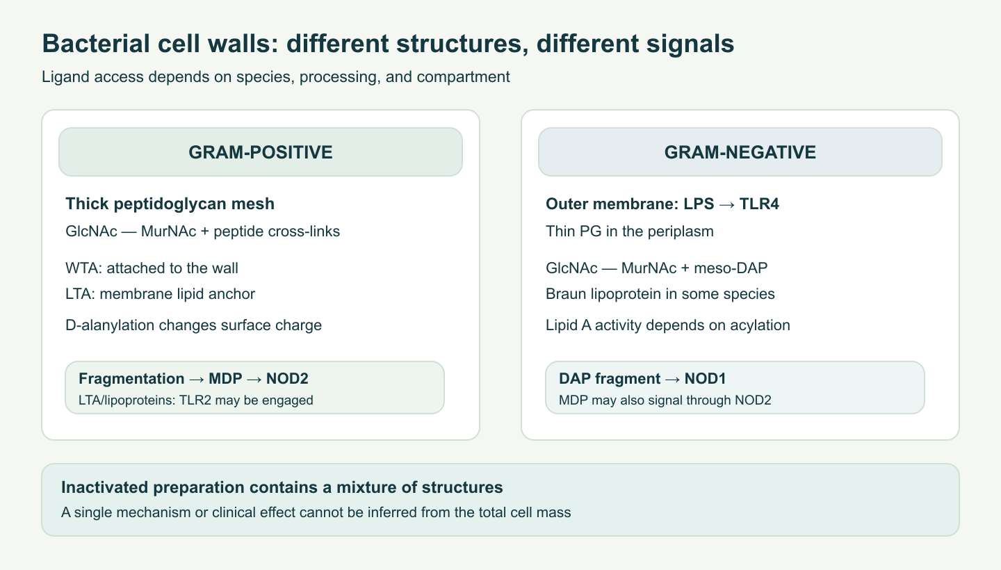
Peptidoglycan (murein) forms a mesh around the bacterial membrane. Its glycan strands consist of alternating N-acetylglucosamine (GlcNAc) and N-acetylmuramic acid (MurNAc), joined by β-1,4-glycosidic bonds. A short peptide stem is attached by an amide bond to the lactyl group of MurNAc. In different bacteria, the third position of the stem contains meso-diaminopimelic acid (meso-DAP) or L-lysine; D-amino acids and interpeptide bridges make the structure distinct from host proteins. Transpeptidases cross-link neighboring stems, usually removing terminal D-Ala from the D-Ala–D-Ala precursor. Strand length, cross-link type, and degree of modification vary substantially by species: not every cell wall should be treated as the same dose of an NOD2 ligand.
In many Gram-positive bacteria, peptidoglycan is thick and coupled to anionic teichoic acids. Wall teichoic acid (WTA) is attached to the cell wall, whereas lipoteichoic acid (LTA) has a membrane lipid anchor; their repeating units often contain glycerol or ribitol phosphate. D-alanylation through the Dlt system reduces negative surface charge and changes binding of cationic antimicrobial peptides. However, LTA structure, acylation, and lipoprotein contamination shape responses in TLR2-dependent systems; it is incorrect to claim that all LTAs induce IL-10 and “suppress inflammation.” The immunostimulatory result from a purified fraction is particularly sensitive to preparation purity.
In Gram-negative bacteria, the peptidoglycan layer is thinner and lies in the periplasm between the inner and outer membranes. The outer leaflet of the outer membrane contains lipopolysaccharide with lipid A, core, and, where present, O antigen. Lipid A is recognized by the TLR4–MD-2 complex, and its acylation changes its activity. In some bacteria, Braun’s lipoprotein connects the outer membrane to the wall, but that architecture should not be generalized to every Gram-negative species without checking. Peptidoglycan fragments with meso-DAP in the appropriate sequence can activate NOD1; the minimal recognized motif is commonly called iE-DAP. This is a distinct pathway, not a synonym for MDP–NOD2.
Wall cleavage also occurs within bacteria during growth and death: autolysins, transpeptidases, amidases, and lytic transglycosylases generate muramyl peptides of different lengths. Host lysozyme cleaves the β-1,4 bond between MurNAc and GlcNAc; PGLYRP2 has N-acetylmuramoyl-L-alanine amidase activity and separates peptide from sugar. Thus, inactivation method and digestive processing change the distribution of ligands. An intact sacculus does not bind cytosolic NOD2 directly: an accessible fragment must be generated and reach the appropriate side of a membrane.
How wall composition changes the ligand dose
The peptidoglycan stem is assembled on a lipid carrier at the bacterial cytoplasmic membrane. GlcNAc is attached to the MurNAc-pentapeptide on lipid II; transglycosylases extend the sugar strands, and transpeptidases form cross-links while removing terminal D-alanine. Mature wall therefore contains a mixture of tetra-, tri-, and other peptide fragments. Mechanical fragmentation, heating, and hydrolysis produce different sets of muramyl peptides even from the same strain. The number of accessible MDP molecules cannot be calculated from total dry biomass without analytical determination and accounting for repeats, cross-links, and degree of degradation.
Gram-positive WTA is attached to the wall through a complex linker, while LTA is embedded in the membrane by its lipid anchor. After a microbe is “killed,” WTA, LTA, lipoprotein fragments, and cytoplasmic DNA may end up in the same preparation. To explain an immune effect specifically through NOD2, useful controls include purified MDP, removal or inactivation of the proposed ligand, NOD2-deficient cells, or gene complementation. A simple rise in IL-6 after bacterial lysate does not identify the receptor: TLR2, TLR4, TLR9, and other sensors may produce the response. This is especially important for preparations from Gram-negative cells, where active lipid A contamination changes the meaning of the result.
Access through the intestine is a multistep problem
An oral dose first encounters gastric acidity, proteases, bile, mucus, and enzymes from both microbes and host. Peptidoglycan does not dissolve as one small molecule: particles vary in size and may become trapped in mucus or contact phagocytic cells across the epithelium under tissue-dependent conditions. In an enterocyte, PepT1 transports suitable small peptide substrates, not an entire cell wall. In a phagocyte, an engulfed particle is in a phagosome and must be processed before the relevant ligand can participate in cytosolic signaling. Luminal concentration, intracellular availability, and plasma concentration are three different quantities. Without assessing them, chemical composition alone cannot establish NOD2 activation in a person after an ordinary dietary dose.
3. MDP: chemistry, transport, and product variants
Canonical muramyl dipeptide is N-acetylmuramyl-L-alanyl-D-isoglutamine. The MurNAc sugar and two amino-acid positions distinguish it from most host molecules. Girardin’s and Inohara’s experiments identified MDP as a shared minimal motif recognized by NOD2, whereas NOD1 preferentially responded to DAP-containing peptides. Stereochemistry and amide-group substitutions substantially change biological activity, but the claim that every analog with a different configuration is “completely inert” is too strong: results depend on system and concentration. MDP is a peptidoglycan motif, not a complete description of the biological activity of a bacterial cell.
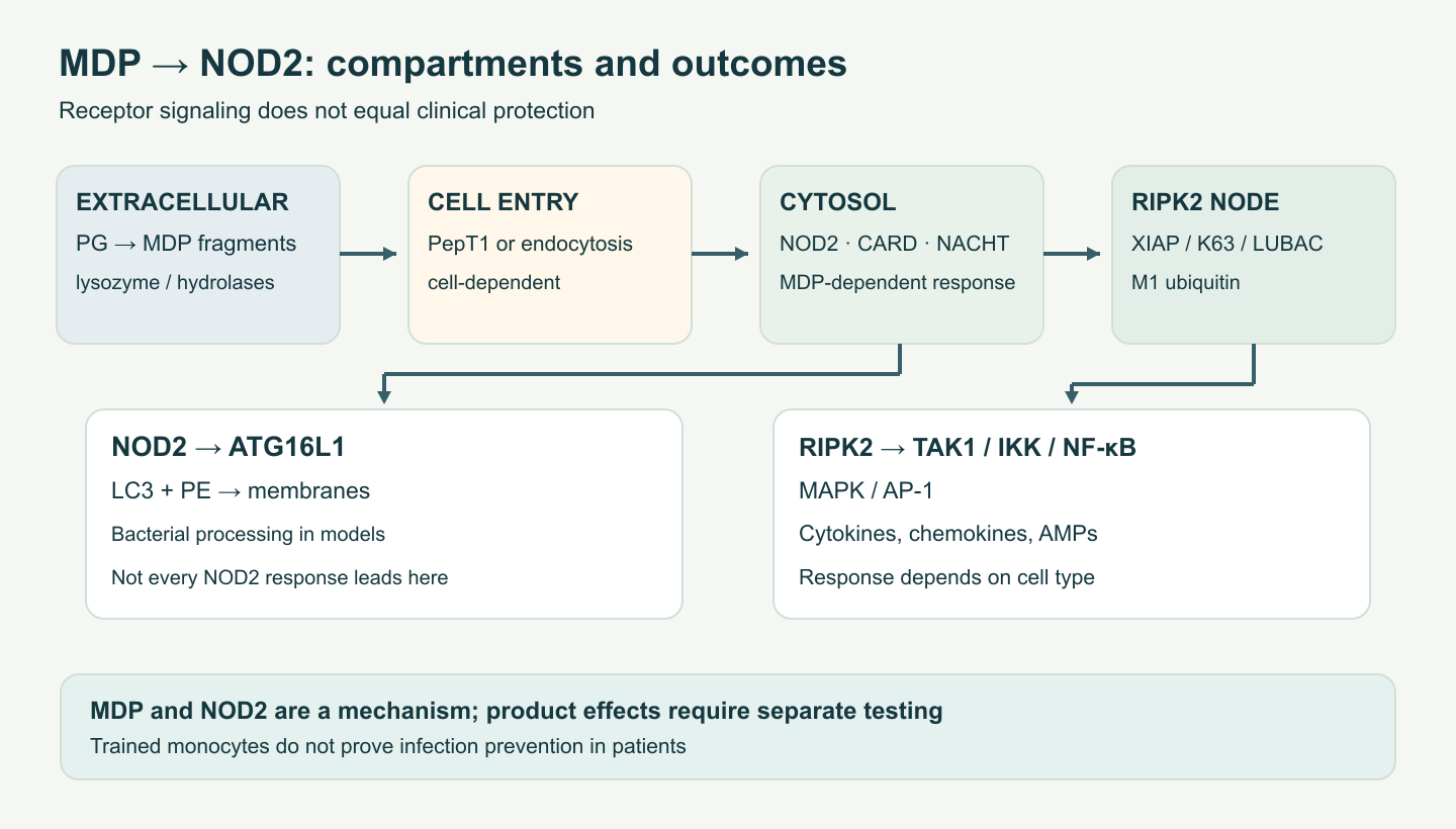
In the intestinal lumen, a small fragment may interact with peptide transporters. Transport of MDP by hPEPT1/SLC15A1 and a subsequent NF-κB/IL-8 response have been shown in experimental epithelial systems. PepT1 uses a proton gradient; expression is generally prominent in the small intestine and may appear in the colon during colitis. This does not mean that all ingested MDP passes through PepT1, reaches myeloid cells, or invariably increases inflammation. Endocytosis, phagocytosis of cell particles, and intracellular degradation in endolysosomes are also possible. The SLC15A3/A4 family participates in microbial peptide handling and intracellular signaling, but it is incorrect to assign SLC15A4 the single obligatory role of exporting MDP in every macrophage; SLC15A4–TASL also functions in the endolysosomal TLR7–9/IRF5 pathway. The compartment must be established experimentally.
Synthetic muramyl peptides differ from MDP and from one another. Glucosaminylmuramyl dipeptide (GMDP) adds GlcNAc to a muramyl peptide; romurtide and mifamurtide have their own lipophilic modifications or delivery systems. Liposomal mifamurtide is a drug formulation of muramyl tripeptide phosphatidylethanolamine studied in oncology alongside chemotherapy; it should not be described as a dietary postbiotic supplement. The presence of a muramyl fragment does not give all derivatives the same affinity, oral bioavailability, dosing schedule, or pyrogenicity profile. Evaluation should rely on each product’s own pharmacokinetic and clinical studies and local regulatory status.
Differences among chemical agonists and drug formulations
Muramyl peptide activity depends on the specific conjugation site and the compound’s ability to enter cells. Adding a lipophilic tail can increase membrane interaction while also changing tissue distribution and accumulation risk. Packaging in a liposome can facilitate uptake by phagocytes but adds its own parameters: vesicle size, lipid composition, encapsulation efficiency, and release rate. Comparing doses in milligrams across chemically different analogs is therefore inappropriate. Even if two compounds produce the same NF-κB reporter signal in vitro, their concentrations in epithelium, macrophages, and blood after oral or intravenous administration may not be comparable.
The pyrogenicity of MDP and its analogs also cannot be reduced to “safe/unsafe.” The response depends on amount, route of administration, endotoxin contamination, baseline immune activation, and formulation. A bioassay of a purified preparation does not automatically predict reactions to a finished product; a history of use in another indication does not establish the safety of a dietary formulation for a different population. Chemically defined analogs can be investigated as medicines or adjuvants, but that is distinct from recommending an uncharacterized inactivated bacterial mixture.
4. NOD2: from cytosolic recognition to transcription
NOD2, also called CARD15/NLRC2, contains two N-terminal CARD domains, a central nucleotide-binding NACHT domain, and C-terminal leucine-rich repeats (LRR). In the absence of the relevant stimulus, the protein is in a regulated state; MDP recognition, nucleotide access, and associated proteins favor oligomerization and recruitment of RIPK2 through CARD–CARD interaction. Structural models explain conformational switching, but published structures do not justify claiming that a particular atom of every MDP directly contacts listed residues of human NOD2 in vivo, or that every nodosome always contains a fixed number of subunits.
RIPK2 is both a kinase and a platform for signaling complexes. XIAP and other E3 ligases promote RIPK2 ubiquitination; K63-linked chains connect the TAK1/TAB complex, while linear M1 chains from LUBAC help recruit NEMO-containing IKK. IKK then phosphorylates IκBα, which is degraded, releasing NF-κB/RelA for nuclear transcription. TAK1-linked p38/JNK and other MAPK cascades engage AP-1. The resulting chemokine, cytokine, and antimicrobial responses depend on context. K63 and M1 polyubiquitin mainly act here as signaling platforms, unlike K48 modification of IκBα before proteasomal degradation. Deubiquitinases CYLD, A20, and OTULIN limit response duration; regulation is not a simple binary “on/off.”
NOD2 can also promote local recruitment of ATG16L1 to a site of contact with an invading bacterium. The ATG12–ATG5–ATG16L1 complex promotes conjugation of LC3 to phosphatidylethanolamine and assembly of autophagic membranes; in relevant models, this supports bacterial processing and antigen presentation. But not every NOD2 activation ends in xenophagy, and not all autophagy is NOD2-dependent. The phenotype depends on pathogen and cell type.
Limits of the signaling diagram
NOD2 is present in different cell types, and its final response depends on the prior inflammatory state. In macrophages and dendritic cells, cytokine production or altered antigen presentation may predominate; in epithelium, chemokine and barrier responses may predominate. Tolerance or enhancement after repeated stimulation depends on timing and the first exposure. Specific ubiquitin residues and chain-assembly order have been studied in molecular systems, but the cellular complex is dynamic; one should not depict an invariant enzyme conveyor belt for every tissue. An RIPK2 inhibitor is also not equivalent to complete NOD2 blockade: the protein has scaffold functions, and its catalytic activity interacts with ubiquitination.
NOD1 has one CARD domain and recognizes DAP-containing fragments. The NOD1/NOD2 distinction matters when interpreting Gram-negative lysate effects: simultaneous activation of NOD1, NOD2, and TLR4 is possible but not guaranteed. NLRP3 is another receptor complex that forms an inflammasome and activates caspase-1; equating NF-κB transcription with mature IL-1β secretion is incorrect. Transcriptional priming of pro-IL-1β and a separate maturation mechanism are generally required. Therefore, increased IL1B mRNA after MDP does not prove a pyrogenic release of mature cytokine without measuring protein and the relevant second signal.
5. The crypt, Paneth cells, and Crohn’s disease
In the ileum, Paneth cells at the base of crypts secrete lysozyme, human α-defensins HD5/HD6, and other antimicrobial molecules; LGR5⁺ stem cells are nearby. NOD2 is expressed in immune and epithelial populations, including Paneth cells. Patient-tissue and model studies link NOD2 variants to changes in defensin responses, microbial interactions, and autophagy. HD5 and HD6 differ in function: HD5 can damage microbial membranes, while HD6 forms nets in experiments that physically trap bacteria. This is not a sterile “safe zone” throughout the crypt and does not guarantee prevention of colonization by a particular pathobiont in a patient.
MDP–NOD2 signaling was linked experimentally to protection of intestinal stem cells against oxidative injury. This cannot be translated into faster mucosal regeneration in people after radiation or cytotoxic drugs. NOD2 and ATG16L1 genes are part of a complex genetic architecture of inflammatory bowel disease; disease activity also depends on epithelium, microbiota, other genes, immune cells, and environment. R702W, G908R, and L1007fs variants raise susceptibility primarily to ileal Crohn’s disease in certain populations, but are not diagnoses and do not give a direct rule for prescribing an NOD2 agonist. Some carriers never develop disease, and most patients do not need their illness explained by one variant.
The loss-of-function sensor paradox in chronic inflammation is not resolved by one cascade. Reduced control of microbial contact, altered Paneth-cell secretion, autophagy, and other innate pathways may sustain inflammation. The original scheme “NOD2 defect → obligatory total TLR4/NLRP3 hyperactivation” oversimplifies alternative mechanisms and does not mean that pharmacological activation of a mutant protein will restore its response. By contrast, certain NOD2 domain variants in Blau syndrome are associated with granulomatous dermatitis, arthritis, and uveitis; this is a rare autoinflammatory disease and a different genetic context, not a manifestation of postbiotic deficiency.
Genotype is not the same as functional phenotype
Initial NOD2 associations with Crohn’s disease were replicated for several variants, but the relative risk varies with population ancestry and allele copy number. A reduced experimental response of a variant to MDP does not prove that its carrier necessarily has low production of all defensins: inflammation, bacterial contact, other genetic variants, and treatment alter tissue phenotype. Ileal biopsy studies are useful for mechanistic hypotheses, but Crohn’s disease is not diagnosed by NOD2. Different effects of some mutations in Blau syndrome further show that variant location, protein structure, and cell type matter.
For clinical study design, this limits the idea of “personalized MDP”: if a receptor mutation reduces signal sensing, increasing the dose may not restore the required spatial and temporal control. If signaling remains intact, excessive enhancement could be undesirable during active inflammation. Genotype-based prescribing would require a clinical study with a prespecified genotype-by-treatment interaction, not biochemical reasoning alone.
6. Trained innate immunity: measuring the actual effect
Trained immunity means a lasting change in innate-cell responses after an initial stimulus: after a rest period, a different stimulus changes cytokine production or antimicrobial function. It differs from clonal memory in T and B cells. In β-glucan and BCG models, investigators observed changes in chromatin accessibility, H3K4me3/H3K27ac, and metabolism. Glycolysis, mTOR–HIF-1α, glutaminolysis, and fumarate may participate in some protocols; other studies examine reprogramming of myeloid precursors in bone marrow. These programs are not identical for every ligand, dose, or tissue.
An experiment involving BCG and NOD2 genotype supported NOD2’s role in an acquired nonspecific monocyte response; protection of mice with severe combined immunodeficiency from experimental candidiasis concerned BCG vaccination, not ingestion of purified MDP or a retail postbiotic. Increased TNF/IL-6 after repeat stimulation ex vivo is not a clinical measure of fewer infections. “Training” may be protective or undesirable in chronic inflammation. Claims of seasonal viral prevention require studies of a specific product with a prespecified rate of confirmed infection, safety assessment, and a control group. This biology does not support a general course of MDP “for immune memory.”
Distinguishing training from acute inflammation
A typical experimental training protocol briefly stimulates human monocytes, removes the stimulus, rests the cells for several days, and gives a second, different stimulus. Investigators measure differences in cytokine production, chromatin-site accessibility, and metabolic flux relative to an untrained control. Immediate TNF release after the first dose is ordinary activation, not proof of memory. In humans, duration is difficult to assess because circulating monocytes turn over; this led to interest in possible modification of hematopoietic precursors, but that cannot automatically be assumed after any oral product.
H3K4me3 and H3K27ac are associated with active regulatory regions, but the presence of a mark alone does not indicate which infection will be prevented. mTOR/HIF-1α-dependent glycolysis and glutaminolysis with fumarate accumulation participated in some model programs; metabolite changes may also accompany acute inflammation. Causality requires pathway interventions and preservation of the relevant clinical effect. Trained innate responses may potentially intensify immunopathology as well as protection; safety should be assessed by symptoms and long-term follow-up, not one cytokine panel.
Which epigenetic changes matter?
During innate-cell training, H3K4me3 at promoters and H3K27ac at active regulatory regions may facilitate repeat transcription, but these are not autonomous “memory switches.” A transcription factor must bind accessible chromatin, and ionic signals and metabolic substrate must support the response. Fumarate can affect 2-oxoglutarate-dependent dioxygenases and change methylation; glycolysis and oxidative phosphorylation can coexist rather than exclude one another. Primary stimulus, rest duration, and secondary challenge differ among protocols, so quantitative comparisons of “training strength” without experimental standardization are unreliable.
Long-lasting shifts in myeloid-precursor differentiation may occur in bone marrow after some strong stimuli. However, showing this in animals after BCG or β-glucan does not confirm that bacterial cell fragments from a gut product reach human hematopoietic stem cells at an effective concentration. Hematopoietic reprogramming may change circulating cell composition and create an appearance of higher cytokine production even without changing the response of an individual cell. Good trials therefore separate changes in cell number, phenotype, and function after a standardized challenge.
7. Human evidence: specific products, outcomes, and limits
The clearest gastroenterology example is a multicenter, double-blind, placebo-controlled trial of heat-inactivated Bifidobacterium bifidum MIMBb75 in adults with IBS diagnosed by Rome III and baseline abdominal pain. Participants took 1 × 10⁹ nonviable cells daily for eight weeks. The primary composite outcome required both ≥30% improvement in pain and adequate relief of overall symptoms in at least four of eight weeks; it was reached by 74/221 (34%) in the product group and 43/222 (19%) in the placebo group. This supports benefit for this form of this strain in this population and on this outcome. It does not prove that the effect was due exclusively to peptidoglycan or NOD2, applies to IBS-D/IBS-M as separate phenotypes, or permits replacing therapy for other diseases. There were no serious product-related adverse events in the trial, but it excluded several severe comorbid conditions and was sponsored by the manufacturer.
Studies of inactivated Lactobacillus acidophilus LB for diarrhea concern different products and ages. They cannot be combined with MIMBb75 into a universal 20–30-hour reduction in diarrhea unless primary design, comparator, and age context are preserved. A separate study of TLR2 and ZO-1 showed an epithelial-barrier mechanism, but was not a trial of purified lipoteichoic acid as a treatment. The LTA composition of a specific strain and lipoprotein contamination can change the response; pharmacological stability and safety need separate characterization.
Liposomal mifamurtide was studied with osteosarcoma chemotherapy in a randomized factorial trial: six-year overall survival was reported to improve from 70% to 78% with addition of muramyl tripeptide, while event-free survival showed a trend without convincing statistical significance. This is a limited oncology context involving a drug compound, not evidence that dietary postbiotics prevent cancer. The NOD2 mechanism does not establish unconditional efficacy of GMDP or other muramyl peptides against recurrent infections, SIBO, or “leaky gut”; doses and indications must be assessed for specific registrations and studies, not transferred among analogs. Guaranteed safety during immunosuppression also does not follow from the absence of viable microbes: pyrogenic and inflammatory reactions remain possible.
A practical question for any finished product is: which strain or source, how was inactivation confirmed, how much of which components are in a dose, is the batch standardized, in whom was the effect studied, and what was the primary outcome? An inactivated bacterial blend with zinc, glutamine, or yeast β-glucans is a combined intervention; results for one component do not prove synergy of the mixture. If use is proposed in a risk group, clinical safety observation in that group matters more than the general label “non-living bacterium.” For live strains as a separate class, see the article on probiotics; for microbiota substrates, see dietary fiber.
Comparing indications without substituting populations
The MIMBb75 IBS trial used Rome III criteria, required pain during enrollment, and excluded several serious diseases. Its selected composite outcome concerns symptoms; it does not measure NOD2 changes, barrier injury, or fewer systemic infections. Therefore, the responder proportion cannot be used as proof of a specific molecular mechanism. Ideally, a follow-up trial would compare the same inactivation process with a non-inactivated strain, maintain the same cell mass, measure wall composition, and test prespecified biomarkers. Even then, proving a mediator would require an additional causal intervention.
The mifamurtide oncology study was a factorial trial alongside multi-agent chemotherapy and surgery; its outcome was survival in osteosarcoma. This signal matters in its specialty but differs from nutrition practice in age, diagnosis, route of administration, and risks. Commercial recommendations to take GMDP “for all viral infections” or preventive blends of inactivated microbes with glutamine, zinc, and β-glucan cannot rely on oncology data. Clinical translation requires a new trial of the relevant blend with an appropriate active control.
Safety and quality are properties of the specific preparation
The theoretical argument that reliable inactivation prevents bacteremia is understandable: a nonviable microbe cannot replicate. But this is only one dimension of risk. Retained surface molecules may activate innate immunity, and isolated muramyl peptides have historically been studied as adjuvants and pyrogenic signals. Breaking the cell wall changes the accessible surface area and may increase, rather than reduce, the acute biological activity of some fractions. In people with inflamed mucosa, fragment access and baseline activation differ from those in healthy volunteers. Separate safety data are therefore useful for critically ill people, premature infants, people with severe immunosuppression, and transplant recipients; absence of viable CFU does not replace these data.
During storage, moisture, aggregation, breakdown of the LTA lipid anchor, chemical modification of sugars, and preservation of other matrix components all matter. A product can be sterile but lose its claimed activity; it can retain activity and still fail microbial-purity requirements. Regulatory requirements differ for foods, supplements, and medicines. A marketing claim that a product is “heat-stable for years at room temperature” should be supported by batch testing and shelf-life data, not inferred from the word “inactivated.” If a clinical effect was demonstrated for one heat treatment, changing the inactivation process may change biological composition, and equivalence must be checked.
Not every dietary component marketed as a “postbiotic” meets the accepted scientific definition. Isolated butyrate, lactic acid, or a purified secreted metabolite without inactivated cells or their components belongs to another category, even if a bacterium produced it. A purified structural fragment without a characterized preparation and clinical benefit also remains investigational. This distinction does not deny potential pharmacology; it prevents results from one chemical molecule being transferred to many products made from mixtures of cell parts.
Review method and limitations
This is a narrative analysis of bacterial chemistry, receptor biology, and selected published clinical studies. On October 9, 2026, targeted checks covered the ISAPP consensus text, primary studies of MDP recognition, NOD2-dependent trained responses after BCG, the randomized trial of inactivated MIMBb75 in IBS, and the oncology study of mifamurtide. No formal systematic search, protocol-based risk-of-bias assessment, or GRADE evaluation was conducted. Main limitations are the strong dependence of mechanism on composition and delivery route, non-equivalent experimental systems, and the lack of direct human trials of purified components for most claimed nutritional uses.
FAQ
What is a postbiotic under the ISAPP definition?
A preparation of inactivated microorganisms and/or their components with demonstrated host health benefit, not every substance of microbial origin.
Can purified MDP be called a postbiotic?
Purified or synthetic MDP alone is a molecular component for research or drug development; the postbiotic label requires the accepted definition and demonstrated benefit of the specific preparation.
An eight-week RCT found more combined responders with the specific heat-inactivated B. bifidum MIMBb75 preparation than placebo; this does not automatically apply to other strains.
Does trained immunity mean protection from all infections?
No. Altered monocyte responses after restimulation and model data with BCG do not establish seasonal prevention with a particular oral postbiotic.
Are postbiotics safe in immunodeficiency?
Proper inactivation prevents replication, but pyrogenicity, composition, and clinical safety must be assessed for the specific product and population.