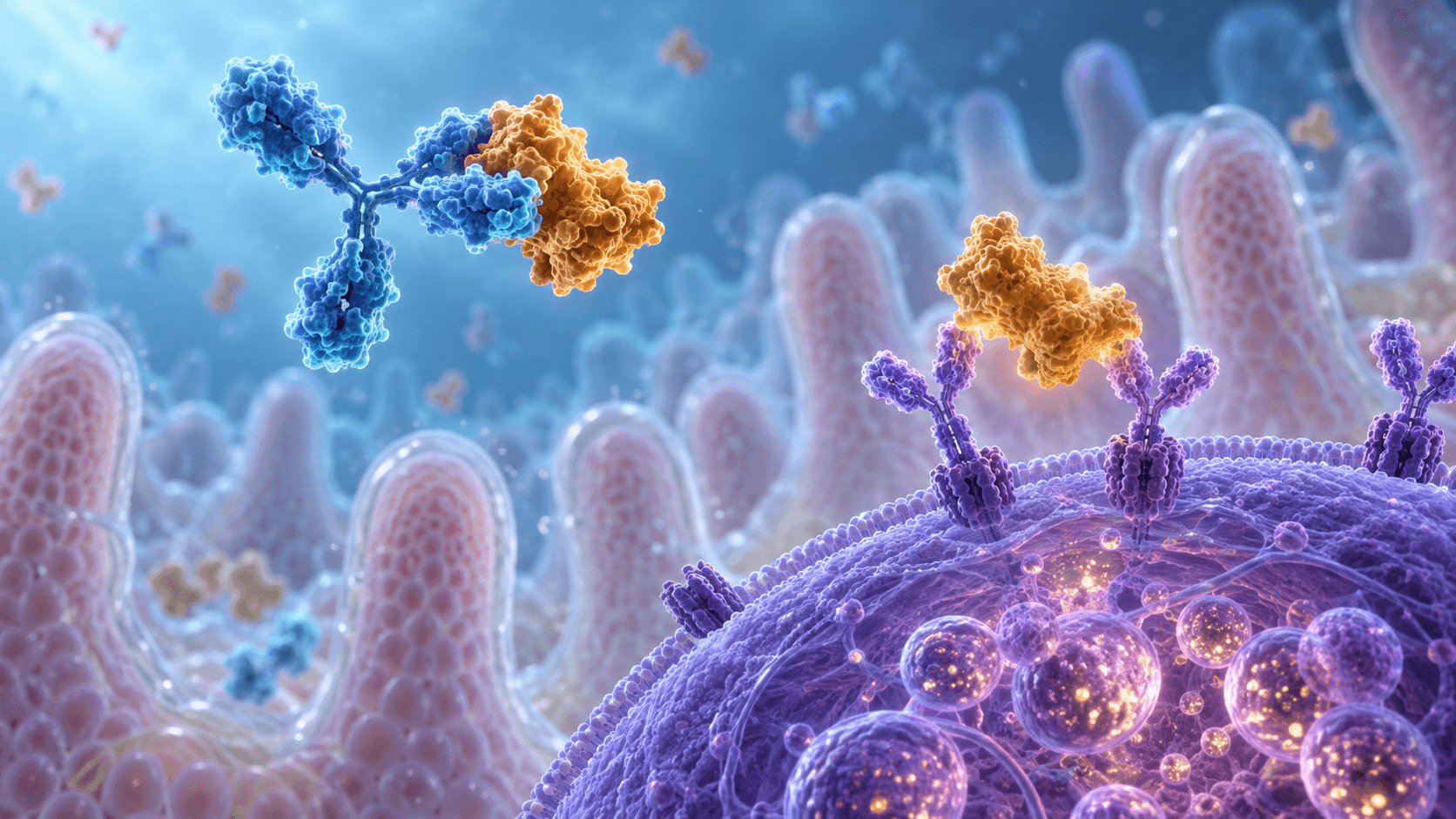
Food-specific IgG4 and IgE allergy: blocking antibodies, degranulation and diagnostic limits
Why food-specific IgG4 neither diagnoses intolerance nor guarantees safety. IgE/FcεRI signalling, IgG4 exchange, BAT, coeliac disease and actual LEAP, EAT, PALISADE and OUtMATCH findings.
A symptom after eating, an antibody to a food protein and a diagnosis are three different things. Bloating after milk may depend on lactose digestion; hives with breathing difficulty after nuts require allergy assessment; chronic difficulty swallowing may indicate eosinophilic oesophagitis. A large antibody list does not unite these conditions as “hidden intolerance” or explain their mechanisms.
IgG4 is particularly easy to misinterpret. This subclass can contribute to blocking IgE-dependent reactions and increase with repeated antigen exposure or immunotherapy. However, a positive food-specific IgG4 result neither diagnoses intolerance nor certifies that a food is safe for an individual. Molecular immunology and clinical research explain both limits.
Food reactions cannot be reduced to two antibodies
In IgE-mediated allergy, the immune system recognises an allergen and effector cells initiate a reaction. Other cellular and tissue mechanisms matter in non-IgE-mediated food allergic disorders; the absence of specific IgE does not exclude them. Nonimmune intolerance includes symptoms caused by inadequate lactose digestion. Toxic reactions to spoiled food form another category.
Coeliac disease is better considered separately as an immune enteropathy with an autoimmune component, rather than ordinary IgE allergy or “IgG4 intolerance”. Noncoeliac wheat sensitivity is likewise not confirmed by food IgG4. Timing helps develop a hypothesis but does not draw an absolute boundary: alpha-gal allergy can be delayed, while FPIES is an acute disorder without the typical IgE presentation.
Tolerance develops in tissues and lymph nodes
The intestinal immune system regularly encounters foreign dietary proteins. Their uptake and presentation can support regulatory responses: dendritic cells in the lamina propria migrate to draining lymph nodes, where retinoid signals and TGF-β favour Foxp3+ Treg differentiation. IL-10, regulatory B cells, epithelial health and microbial metabolites participate. The process depends on age, inflammation, the route of antigen entry and cellular context.
Retinal dehydrogenases use NAD+ to produce retinoic acid from retinal. Retinoic acid binds nuclear RAR/RXR receptors and supports intestinal homing molecules, including α4β7 and CCR9. This network cannot be reduced to “IL-10 exclusively switches everything to IgG4”. IL-4/IL-13, CD40 and the regulatory environment jointly affect class switching; IgA and IgG4 production have different requirements. Secretory IgA acts mainly at mucosal surfaces, whereas serum IgG4 captures only part of the response.
Why IgE binds a cell before a reaction occurs
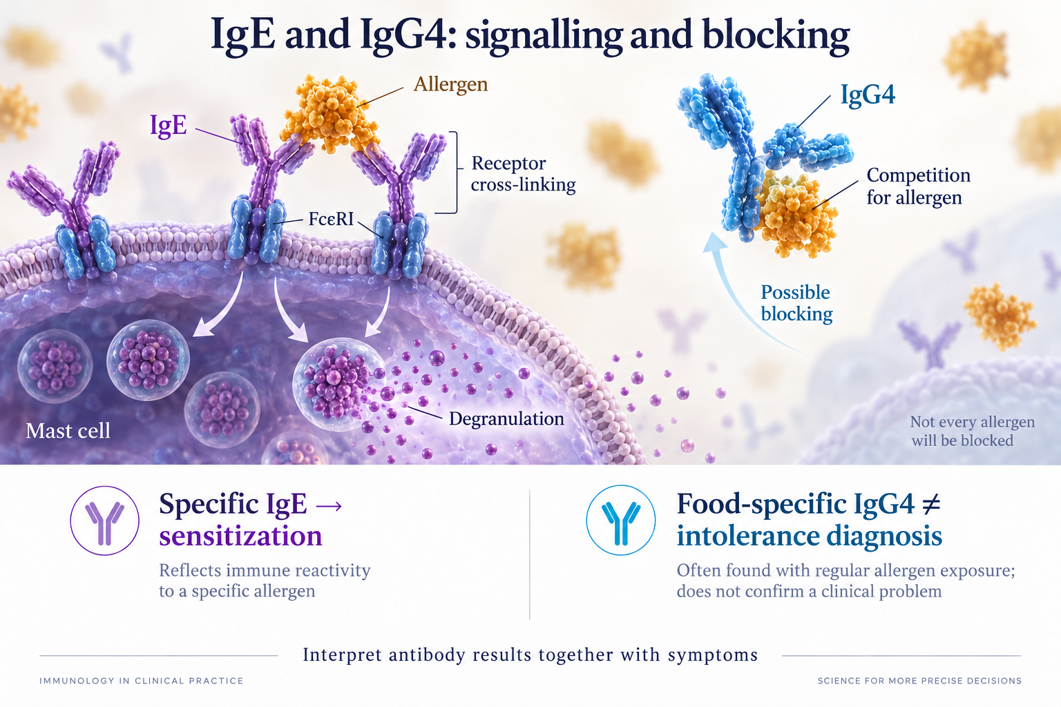
IgE contains ε heavy chains with four constant domains and lacks the typical IgG hinge. Its Fc portion binds the α chain of high-affinity FcεRI; in mast cells and basophils the complex usually also contains β and two γ chains. Binding affinity is approximately subnanomolar, although its constant depends on measurement conditions. Free and cell-bound IgE have different kinetics; the lifetime of the complex does not automatically determine how long clinical allergy lasts.
Sensitization means specific IgE or a positive skin test is present. Reaction depends on epitopes, allergen availability, the density and specificity of cell-bound IgE, and the cell's activation threshold. Binding a single IgE molecule to its receptor ordinarily does not constitute anaphylaxis. The allergen must produce sufficient receptor cross-linking, and tissue and systemic conditions determine the consequences.
From epithelial alarmins to IgE synthesis
A damaged epithelial barrier can release TSLP, IL-33 and IL-25, supporting type 2 responses. Cutaneous exposure to food proteins during eczema and intestinal exposure may have different immunological consequences. Dendritic cells and T-helper cells convey signals to B cells; IL-4Rα/STAT6 and CD40 contribute to the class-switching programme.
Activation-induced cytidine deaminase, AID, initiates DNA modifications in immunoglobulin switch regions. Repair and recombination connect the antigen-binding segment to a different constant region; plasma cells then secrete the new antibody class. This is cellular differentiation, not instantaneous conversion of an existing IgG4 molecule into IgE. Somatic hypermutation and class switching both involve AID but remain distinct processes.
Degranulation: membrane, endoplasmic reticulum and granules
FcεRI cross-linking causes phosphorylation of ITAM motifs in β/γ chains, Syk recruitment and assembly of signalling complexes with LAT. Kinases use ATP as phosphate donor. PLCγ hydrolyses membrane PI(4,5)P2 into IP3 and diacylglycerol: IP3 mobilises Ca2+ from the endoplasmic reticulum, and DAG contributes to PKC activation. Depletion of intracellular stores sustains Ca2+ entry through STIM1/ORAI1.
Calcium and cytoskeletal changes help secretory granules fuse with the plasma membrane. Histamine and proteases, including tryptase, are released; vascular, skin, airway and intestinal effects depend on the target organs. This network contains amplification and inhibition, so a linear Lyn→Syk→Ca2+ diagram is a useful map rather than a complete account of activation.
Lipid mediators are synthesised in parallel
Cytosolic phospholipase A2 releases arachidonic acid from membrane phospholipids. 5-lipoxygenase and FLAP participate in forming the leukotriene precursor LTA4; LTC4 synthase adds glutathione, after which extracellular processing yields LTD4 and LTE4. The cyclooxygenase pathway produces PGD2 through PGH2 and prostaglandin D synthase. Lipoxygenase contains iron, cyclooxygenases use haem, and oxidative steps require oxygen.
Lipid mediators can be released during the early response; their entire synthesis should not be assigned only to a 2–6-hour interval. Later cellular inflammation involves newly produced cytokines and recruitment of other leukocytes. It is not identical to biphasic anaphylaxis, when symptoms recur after the initial episode resolves. Suspected anaphylaxis needs immediate medical assistance; antihistamines do not replace intramuscular epinephrine under an individual emergency plan.
IgG4: the hinge and half-molecule exchange
IgG4 has a conventional IgG architecture with a hinge, but Ser228 and CH3-domain interactions, including Arg409, facilitate half-molecule exchange. A heavy chain together with its attached light chain is exchanged, rather than a free Fab cut off by a protease. Disulfide rearrangement and dissociation of the noncovalent CH3 interface allow halves from different IgG4 molecules to reunite.
The resulting bispecific molecules are functionally monovalent for each of two different antigens. Exchange reduces their ability to form certain cross-linked complexes, and Fc properties make IgG4 a weak activator of classical complement. However, “noninflammatory” does not mean every IgG4 is harmless: pathogenic IgG4 can disrupt target-protein function in some autoimmune diseases. Experimental reducing conditions cannot be presented as a universal glutathione concentration controlling exchange in all tissues.
Competitive blocking depends on the epitope
An IgG4 antibody can occupy an allergen site recognised by IgE or sterically restrict access to a neighbouring site. Its effect depends on concentration, affinity, epitope distribution and the timing of molecular encounters. Other IgG subclasses can have similar blocking functions; these are not exclusive to IgG4.
For one site and two competitors, equilibrium binding can be illustrated by [IgG4]/Kd,G4 divided by 1+[IgG4]/Kd,G4+[IgE]/Kd,E. This is the law of mass action under idealised conditions, not a clinical calculator. Allergen multivalency, membrane IgE, epithelial transport and differing epitopes violate its assumptions. Universal claims that “99.8% of allergen is blocked” or a particular IgE/IgG4 ratio guarantees tolerance do not follow from this formula.
FcγRIIb and SHIP-1: inhibition with conditions
In experimental systems, allergen–antibody complexes can engage activating FcεRI together with inhibitory FcγRIIb. Its phosphorylated ITIM recruits SHIP-1, an inositol 5-phosphatase converting PI(3,4,5)P3 to PI(3,4)P2. This changes lipid docking sites for signalling proteins and the calcium response. PI(3,4)P2 is not simply inactive waste; it has its own signalling functions.
Inhibition depends on the antibody, complex size, receptor expression and cell type. Basophil findings cannot be transferred unconditionally to every tissue mast cell. Primary studies show both FcγRIIb-dependent mechanisms and settings in which blocking this receptor did not remove the effect of post-treatment serum. Neither complete degranulation shutdown nor a single mechanism explaining immunotherapy efficacy is established.
Why food IgG4 does not diagnose intolerance
An immunoassay may correctly measure antibody binding to a food extract and still lack clinical diagnostic value. Diagnosis requires evidence that a result distinguishes people with a particular disease from those without it, predicts a meaningful outcome and improves decisions. Colour-coded IgG/IgG4 “red lists” do not meet these requirements for food intolerance.
EAACI does not recommend food-specific IgG4 as a diagnostic tool. These antibodies occur in people who eat the food without symptoms and can coexist with allergy. A positive test does not establish inflammation, and a negative result does not exclude IgE allergy. Not every commercial alternative test measures IgG; cellular methods have different methodologies and do not become validated by sharing the “intolerance” label.
Unjustified removal of many foods can reduce protein, calcium, iron and energy intake, compromise a child's growth and increase anxiety about eating. However, it is wrong to claim that every person inevitably loses IgG4 and develops anaphylaxis after a few months of avoidance. Reintroduction depends on reaction history, current sensitization and clinical assessment, not an attempt to restore an antibody titre.
Lactose and fructose: enzyme and transporter
Brush-border lactase-phlorizin hydrolase uses water to split lactose into glucose and galactose. With inadequate activity, some lactose remains in the lumen, retains water and is fermented by microbiota into gases and organic acids. Symptoms depend on quantity, accompanying food and intestinal sensitivity. Malabsorption does not always cause intolerance; hydrogen breath tests are interpreted alongside symptoms.
The −13910 C/T variant in the MCM6 regulatory region is informative in certain populations but does not account for every lactase-persistence variant or secondary mucosal injury. Fructose absorption depends on apical GLUT5 capacity and subsequent transport, rather than antibodies. Fructose malabsorption must not be confused with hereditary fructose intolerance due to aldolase B deficiency, which disrupts intracellular fructose-1-phosphate metabolism.
Histamine: DAO, HNMT and diagnostic uncertainty
DAO, encoded by AOC1, is a copper-containing amine oxidase with a topaquinone cofactor. Oxidative deamination of histamine uses O2 and water to form imidazole acetaldehyde, ammonia and H2O2; the aldehyde subsequently becomes an acid. This is not hydrolysis or an enzyme necessarily requiring zinc and copper together. Intracellular HNMT transfers a methyl group from S-adenosylmethionine to histamine, producing N-methylhistamine and S-adenosylhomocysteine.
These reactions do not diagnose “histamine intolerance” from low serum DAO. There is no reliable universal laboratory criterion, symptoms are nonspecific, and food histamine content varies. A presumed individual reaction should be distinguished from toxic spoiled-fish exposure, allergy and mast-cell disorders. Small oral-DAO studies do not establish treatment of IgE allergy or protection against anaphylaxis; local enzyme activity depends on the preparation and protein survival during digestion.
Wheat: coeliac disease, fructans and other mechanisms
Transglutaminase 2 deamidates selected glutamine residues in gluten peptides to glutamate in a Ca2+-dependent process. This improves presentation of some peptides through HLA-DQ2/DQ8 and T-cell activation. IL-15 and intraepithelial cytotoxic cells contribute to epithelial injury. Not every HLA carrier develops coeliac disease, and its manifestations do not necessarily imply complete atrophy of all villi.
Initial serology ordinarily includes anti-tTG IgA and total IgA while gluten is being consumed; appropriate IgG tests are used in IgA deficiency. These are specific coeliac tests, not a food-IgG panel. Biopsy decisions and exceptions depend on age and clinical criteria. Fructans in FODMAPs and ATIs are discussed in noncoeliac wheat sensitivity; experimental ATI-mediated TLR4 activation does not prove causation in every patient. Coeliac disease and wheat allergy are excluded first, followed by assessment of symptom reproducibility.
Delayed and tissue-based disorders need separate assessment
Alpha-gal is the carbohydrate epitope galactose-α-1,3-galactose. IgE to it can emerge after tick bites and cause reactions to mammalian products several hours later, often around 3–6 hours. Lipid digestion and glycolipid transport help explain the delay, but timing varies and depends on cofactors.
FPIES typically causes repeated vomiting after 1–4 hours, sometimes with lethargy, dehydration and shock, without usual skin or respiratory features. Its mechanism is more complex than “one T cell→TNF”. In eosinophilic oesophagitis, dysphagia and food impaction are assessed together with biopsies; approximately 15 eosinophils per high-power field is interpreted clinically. Neither an IgG4 panel nor a negative IgE result replaces diagnosis of these conditions.
History matters more than panel size
The clinical pathway begins with a specific food, preparation and quantity, reaction timing, reproducibility and symptom combination. Exercise, alcohol, NSAIDs, asthma and infections are considered. Targeted skin tests and specific IgE address plausible hypotheses. Without a reaction history, broad panels increase incidental findings mistakenly labelled as disease.
Skin-prick testing is usually read after 15–20 minutes with positive and negative controls; a wheal approximately 3 mm larger than the negative control indicates sensitization. Serum sIgE quantifies an antibody; an analytical method's detection boundary differs from a clinical probability threshold. There are no universal values for future reaction severity. Published positive-predictive thresholds depend on age, population, allergen and platform.
Component testing refines assessment without guarantees
Peanut Ara h 2, hazelnut Cor a 14 and cashew Ana o 3 help identify some clinically relevant profiles rather than simple cross-sensitization. Ara h 8 and Cor a 1 belong to PR-10 and are often associated with pollen-food syndrome. Gal d 1 is ovomucoid and Bos d 8 is casein; heat stability influences reactions to processed food without guaranteeing their presence or absence.
Other important families include nsLTPs, tropomyosins and fish parvalbumins. Disulfide bonds, coiled-coil structures and Ca2+-binding EF-hand motifs help explain stability and cross-recognition. Nevertheless, “all fish species are dangerous” and “boiling completely neutralises milk” are incorrect universal conclusions. In adults with suspected hazelnut allergy, one study found that component measurements could not replace food challenge.
Multiplex ISAC/ALEX systems and single-analyte methods differ in composition, units and analytical characteristics. Inhibiting cross-reactive carbohydrate determinants can reduce some nonspecific findings but does not correct every false diagnosis. The number of allergens on a chip and a percentage of “eliminated errors” are not universal characteristics of allergy diagnosis.
BAT and food challenge answer different questions
BAT assesses basophil function ex vivo, usually using CD63 and CD203c after allergen stimulation. Viability and response controls, sample handling and cells unresponsive to IgE-dependent stimulation must be considered. Results depend on allergen dose and method; one laboratory's performance cannot be generalised to all foods.
Santos's peanut study examined 43 allergic, 36 sensitized but tolerant and 25 nonsensitized children, with independent validation in 65 children. At selected cutoffs, BAT showed 97% accuracy, 95% positive predictive value and 98% negative predictive value. This was a diagnostic study in a particular population, not a promise of identical performance from every BAT panel.
A supervised oral food challenge assesses clinical reaction. Double blinding with placebo is useful for ambiguous subjective symptoms and research; an open challenge can suffice in practice. A trained team with reaction-treatment facilities conducts it. An intensive-care room for every challenge and identical dosing intervals for every patient are not universal requirements. Home replication of a challenge in suspected severe allergy is inappropriate.
LEAP and EAT: prevention with different results
LEAP randomized 640 infants with severe eczema and/or egg allergy, starting at 4–11 months, to peanut consumption or avoidance until 60 months. Target intake was at least 6 g peanut protein weekly in an age-appropriate form. The primary outcome was peanut allergy at five years: among 530 infants with an initially negative skin test, 13.7% with avoidance versus 1.9% with consumption; among 98 with a 1–4 mm wheal, 35.3% versus 10.6%. IgG4 increased more with consumption, but this establishes neither lifelong safety nor treatment efficacy for existing allergy.
EAT studied 1,303 exclusively breast-fed infants from the general population at three months: early introduction of six foods versus standard introduction around six months. The primary outcome was allergy to one or more study foods at 1–3 years. Intention-to-treat results were negative: 5.6% versus 7.1%, p=0.32. Per-protocol results were 2.4% versus 7.3%, p=0.01; milk, sesame, fish and wheat showed no significant effects. Associations with 2 g weekly peanut/egg protein and difficulties adhering to the regimen must not become “early exposure always prevents allergy”.
Oral immunotherapy raises thresholds without erasing risk
OIT administers measured allergen under medical supervision. Cellular reactivity, specific IgG4 and T-cell responses change during treatment, but IgG4 increases do not establish lasting tolerance after withdrawal. Desensitization during continued dosing, persistence after interruption and natural tolerance are different endpoints.
PALISADE was a double-blind placebo-controlled AR101 trial involving 551 participants aged 4–55, with the principal analysis in 496 children/adolescents aged 4–17. After escalation, 300 mg peanut protein daily was maintained for approximately 24 weeks. At exit challenge, 67.2% versus 4.0% tolerated at least 600 mg without dose-limiting symptoms: difference 63.2 percentage points, 95% CI 53.0–73.3. Adverse events affected over 95% of children/adolescents; efficacy was not demonstrated in adults. This is neither permission to eat peanut freely nor a guarantee against anaphylaxis.
Sublingual immunotherapy provides a different mucosal exposure, generally at smaller quantities. It should not be equated with OIT efficacy or assumed automatically safe. Selection depends on the preparation, protocol and treatment goal. Trial doses describe research, not instructions for self-treatment.
Omalizumab: a separate mechanism and a specific endpoint
Omalizumab binds free IgE and prevents interaction with FcεRI, gradually reducing available IgE and receptor expression. It is not food-specific IgG4 and does not establish IgG4 as the sole protective mechanism.
OUtMATCH's first stage examined omalizumab as monotherapy, not a proven OIT combination. It randomized 180 people allergic to peanut and at least two other foods; analysis included 177 participants aged 1–17. Subcutaneous doses based on weight and baseline IgE were given every 2–4 weeks for 16–20 weeks. The primary 600 mg peanut-protein threshold without dose-limiting symptoms was reached by 79/118 (67%) versus 4/59 (7%); for 1,000 mg cashew, milk and egg, results were 41/3%, 66/10% and 67/0%. Injection-site reactions were more frequent; cure or tolerance of every allergen by every participant was not established.
IgG4-related disease is another clinical problem
IgG4-related disease is a fibroinflammatory syndrome with organ involvement, characteristic histology and complex diagnosis. Total serum IgG4 can help, but normal levels do not exclude it and high levels do not prove it. IgG4+ plasma-cell counts and ratios to IgG are interpreted according to the organ and morphology, including storiform fibrosis and obliterative phlebitis.
A food-specific panel does not diagnose this syndrome. The reverse claim that “any high IgG4 is beneficial” is also incorrect. Antibody function depends on its antigen: blocking an allergen and disrupting one's own proteins are different biological situations.
Barrier support and supplements: mechanism cannot replace a trial
Butyrate can influence histone deacetylases and regulatory T cells, and its production is linked to microbial fibre fermentation. Histone acetylation is not DNA demethylation at FOXP3, however, and an oral dose does not define a lymph-node concentration. Vitamin D acts through VDR; retinoid signals, cellular energetics and epithelial state contribute to tolerance. These mechanisms do not demonstrate that vitamin D or butyrate supplements treat food allergy.
Quercetin, luteolin and EGCG affected mast-cell signalling in experimental models. Inhibiting Syk, Ca2+ or cytokines at a particular concentration cannot be extrapolated to protection after a capsule. Glutamine, zinc-carnosine and palmitoylethanolamide likewise lack a universal regimen restoring food tolerance. PEA is mainly associated with PPARα and indirect lipid signalling; direct CB2 agonism requires different evidence.
Probiotic effects depend on strain, substrate and host; a species name does not guarantee an absence of histamine production. PHGG, arabinogalactan and other fibres have variable tolerance and can cause gas. A universal 70% rise in MUC2 and IgG4 switching in every patient are unsubstantiated. Intestinal alkaline phosphatase and LPS detoxification explain one barrier mechanism, without establishing food-allergy treatment.
Returning foods needs an appropriate basis
Food ladders with baked milk or egg suit selected clinical situations and patients. Heating and the food matrix change epitope accessibility, but oven temperature is not protein temperature, and linear epitopes may remain. Neither 180 °C for 30 minutes nor 2–4-week intervals guarantee a hundredfold decrease in allergenicity or freedom from systemic reactions.
When a food was removed only because of IgG4, first establish whether symptoms and sensitization were actually present. A history of immediate reactions, asthma or suspected alpha-gal, FPIES or EoE requires a specialist-defined pathway. The aim is a diverse tolerated diet and treatment of a specific disease. Antibodies address limited questions; food IgG4 is not an intolerance diagnosis, and its increase does not replace clinical safety assessment.
FAQ
Can IgG4 justify food exclusion?
This result alone is not a diagnostic basis. Symptom history and assessment of a specific disease are needed.
Does higher IgG4 after OIT mean cure?
No. Desensitization during treatment and tolerance after withdrawal are different outcomes.
Does low DAO confirm histamine intolerance?
There is no reliable universal laboratory criterion; biochemistry does not replace differential assessment.
Sources
- Testing for IgG4 against foods is not recommended as a diagnostic tool: EAACI Task Force Report
- EAACI guidelines on the diagnosis of IgE-mediated food allergy
- Anti-inflammatory activity of human IgG4 antibodies by dynamic Fab arm exchange
- Dynamics of inter-heavy chain interactions in human immunoglobulin G subclasses studied by kinetic Fab arm exchange
- On the role of allergen-specific IgG subclasses for blocking human basophil activation
- Oral immunotherapy induces IgG antibodies that act through FcγRIIb to suppress IgE-mediated hypersensitivity
- Changes in passively-sensitized basophil activation to αS1-casein after oral immunotherapy
- Randomized trial of peanut consumption in infants at risk for peanut allergy
- Impact of peanut consumption in the LEAP Study: Feasibility, growth, and nutrition
- Randomized Trial of Introduction of Allergenic Foods in Breast-Fed Infants
- AR101 Oral Immunotherapy for Peanut Allergy
- Omalizumab for the Treatment of Multiple Food Allergies
- Structure and inhibition of human diamine oxidase
- Guideline on management of suspected adverse reactions to ingested histamine
- Basophil activation test discriminates between allergy and tolerance in peanut-sensitized children
- Measurement of IgE to hazelnut allergen components cannot replace hazelnut challenge in Dutch adults
- Compartmentalized gut lymph node drainage dictates adaptive immune responses
- Retinoic acid-producing DCs and gut-tropic FOXP3(+) regulatory T cells in the induction of oral tolerance
- Gut CD103+ dendritic cells express indoleamine 2,3-dioxygenase which influences T regulatory/T effector cell balance and oral tolerance induction
- Blocking antibodies induced by peanut oral and sublingual immunotherapy suppress basophil activation and are associated with sustained unresponsiveness
- European Society for the Study of Coeliac Disease 2025 Updated Guidelines on the Diagnosis and Management of Coeliac Disease in Adults. Part 1: Diagnostic Approach
- Sex as a predictor of clinical phenotype and determinant of immune response in IgG4-related disease
- Dietary Bioactive Compounds and Their Role in Allergy Prevention: A Comprehensive Review
- The soluble isoform of human FcεRI is an endogenous inhibitor of IgE-mediated mast cell responses
- Basophil activation test: food challenge in a test tube or specialist research tool?