
The endothelial glycocalyx: hyaluronan, shear stress, and the vascular barrier
HS, HA, syndecan and glypican architecture; shear signaling, eNOS, SOD3, and antithrombin. Revised Starling forces, rheology, renal and brain barriers, indirect PBR and biomarkers, and positive and negative repair trials.
The glycocalyx is a hydrated network of carbohydrate chains, proteins, and associated molecules on the blood-facing surface of endothelial cells. It influences how cells and solutes approach the vessel wall, participates in mechanical signaling, and provides a local environment for enzymes and coagulation regulators. It is a dynamic component of the vascular barrier, with substantial differences between organs and measurement methods.
Molecular mechanisms, experimental measurements, and treatment outcomes must be considered separately. Enzymatically removing heparan sulfate from cultured cells can change nitric oxide production; circulating syndecan-1 can accompany severe illness; sublingual microscopy can detect changes in the region accessible to red cells. None of these findings alone establishes that a supplement rebuilds vessels or prevents cardiovascular events.
1. Building the endothelial surface layer
Glycocalyx and ESL
In its narrower sense, the glycocalyx comprises membrane-associated glycoproteins, proteoglycans, and glycolipids. The broader endothelial surface layer, ESL, also includes water and reversibly associated plasma components. The terms overlap in the literature. Apparent thickness depends on species, vessel, flow, and method: electron microscopy after fixation, fluorescent-tracer exclusion, and analysis of red-cell movement interrogate different properties.
Vink and Duling (1996) demonstrated a near-wall region inaccessible to certain tracers and red cells in hamster cremaster capillaries. These observations support a functional surface layer, without establishing a universal human thickness. Measurements ranging from hundreds of nanometers to micrometers cannot be merged into one normal interval. Neither a fixed water fraction nor a single pore size or molecular-weight cutoff applies to every vessel. The network deforms, exchanges components, and has heterogeneous charge and hydraulic resistance.
Syndecans and glypicans
Syndecans are transmembrane protein scaffolds. Their extracellular domains carry linear glycosaminoglycan chains, principally heparan sulfate and, for some syndecans, chondroitin sulfate. A carbohydrate linker attaches these chains to serine residues. Several chains on a core protein do not mean that the polysaccharides themselves are branched. The transmembrane region helps organize complexes, while the short intracellular C1–V–C2 tail associates with adaptors, including PDZ proteins and cytoskeletal connections. The relevant partners depend on the syndecan and cellular state.
Syndecan-1 and syndecan-4 differ in both expression and complex assembly. A transmembrane GXXXG motif contributes to self-association, but its strength depends on the surrounding sequence (Dews and MacKenzie, 2007). Structural work demonstrated binding of the syndecan-4 C-terminal motif to a syntenin-1 PDZ domain (Choi et al., 2016). These findings clarify scaffold–adaptor connections without establishing identical mechanosignaling in every tissue.
Glypicans are attached to the outside of the membrane through a GPI anchor and have no cytoplasmic tail of their own. Communication with intracellular machinery therefore involves neighboring proteins and membrane complexes. Glypican-1 has been studied in mechanosensing and membrane microdomain organization, but it is not the only possible endothelial glypican. Ebong et al. (2014) found that glypican-1 and syndecan-1 interventions affected flow-induced NO and cellular remodeling differently. The findings support specialized functions rather than one immutable fiber-to-caveola-to-enzyme sequence.
Sulfated glycosaminoglycans
Heparan sulfate, HS, consists of repeating disaccharides containing glucosamine and a uronic acid. N-deacetylation/N-sulfation, conversion of glucuronic to iduronic acid, and O-sulfation create domains with different protein affinities. Sulfate position and chain conformation matter beyond the total negative charge. Synthesis and modification involve the cellular secretory machinery: EXT enzymes, NDST enzymes, and sulfotransferases do not simply attach an orally consumed polysaccharide to the endothelial surface.
Chondroitin sulfate, CS, contains N-acetylgalactosamine and glucuronic acid; dermatan sulfate, DS, includes iduronic acid. Their abundance and sulfation patterns vary between tissues and core proteins. Carboxyl and sulfate groups contribute to hydration, ion distribution, and protein interactions. Nevertheless, negatively charged albumin can associate with the surface layer. A blanket rule that every negatively charged molecule is excluded fails to capture the chemistry and structure of this network.
Hyaluronan and sialic acids
Hyaluronan, HA, is an unsulfated linear polymer of glucuronic acid and N-acetylglucosamine. Unlike HS and CS, it is not synthesized as a covalent chain on a protein scaffold. Membrane synthases HAS1–3 use activated UDP sugars and extrude the growing chain. CD44 and other hyaladherins retain and organize HA at the surface. Maloney et al. (2022) resolved synthesis and translocation mechanisms using a viral HAS, providing molecular evidence for the principle rather than a trial in human endothelium or of oral N-acetylglucosamine.
HA molecular weight affects its physical properties, but there is no universal switch in which every large chain is protective and every short fragment inflammatory. Origin, purity, associated proteins, and cellular context matter. Sialic acids, including Neu5Ac, occupy terminal positions on glycoprotein and glycolipid carbohydrates. They contribute to surface charge and recognition but are not themselves glycosaminoglycans.
Albumin and plasma-associated components
The ESL exchanges albumin, enzymes, growth factors, and coagulation regulators with plasma. Albumin, approximately 66.5 kDa, contributes to plasma oncotic properties and carries many ligands, including sphingosine-1-phosphate, S1P. ApoM within HDL is another important S1P carrier. A freely circulating protein, a reversibly associated surface protein, and an enzyme anchored to HS have different functions. This biology does not establish that everyone with low albumin needs an infusion to repair the glycocalyx.
2. From blood flow to biological signaling
Shear stress and pressure
Pressure acts mainly perpendicular to the surface; viscous shear acts tangentially. For a Newtonian fluid, the local relation is:
Here is wall shear stress in pascals, is dynamic viscosity in Pa·s, is velocity along the wall in m/s, and is distance from it in meters. One pascal equals 10 dyn/cm². For steady laminar flow in a rigid circular tube, the Poiseuille model gives:
is volumetric flow rate and is radius. These assumptions do not automatically hold in a capillary containing individual deforming red cells, a pulsatile vessel, or a damaged porous layer. The glycocalyx itself changes the velocity profile and load transmission.
A network of mechanosensors
Weinbaum et al. (2003) modeled how flow transfers force through surface-network elements to membrane and cytoskeleton. In cultured endothelium, Florian et al. (2003) found that removing HS with bacterial heparinase III suppressed flow-induced NO while preserving the response to bradykinin. Pahakis et al. (2007) showed component-specific effects: depletion of HS, HA, or sialic residues impaired the NO response, whereas flow-induced prostacyclin remained. The glycocalyx therefore contributes to particular pathways; its damage does not abolish all flow sensing.
Other participants include the PECAM-1–VE-cadherin–VEGFR2 complex, integrins, cytoskeletal structures, Piezo1 and TRPV4 channels, and membrane microdomains. Their interactions depend on endothelial phenotype and exposure duration. A scientific diagram represents functional relationships rather than exact molecular geometry or a single obligatory signaling route.
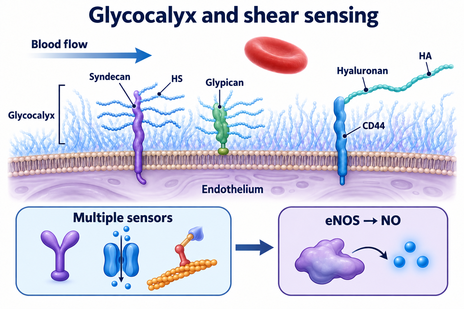
Figure1. Linear syndecan-associated chains, the external GPI anchor of glypican, and HA retained by CD44 are schematic. The multiple-sensors box represents cooperative mechanosignaling rather than one exclusive direct route; eNOS also remains calcium–calmodulin regulated. Molecular geometry and scale are not reproduced (Florian et al., 2003; Pahakis et al., 2007; Ebong et al., 2014).
eNOS, NO, and calcium
Endothelial nitric oxide synthase, eNOS, uses L-arginine, oxygen, and reducing equivalents from NADPH. Coupled catalysis requires heme, BH4, FAD, FMN, and calmodulin. Phosphorylation of human Ser1177, including by Akt, can increase activity and alter sensitivity to calcium–calmodulin regulation (Dimmeler et al., 1999). This does not mean complete calcium independence or physical removal of an autoinhibitory domain. The homologous residue has different numbering in some species.
NO stimulates soluble guanylyl cyclase and cGMP signaling in smooth muscle, contributing to relaxation; it also influences platelets and inflammatory interactions. BH4 deficiency and other coupling disturbances can redirect eNOS toward superoxide production. Consequently, eNOS abundance and glycocalyx integrity do not themselves determine NO bioavailability. The mechanism is developed further in the article on eNOS uncoupling.
Sustained adaptation and disturbed flow
Sustained directional flow can promote KLF2/KLF4 transcriptional programs, anticoagulant properties, and restrained endothelial inflammation. Dekker et al. (2002) demonstrated KLF2 induction under prolonged shear in cultured cells. At branches and curves, flow may be weak, reversing, or spatially heterogeneous. Disturbed or oscillatory flow is not necessarily turbulent.
Nor does every oscillation stop NO production: the specific acute oscillatory condition tested by Florian et al. (2003) also stimulated an NO response. Waveform, mean stress, exposure duration, and chronic gene expression are distinct levels of analysis. Local hemodynamics help explain atherosclerotic distribution but do not replace the causal importance of apoB-containing lipoproteins and their intimal retention.
SOD3 and antithrombin
Extracellular superoxide dismutase, SOD3, is a Cu/Zn enzyme with a C-terminal region that binds sulfated polysaccharides (Lookene et al., 2000). Local retention helps limit the reaction between superoxide and NO:
Hydrogen peroxide also needs downstream handling by other systems. Loss of binding sites can redistribute SOD3 between tissue and plasma. Circulating SOD3 therefore need not decrease with surface injury, and it is not a universal thickness test.
Antithrombin recognizes a particular sulfated sequence rather than all HS. A pentasaccharide motif promotes allosteric activation against factors Xa and IXa; efficient acceleration of thrombin inhibition requires a longer chain able to bridge protease and inhibitor (Johnson et al., 2006). The commonly discussed length of approximately 18 sugar residues concerns certain heparin chains and is not an exception-free threshold for every synthetic oligosaccharide. Loss of surface anticoagulant properties may contribute to thrombosis without explaining the entire coagulation cascade.
3. Injury and shedding
Enzymes, proteolysis, and oxidation
Heparanase, HPSE, cleaves HS. Processing removes an internal linker from its precursor, mainly through cathepsin L, producing an active complex of approximately 8- and 50-kDa subunits (Abboud-Jarrous et al., 2008). Human heparanase differs from bacterial heparinase III used experimentally. HPSE should not be described as an enzyme that cleaves every GAG.
HA turnover involves synthesis, uptake, and degradation. HYAL1 and HYAL2 differ in location, activity conditions, and association with intracellular processing; degradation is not one extracellular blade producing fragments of a fixed size. Metalloproteinases and ADAM proteases can shed proteoglycan ectodomains. This cuts the protein scaffold, unlike hydrolysis of a polysaccharide chain. Enzyme contribution and retention of membrane remnants are context dependent.
An external GPI anchor has been demonstrated for HYAL2, but location is not equivalent to high catalytic activity: Rai et al. (2001) established a surface protein without detecting HA cleavage in their system. HA degradation also involves uptake and acidic intracellular processing, including HYAL1. Localization, pH, and protein abundance require separate consideration.
Reactive oxygen and nitrogen species can modify carbohydrates, proteins, and enzyme regulation. There is nevertheless no common rule for losing a fixed fraction of every vascular glycocalyx within seconds. An oxidative mechanism does not establish efficacy for every antioxidant supplement.
HA fragments and inflammation
The hypothesis that HA fragments act as danger signals depends on the experimental system. Dong et al. (2016) found that endotoxin-free fragments did not produce the expected activation in the macrophages and dendritic cells examined; some positive effects of preparations were attributable to endotoxin contamination. This is an essential methodological qualification. It does not exclude every action of endogenous HA in injured tissues, but it prevents classifying every short chain as an obligatory TLR2/TLR4 agonist.
Hyperglycemia and diabetes
In Nieuwdorp et al. (2006), acute hyperglycemia in a study of ten healthy men was associated with a reduction in estimated glycocalyx volume from approximately 1.7 to 0.8 liters. This was a tracer-distribution estimate, not direct measurement of all physical gel in the body. Mannitol did not reproduce the change, challenging an exclusively osmotic-collapse explanation. Intravenous N-acetylcysteine attenuated the change. A small acute experiment does not demonstrate long-term benefit from oral NAC in diabetes.
Chronic hyperglycemia involves oxidative stress, advanced glycation products and RAGE signaling, inflammation, altered synthesis, and increased component turnover. Contributions differ between vascular beds. Postmeal glucose fluctuations also matter: glycemic variability and endothelial injury cannot be captured by a single glycocalyx measurement and are not equivalent to average glucose concentration.
Sepsis, coagulation, and atherosclerosis
Schmidt et al. (2012) implicated heparanase activation in pulmonary glycocalyx loss, neutrophil adhesion, and lung injury in experimental sepsis. Genetic or pharmacological interventions protected animals; human samples supported relevance without constituting a treatment trial. Septic vascular leakage combines surface-layer changes, endothelial-junction dysfunction, inflammation, and hemodynamics. It is not synonymous with rare idiopathic systemic capillary leak syndrome.
Disseminated intravascular coagulation involves tissue factor, thrombin, platelets, NETs, anticoagulant pathways, and fibrinolysis. Shedding does not necessarily denude endothelium or activate a single indispensable coagulation factor. In atherosclerosis, an altered surface network may influence lipoprotein transport and leukocyte interactions, while LDL also crosses endothelium through regulated transport. Glycocalyx injury is not established as the sole entry window required for every plaque.
4. Fluid exchange, blood rheology, and organ barriers
The revised Starling principle
For many continuous microvessels, a useful representation incorporates local subglycocalyx oncotic pressure:
is volume filtration rate, is exchange area, and is hydraulic conductivity. and are capillary and interstitial hydrostatic pressure; is plasma oncotic pressure and is local subglycocalyx oncotic pressure. is the dimensionless reflection coefficient for the protein considered. With pressures in Pa and in m/(s·Pa), is in m/s. A positive result denotes outward fluid movement.
Subglycocalyx protein concentration depends on transport restriction and local washout; it is not automatically the concentration of the bulk interstitium. Adamson et al. (2004) demonstrated a substantial effective oncotic gradient in rat microvessels even with equal albumin concentrations in plasma and interstitial fluid. Nevertheless, is not always zero and is not one fixed constant across tissues or diseases.
Steady-state filtration and lymphatic return are important in most continuous vascular beds. A sudden fall in capillary pressure can produce transient reabsorption, and specialized tissues, including kidney and intestine, require separate analysis. Both obligatory venous-end absorption in every capillary and an absolute prohibition on vascular absorption are inaccurate. Endothelial junctions and other barrier components cooperate with the glycocalyx.
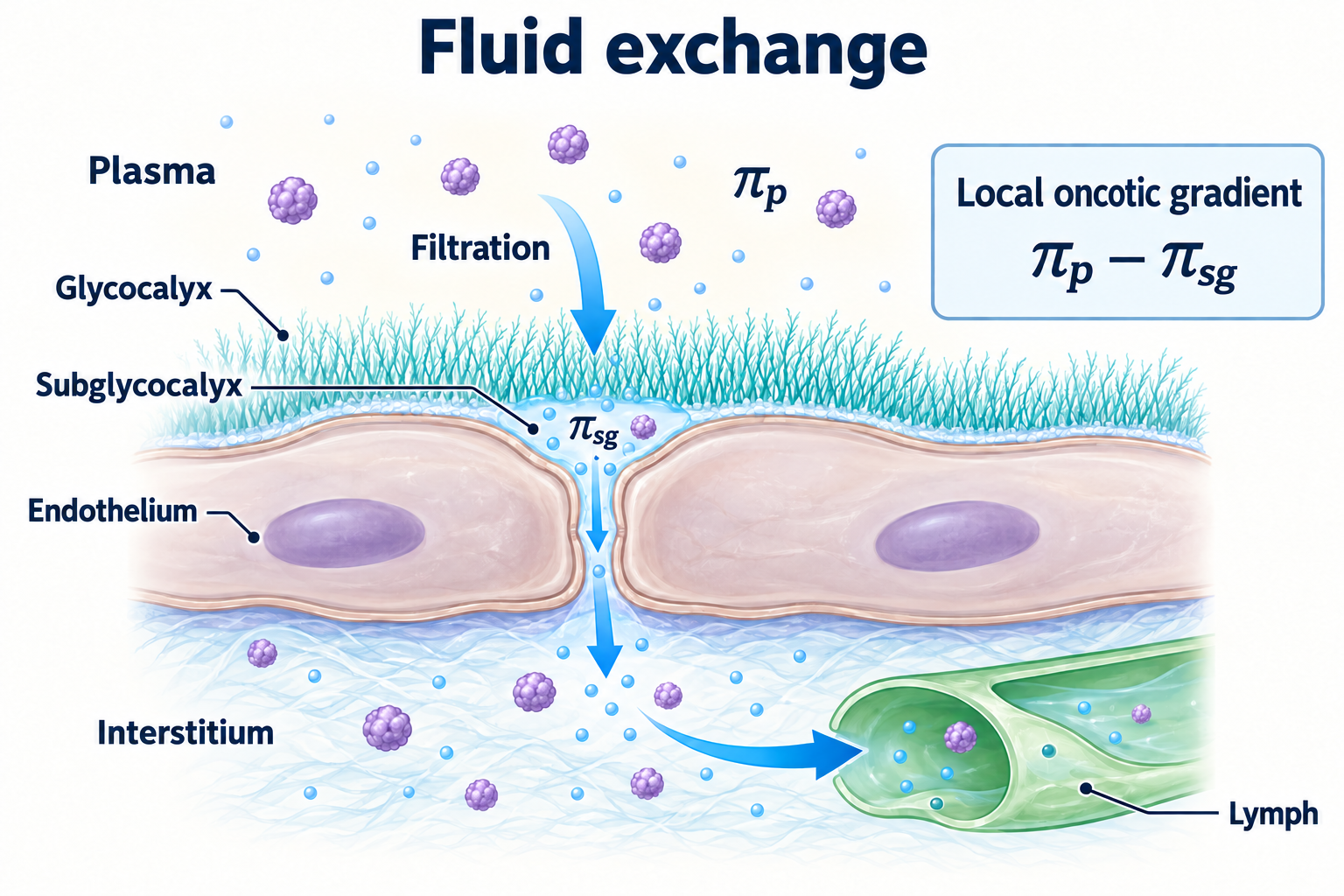
Figure2. A qualitative model of steady outward filtration through the surface barrier to interstitium and then lymph. The subglycocalyx zone is local extracellular space at the intercellular cleft entrance; its oncotic pressure is neither automatically bulk interstitial pressure nor invariably zero. Transient reabsorption and specialized vessels are not depicted; these are not measured patient data (Adamson et al., 2004).
Red cells and apparent viscosity
Red cells migrate toward the vessel axis, deform, and move at a different mean velocity from bulk blood. The Fåhræus effect describes tube hematocrit being lower than discharge hematocrit . Under steady conservation of red-cell flux:
is mean bulk-flow velocity and is mean red-cell velocity. The Fåhræus–Lindqvist effect concerns declining apparent viscosity as diameter decreases over a particular range; resistance rises again in extremely narrow passages. These are different phenomena and cannot be reduced to glycocalyx lubrication.
The surface layer limits available lumen, affects near-wall flow, and contributes to resistance. Following experimental photodamage, Vink and Duling (1996) observed an approximately 60% increase in tube hematocrit. This does not establish a fixed hematocrit for all human capillaries or a severalfold viscosity change after any injury.
Volume loading and edema
Excess intravascular volume loading can increase atrial natriuretic peptide release and accompany glycocalyx-component shedding. Chappell et al. (2014) studied this in a perioperative setting. Permeability, hydrostatic pressure, protein gradients, and lymphatic drainage together determine edema. The observations support attention to fluid balance, but not withholding necessary shock resuscitation or claiming that every infusion destroys the layer. Fluid type, amount, and rate must address the clinical situation.
Kidney and blood–brain barrier
The glomerular filtration barrier comprises fenestrated endothelium with its surface layer, basement membrane, and podocytes. In cultured human glomerular endothelial cells, HS removal increased albumin passage without necessarily changing electrical resistance (Singh et al., 2007). This supports a surface-network contribution but does not make albuminuria a direct measure of charge or thickness. Selectivity depends on size, structure, and interactions throughout the barrier; comparing albumin radius with an assumed pore diameter is not a valid size-exclusion calculation.
The brain barrier depends on tight junctions, suppressed transcytosis, basement membrane, pericytes, and astrocytes. Endothelial glycocalyx is one contributor. Zhu et al. (2018) linked enzymatic degradation to greater barrier dysfunction and edema after asphyxial cardiac arrest in rats. The study does not establish drug efficacy in humans or explain all Alzheimer disease. A universal uniquely thick cerebral glycocalyx with maximal HS sulfation has not been established.
5. What measurements can tell us
Sublingual imaging and PBR
SDF/IDF video microscopy primarily records red cells rather than molecular chains. GlycoCheck estimates the perfused boundary region, PBR, the region accessible to variation in red-cell-column position. Conceptually:
is the algorithmically estimated perfused diameter and is median red-cell-column width. Both and PBR are expressed in micrometers. Perfused diameter is not necessarily anatomical diameter and should not be replaced with a single maximum width in a frame. A higher PBR is generally interpreted as greater near-wall accessibility and less effective red-cell exclusion. It is an indirect functional estimate, not direct thickness measurement, an image of chemical composition, or a diagnosis of systemic destruction.
Algorithm version, vessel-size selection, movement, focus, probe pressure, blood flow, and the fraction of time containing red cells matter. Eickhoff et al. (2020), in 42 healthy smokers, demonstrated examination-condition effects and improved reproducibility with repeated measurements. Bol et al. (2022), in 49 healthy volunteers, found poor agreement for single measurements and improvement when three were averaged. Changes in an individual may be smaller than method variability. Results across software and populations cannot be assembled into universal normal/severe-injury bands; the sublingual region is not a direct map of every organ.
Circulating markers and albuminuria
| Measure | Possible information | Main limitation |
|---|---|---|
| Syndecan-1 | Ectodomain shedding, inflammation, and tissue injury | Also present in epithelium and other cells; clearance and assay matter |
| Heparan sulfate | HS turnover and cleavage | Other tissues and matrix contribute; fragments differ |
| Hyaluronan | HA synthesis, breakdown, and release | Liver function, matrix turnover, and clearance affect concentration |
| Endocan | Endothelial secretion and inflammatory signaling | A secreted DS proteoglycan, not exclusively a shed membrane anchor |
| Urine albumin/creatinine, ACR | Impaired renal albumin retention | A2 begins at 30 mg/g; one result does not identify the mechanism |
| Plasma SOD3 | Production, binding, and redistribution | Detachment can increase concentration; no universal glycocalyx-injury threshold |
| PBR | Red-cell accessibility to the near-wall perfused region | Indirect sublingual estimate with measurement variability |
Such measures can serve research and selected clinical questions, but do not automatically form a validated panel for selecting a repair regimen. ACR requires attention to repeat confirmation and transient causes of elevation; circulating fragments require assay standardization and clinical context.
6. Repair hypotheses and clinical outcomes
Sulodexide and conflicting evidence
Sulodexide contains glycosaminoglycan fractions, commonly described as approximately 80% fast-moving heparin fraction and 20% dermatan sulfate. It is not interchangeable with every low-molecular-weight heparin and does not simply stick electrostatically to a negatively charged surface as replacement material. Proposed mechanisms include effects on degradation enzymes, inflammation, and coagulation; synthesis of new HS remains a cellular process.
| Study | Design and finding | Interpretation |
|---|---|---|
| Gambaro et al., 2002, Di.N.A.S. | 223 people with type 1 or 2 diabetes; placebo or 50/100/200 mg/day for four months; reduced albuminuria | Positive surrogate result, not proof of fewer cardiovascular events |
| Broekhuizen et al., 2010 | 10 men with type 2 diabetes and 10 controls assessed before/after two months of 200 mg/day; indirect vascular measurements changed | Small study without parallel placebo; methods are not identical to modern PBR |
| Lewis et al., 2011, SUN-micro | 1056 people with type 2 diabetes; large randomized trial did not improve the primary albuminuria endpoint | Early favorable findings were not reliably reproduced |
| Packham et al., 2012, SUN-macro | 1248 people with diabetic nephropathy; no proteinuria improvement; early termination limited power for hard renal outcomes | Benefit remains unproven; uncertainty in the hard endpoint is not demonstrated equivalence |
These doses describe trials rather than instructions to the reader. Product activity units, LE/LRU, and milligrams cannot be converted through one universal factor. Indications in a particular jurisdiction do not establish general glycocalyx therapy. Contraindications, bleeding, and combinations with antithrombotic drugs remain relevant.
Sugar precursors and HA
UDP-GlcNAc is required for endogenous HA and HS production. However, N-acetylglucosamine in a product does not demonstrate delivery of sufficient activated substrate to endothelial cells, incorporation into the vascular network, or improved clinical outcomes. The same distinction applies to glucosamine, chondroitin, and oral HA: joint or skin findings do not establish vascular-layer repair. Molecular weight, route, and experimental model are crucial for high-molecular-weight HA; intravenous animal experiments do not yield a home treatment schedule.
Enzyme inhibitors and antioxidants
Roneparstat and necuparanib have been studied as modified heparin-like compounds, particularly in oncology. Heparanase inhibition is a research strategy. Reduced anticoagulant activity does not mean bleeding is impossible or broad use is proven safe. Doxycycline can affect metalloproteinases, but established uses and studies in other tissues do not make it standard glycocalyx therapy; it has its own adverse effects.
A phase I roneparstat study in multiple myeloma primarily assessed tolerability; a randomized phase II necuparanib study in pancreatic cancer did not improve overall survival and stopped for futility. These oncology results do not establish clinical glycocalyx restoration (Galli et al., 2018; O’Reilly et al., 2020).
The acute intravenous NAC experiment does not support a universal oral course. Vitamin C also illustrates why antioxidant biochemistry must be tested clinically. In LOVIT (Lamontagne et al., 2022), 872 patients with sepsis were randomized and 863 entered the primary analysis. Death or persistent organ dysfunction at day 28 occurred more often with high-dose intravenous vitamin C: 44.5% versus 38.5%, RR 1.21, 95% CI 1.04–1.40. This is not evidence of harm from ordinary dietary vitamin C, but it contradicts an unconditional promise of benefit from this infusion in sepsis.
Polyphenols may modify AMPK, SIRT1, and inflammatory signaling in experimental models. Recommendations require information about formulation, dose, bioavailability, and meaningful outcomes. Universal courses of antioxidants, building blocks, and enzyme inhibitors cannot be inferred from a molecular diagram.
S1P, albumin, and established vascular care
S1P is a signaling sphingolipid, also termed a lysosphingolipid. Zeng et al. (2014) found that S1P1 signaling limited surface-component loss in a protein-depleted endothelial cell model. Receptor subtype, carrier, and desensitization affect the response. Physiological S1P activity does not establish that every pharmacological agonist safely seals vessels.
Albumin should likewise not be prescribed solely from this mechanism. ALBIOS (Caironi et al., 2014) randomized 1818 patients with severe sepsis and found no overall survival improvement from albumin plus crystalloids: day-28 mortality was 31.8% versus 32.0%. This does not abolish particular clinical indications for albumin, but does not support universal infusion to repair the ESL.
Statins and renin–angiotensin system drugs are selected for established cardiovascular or renal indications. Possible effects on the endothelial network do not replace assessment of LDL, blood pressure, kidney function, and comorbidity. An elevated PBR alone is insufficient grounds for prescribing a drug.
Sodium, hydration, activity, and practical decisions
Oberleithner et al. (2011) found changes in surface-layer mechanics and composition after prolonged elevation of sodium in an experimental preparation of human umbilical artery endothelium in the presence of aldosterone. This was not a randomized dietary-salt trial and does not show that an ordinary salty meal immediately produces the same blood sodium concentration. Sodium and salt mass must also be distinguished: approximately 2 g of sodium corresponds to 5 g of NaCl.
Blood-pressure and metabolic management, smoking cessation, and suitable physical activity have clinical value beyond any promised percentage increase in glycocalyx thickness over a few weeks. Normal hydration does not imply a fixed water target for every person; heart and kidney failure require individualized decisions. Physiological natriuretic signaling is not a reason to restrict necessary drinking.
The practical pathway begins with a clinical problem, such as persistent albuminuria, hypertension, diabetes, edema, or severe infection. Its cause is assessed and an intervention with demonstrated outcomes selected. Research PBR or shedding markers may add information without replacing diagnosis, risk assessment, or evaluation of treatment. These data establish no universal combination of sulodexide, NAC, N-acetylglucosamine, HA, and vitamins for every vascular bed.
How the evidence was assessed
This scientific discussion uses targeted checks of primary mechanistic studies, human experiments, and treatment trials, current to 8 October 2026. Study design, species and tissue, measurement method, endpoint, bibliographic metadata, and negative results were considered. This is not a systematic review, GRADE assessment, or external peer review. Full text was not accessible for every study: some primary publications were checked through indexed abstracts and available publisher pages. In those cases, conclusions remain limited to reported findings. Experimental mechanisms, surrogate measures, and clinical outcomes are treated as distinct forms of evidence.
FAQ
Does PBR measure glycocalyx thickness directly?
No. It indirectly estimates red-cell penetration and depends on algorithm, flow, and examination conditions. It does not provide direct thickness for all organs.
Does sulodexide reliably repair every vascular bed?
No. Early surrogate findings and a small before/after study were not reproduced in larger diabetes trials. Particular drug indications require separate assessment.
Are all HA fragments inflammatory?
No. Size, associated proteins, purity, and model matter; endotoxin contamination can create an apparent direct inflammatory effect.
Does every venous capillary end absorb fluid?
No. Steady-state filtration and lymphatic return dominate many continuous beds, with transient reabsorption and specialized beds requiring qualification.
Should everyone take NAC, HA, and N-acetylglucosamine?
These data establish no universal course. Molecular precursors and acute intravenous experiments do not prove benefit from long-term oral combinations.
What should guide clinical decisions?
Identify the cause of albuminuria, edema, diabetes, or another problem and select treatment with demonstrated outcomes, rather than prescribing from one research marker.
Sources
- Vink H, Duling BR (1996). Identification of distinct luminal domains for macromolecules, erythrocytes, and leukocytes within mammalian capillaries. Circ Res. 79:581–589.
- Weinbaum S et al. (2003). Mechanotransduction and flow across the endothelial glycocalyx. PNAS. 100:7988–7995.
- Florian JA et al. (2003). Heparan sulfate proteoglycan is a mechanosensor on endothelial cells. Circ Res. 93:e136–e142.
- Ebong EE et al. (2014). The role of the glycocalyx in flow-induced endothelial cell remodeling and nitric oxide production. Integr Biol. 6:338–347.
- Pahakis MY et al. (2007). The role of endothelial glycocalyx components in mechanotransduction of fluid shear stress. Biochem Biophys Res Commun. 355:228–233.
- Dimmeler S et al. (1999). Activation of nitric oxide synthase in endothelial cells by Akt-dependent phosphorylation. Nature. 399:601–605.
- Dekker RJ et al. (2002). Prolonged fluid shear stress induces a distinct set of endothelial cell genes, most specifically lung Krüppel-like factor (KLF2). Blood. 100:1689–1698.
- Lookene A, Stenlund P, Tibell LA (2000). Characterization of heparin binding of human extracellular superoxide dismutase. Biochemistry. 39:230–236.
- Johnson DJD et al. (2006). Antithrombin-S195A factor Xa-heparin structure reveals the allosteric mechanism of antithrombin activation. EMBO J. 25:2029–2037.
- Abboud-Jarrous G et al. (2008). Cathepsin L Is Responsible for Processing and Activation of Proheparanase through Multiple Cleavages of a Linker Segment. J Biol Chem. 283:18167–18176.
- Maloney FP et al. (2022). Structure, substrate-recognition, and initiation of hyaluronan synthase. Nature. 604:195–201.
- Dong Y et al. (2016). Endotoxin free hyaluronan and hyaluronan fragments do not stimulate TNF-α, interleukin-12 or upregulate co-stimulatory molecules in dendritic cells or macrophages. Sci Rep. 6:36928.
- Nieuwdorp M et al. (2006). Loss of endothelial glycocalyx during acute hyperglycemia coincides with endothelial dysfunction and coagulation activation in vivo. Diabetes. 55:480–486.
- Schmidt EP et al. (2012). The pulmonary endothelial glycocalyx regulates neutrophil adhesion and lung injury during experimental sepsis. Nat Med. 18:1217–1223.
- Adamson RH et al. (2004). Oncotic pressures opposing filtration across non-fenestrated rat microvessels. J Physiol. 557:889–907.
- Chappell D et al. (2014). Hypervolemia increases release of atrial natriuretic peptide and shedding of the endothelial glycocalyx. Crit Care. 18:538.
- Singh A et al. (2007). Glomerular endothelial glycocalyx constitutes a barrier to protein permeability. J Am Soc Nephrol. 18:2885–2893.
- Zhu J et al. (2018). Glycocalyx degradation leads to blood-brain barrier dysfunction and brain edema after asphyxia cardiac arrest in rats. J Cereb Blood Flow Metab. 38:1979–1992.
- Eickhoff MK et al. (2020). Assessment of the sublingual microcirculation with the GlycoCheck system: Reproducibility and examination conditions. PLoS One. 15:e0243737.
- Bol ME et al. (2022). Variability of microcirculatory measurements in healthy volunteers. Sci Rep. 12:19887.
- Gambaro G et al. (2002). Oral sulodexide reduces albuminuria in microalbuminuric and macroalbuminuric type 1 and type 2 diabetic patients: the Di.N.A.S. randomized trial. J Am Soc Nephrol. 13:1615–1625.
- Broekhuizen LN et al. (2010). Effect of sulodexide on endothelial glycocalyx and vascular permeability in patients with type 2 diabetes mellitus. Diabetologia. 53:2646–2655.
- Lewis EJ et al. (2011). Sulodexide for kidney protection in type 2 diabetes patients with microalbuminuria: a randomized controlled trial. Am J Kidney Dis. 58:729–736.
- Packham DK et al. (2012). Sulodexide fails to demonstrate renoprotection in overt type 2 diabetic nephropathy. J Am Soc Nephrol. 23:123–130.
- Lamontagne F et al. (2022). Intravenous Vitamin C in Adults with Sepsis in the Intensive Care Unit. N Engl J Med. 386:2387–2398.
- Caironi P et al. (2014). Albumin Replacement in Patients with Severe Sepsis or Septic Shock. N Engl J Med. 370:1412–1421.
- Oberleithner H et al. (2011). Salt overload damages the glycocalyx sodium barrier of vascular endothelium. Pflugers Arch. 462:519–528.
- Zeng Y et al. (2014). Sphingosine-1-phosphate protects endothelial glycocalyx by inhibiting syndecan-1 shedding. Am J Physiol Heart Circ Physiol. 306:H363–H372.
- Dews IC, MacKenzie KR (2007). Transmembrane domains of the syndecan family of growth factor coreceptors display a hierarchy of homotypic and heterotypic interactions. PNAS. 104:20782–20787.
- Choi Y et al. (2016). New structural insight of C-terminal region of Syntenin-1, enhancing the molecular dimerization and inhibitory function related on Syndecan-4 signaling. Sci Rep. 6:36818.
- Rai SK et al. (2001). Candidate tumor suppressor HYAL2 is a GPI-anchored cell-surface receptor for jaagsiekte sheep retrovirus, the envelope protein of which mediates oncogenic transformation. PNAS. 98:4443–4448.
- Galli M et al. (2018). Phase I study of the heparanase inhibitor roneparstat: an innovative approach for multiple myeloma therapy. Haematologica. 103:e469–e472.
- O’Reilly EM et al. (2020). Randomised phase II trial of gemcitabine and nab-paclitaxel with necuparanib or placebo in untreated metastatic pancreas ductal adenocarcinoma. Eur J Cancer. 132:112–121.