
Mast Cell Activation Syndrome: Mediators, Diagnosis, and Limits of Therapy
How to distinguish MCAS from mastocytosis and allergy; event-related tryptase, histamine, PGD₂, leukotrienes, diagnostic criteria, treatment evidence, and safety limits.
Mast cells can release preformed granule mediators within minutes and then synthesize lipid mediators and cytokines. Flushing, itching, bronchospasm, diarrhea, or a fall in blood pressure may occur during such a response, but each symptom alone is nonspecific. Mast cell activation syndrome (MCAS) requires evidence of recurrent systemic episodes, an objective mediator increase relative to the person’s own baseline, and clinically meaningful improvement with targeted therapy. It is not a synonym for any chronic “histamine” complaint, mastocytosis, or a positive response to an antihistamine.
1. Terms, criteria, and classification
Mastocytosis is characterized by clonal accumulation of abnormal mast cells in tissue according to established morphologic, immunologic, and molecular criteria; systemic mastocytosis can also cause mediator episodes. MCAS describes a syndrome of systemic activation that may occur with clonal disease, with an identifiable secondary trigger (for example, IgE-mediated allergy), or remain idiopathic after evaluation. Absence of an infiltrate in one biopsy does not prove idiopathic MCAS, and an established food allergy does not exclude systemic activation during a severe reaction. When known, classify both the cause and the episode phenotype.
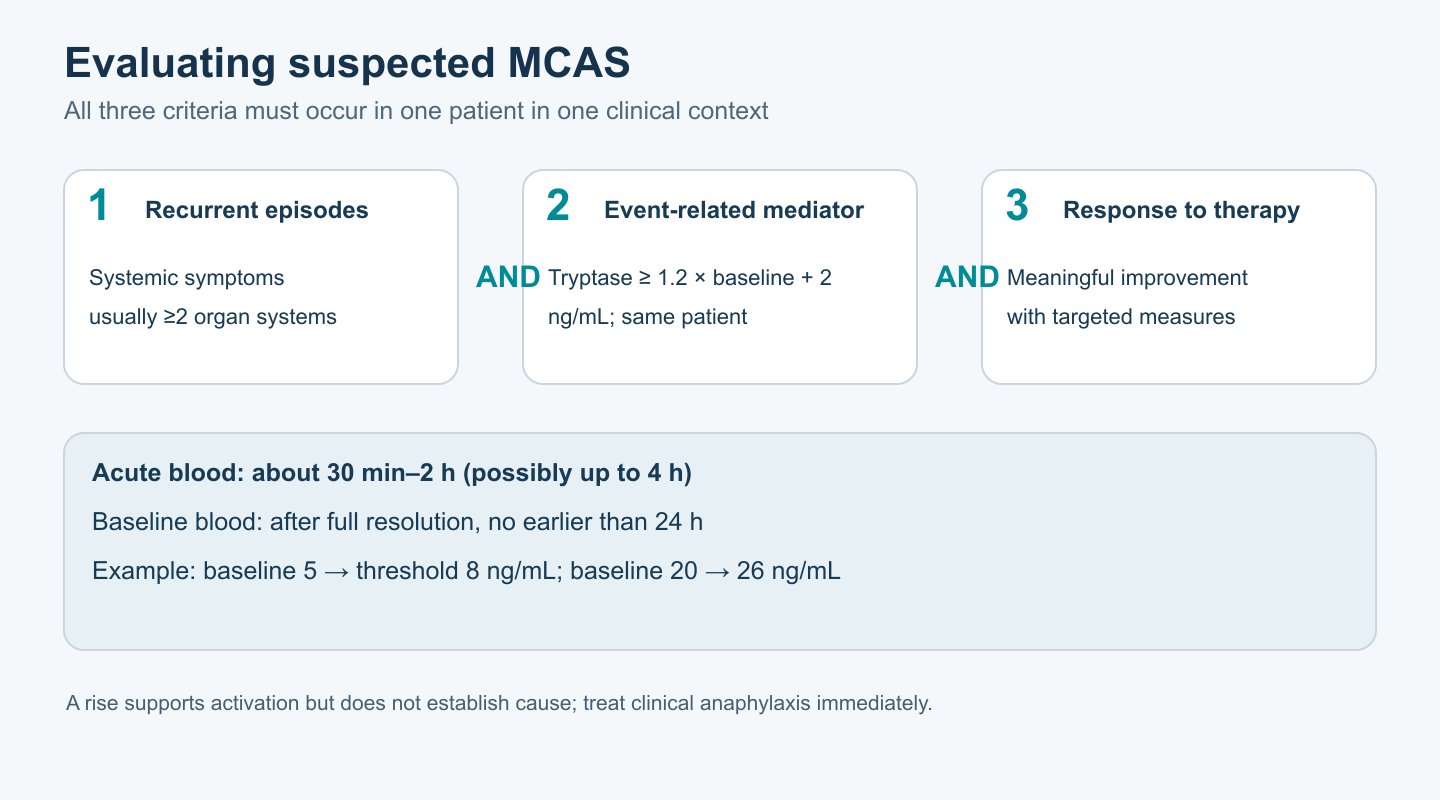
A widely used international consensus requires three elements at the same time. First, typical recurrent episodes of systemic mediator-related symptoms, usually involving at least two organ systems and often resembling anaphylaxis clinically: skin/mucosa, respiratory, cardiovascular, and gastrointestinal systems. Second, a substantial event-related mediator increase, most validly measured by total serum tryptase using an individual formula. Third, a meaningful response to measures that block mediators or mast-cell activation. The third element supports the interpretation, but response to an H1 blocker is nonspecific and cannot replace the first two. A discretionary “50% improvement” threshold is not part of the generally accepted diagnostic criteria.
Broader proposals that include persistent, heterogeneous symptoms, multiple nonspecific markers, and increased mast-cell numbers in mucosa are discussed, but they are not equivalent to the validated consensus definition. Sensitivity and specificity comparisons between two “schools” reported as 95%, 50%, or 70% without a shared diagnostic reference standard and an independent population are misleading. An expanded scheme should not be used to diagnose MCAS automatically after one episode of flushing, POTS, IBS, or finding local mast cells. Conversely, a late or incorrectly collected specimen after a convincing episode calls for properly timed repeat assessment, not categorical rejection of possible anaphylaxis.
| Condition | What must be established | Common error |
|---|---|---|
| Systemic mastocytosis | Clonal morphologic/molecular criteria and organ distribution | Equating baseline tryptase with a diagnosis |
| Primary clonal MCAS | Systemic episodes plus evidence of mast-cell clonality | Treating any activating mutation as obligatory |
| Secondary MCAS | The same activation criteria and an identified trigger, often allergic | Calling allergy and MCAS mutually exclusive |
| Idiopathic MCAS | Confirmed systemic activation without an identified clonal or secondary cause | Diagnosing it from absence of a KIT variant without biochemical evidence |
| Hereditary α-tryptasemia | Extra copies of α-tryptase–coding TPSAB1 and elevated baseline tryptase | Interpreting a high baseline as an acute peak |
Hereditary α-tryptasemia (HαT) is associated with an increased copy number of the α-tryptase–coding sequence in TPSAB1. Family and population studies have linked it to elevated total baseline tryptase and variable symptoms, but copy number does not determine the same linear severity in every carrier. HαT may coexist with mastocytosis or allergy and may modify reaction risk in some cases; it should not be turned into an MCAS diagnosis or used to attribute POTS/hEDS to all carriers. The event-related rise formula remains individual and is applied relative to that person’s own, possibly elevated, baseline.
Differential diagnosis depends on the nature of the episode. IgE-mediated anaphylaxis after a specific food, medication, or sting; non-IgE drug reactions; chronic urticaria; asthma; vasovagal syncope; orthostatic hypotension; neuroendocrine and catecholamine-secreting tumors; gastrointestinal disease; infections; and medication effects can cause similar complaints. When the clinical picture warrants it, tests may include 5-HIAA for a serotonin-secreting neuroendocrine tumor or metanephrines when pheochromocytoma is suspected; these tests are not needed for everyone with itching. Systemic anaphylaxis is an acute clinical syndrome and can also be a manifestation of MCAS when recurrent episodes are confirmed; treating them as mutually exclusive by definition is incorrect.
2. Mediators by time of formation and their receptors
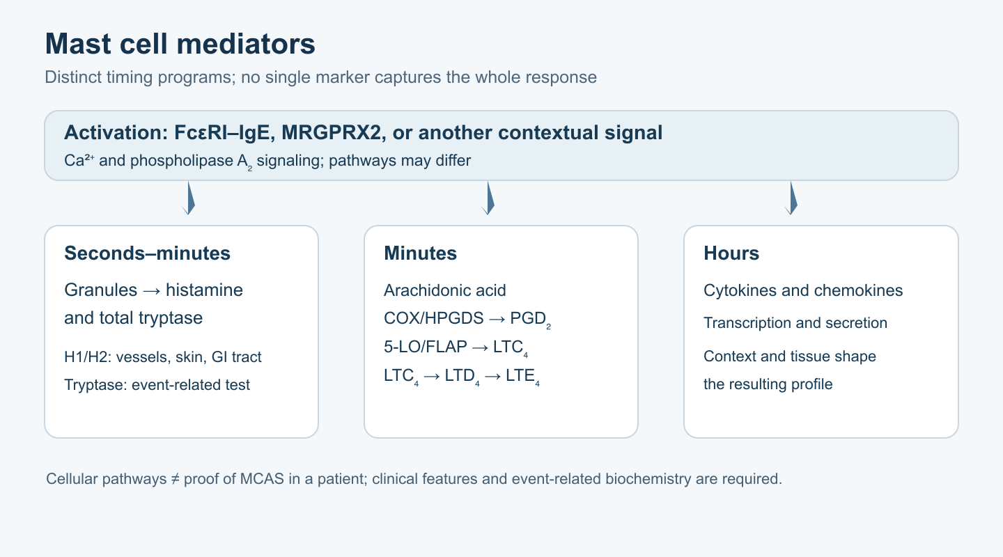
Mast-cell granules already contain histamine, tryptase, and other proteases in a heparin-containing matrix. Allergen binding to FcεRI-associated IgE initiates tyrosine-kinase and calcium signaling, granule redistribution, and exocytosis. Within minutes, phospholipase A₂ releases arachidonic acid from membranes, after which cyclooxygenase and 5-lipoxygenase pathways generate prostaglandin D₂ and leukotrienes. Hours later, cells may change cytokine and chemokine transcription. The extent of granule degranulation, lipid response, and cytokine program need not run in parallel; this is why one laboratory marker cannot describe the whole state.
Tryptase
Mature β-tryptase is a serine protease that can form a heparin-stabilized tetramer with active sites inside the complex. Serum assays generally measure total tryptase, including constitutively released protryptases and active forms, rather than a direct “fraction of opened granules.” Baseline concentration depends on mast-cell number, TPSAB1 genetics, and other conditions; a single value within the reference interval does not exclude a substantial relative rise, and an elevated value outside an episode does not establish MCAS. Tryptase interaction with PAR2 and tissue remodeling is known mainly from experimental systems; one should not attribute every systemic symptom to direct PAR2 cleavage or transfer a specific in vitro kinetic result to a clinical blood test.
During an acute episode, total serum tryptase usually rises after symptom onset; the most informative draw is approximately 30 minutes to 2 hours after onset. A sample taken later, up to four hours, may still be informative, but sensitivity decreases. Baseline is measured before an episode or at least 24 hours after complete resolution. The consensus formula is: acute tryptase ≥ 1.2 × personal baseline tryptase + 2 ng/mL. With a baseline of 5 ng/mL, the threshold is 8 ng/mL, even if both numbers are within the laboratory’s standard range. With a baseline of 20 ng/mL, the threshold is 26 ng/mL; simply being “above 20” is insufficient. A rise supports systemic mast-cell activation during the event but does not establish its cause: it also occurs in severe allergic anaphylaxis. A negative result, especially after a late draw or a mild episode, does not exclude all possible pathology and must not delay emergency care.
Histamine and receptors
Histidine decarboxylase forms histamine from L-histidine with pyridoxal phosphate; histamine is stored in granules with polysaccharides. The H1 receptor usually signals through Gq/PLC/IP₃/Ca²⁺ and affects itching, vascular permeability, bronchial responses, and sensory responses; H2 signals through Gs/cAMP and participates in gastric acid secretion and vascular effects. H3 is predominantly neuronal and presynaptic, while H4 is found on immune cells. The presence of a receptor does not justify declaring every episode of “brain fog” or tachycardia a direct consequence of a particular receptor subtype in MCAS.
In tissues, histamine-N-methyltransferase (HNMT) uses S-adenosylmethionine to form N-methylhistamine; further oxidation produces other metabolites. Diamine oxidase (DAO/AOC1) acts mainly in extracellular compartments and intestinal mucosa on dietary histamine and other histamine accessible to it. These pathways differ in location and are not interchangeable. Plasma histamine is short-lived and sensitive to preanalytical handling; urinary N-methylhistamine may support evaluation of an episode, but its sources and renal clearance require cautious interpretation. Low DAO activity or a subjective food reaction does not confirm MCAS.
PGD₂ and cysteinyl leukotrienes
Cyclooxygenases COX-1/COX-2 form PGH₂ from arachidonic acid, followed by hematopoietic PGD synthase forming PGD₂. It acts through DP1 and DP2/CRTH2, expressed on different cells and potentially affecting vascular responses and inflammatory migration. Urinary PGD₂ metabolites, including 11β-PGF₂α and 2,3-dinor-11β-PGF₂α, are measured as adjunct markers in some laboratories, but other cells also produce prostaglandins and exact reference limits depend on the method. An isolated elevated result does not establish a mast-cell cause.
In another pathway, 5-lipoxygenase and FLAP form LTA₄; LTC₄ synthase conjugates it with reduced glutathione (GSH) to form LTC₄. After it leaves the cell, γ-glutamyltransferases remove glutamate (LTC₄ → LTD₄), and dipeptidases remove glycine (LTD₄ → LTE₄). LTD₄ and LTC₄ act through CysLT1/CysLT2 and contribute to bronchoconstriction and vascular permeability. Urinary LTE₄ may support assessment of a lipid response, but the leukotriene pathway is activated by cells other than mast cells and is affected by asthma and medications. Therefore, an isolated LTE₄ increase is not equivalent to an MCAS diagnosis. Claims of an exact thousand-fold “potency” of leukotrienes relative to histamine cannot be transferred across organs and experimental conditions.
Heparin, chymase, CPA3, PAF, and cytokines deserve mechanistic attention. Chymase participates in local processing of peptides and matrix; PAF can increase vascular leakage; cytokines may appear later. But listing mediators does not make them routinely validated MCAS tests. Granule-derived heparin is not established by a subjective tendency to bruise without a standard hematologic evaluation; protease concentrations differ among tissue mast-cell phenotypes.
3. How mast cells are activated
The classic FcεRI signal after antigen binds cell-bound IgE should be distinguished from direct drug reactions and cytokine responses to microbial patterns. MRGPRX2 is a receptor described on human skin mast cells that responds experimentally to several cationic drugs and endogenous peptides. It helps explain some allergy-like reactions without IgE, but it does not confirm that every flare after food or stress is caused by MRGPRX2. Concentration, tissue type, and ligand structure matter; an MRGPRX2 antagonist is not yet a universally validated MCAS therapy.
The sequence of FcεRI events matters: an allergen cross-links two or more surface IgE–FcεRI complexes; Src kinases and SYK phosphorylate adaptors; PLCγ generates IP₃ and diacylglycerol; calcium enters from intracellular stores and across the plasma membrane. This drives granule–membrane fusion and also changes cytosolic PLA₂ activity and transcriptional pathways. Specific IgE without exposure to the corresponding allergen does not prove that it caused a particular episode. Conversely, some drugs can trigger similar symptoms through MRGPRX2 without prior IgE sensitization; a negative standard allergy test by itself does not make such a reaction MCAS.
TLR2/TLR4/TLR3 can alter cytokine synthesis in mast-cell models without necessarily causing equally strong immediate degranulation. Complement C3a and C5a interact with their respective receptors in ways that depend on species and cellular phenotype. Mechanical stimuli, temperature, and neuropeptides are also discussed, but much of the evidence on PIEZO1, specific TRP channels, CRH, and neurotensin comes from in vitro or animal work. It does not yield a clinical test for “thermal MCAS” or a causal explanation for every episode during emotional stress. Record an identified provoking factor in the episode history; assess the mechanism separately.
The diversity of pathways explains why an H1 medication may reduce itching without resolving bronchospasm or a vascular reaction: different mediators act at the same time. It also explains why many symptoms need not be mast-cell mediated. Dietary, autonomic, neuroendocrine, and anxiety-related conditions may cause similar sensations without biochemical evidence of systemic degranulation. Define the phenotype from the clinical episode and correctly collected tests, not from the number of possible biochemical pathways on a diagram.
4. Clinical manifestations and disputed combinations
Urticaria, flushing, or angioedema together with wheezing, hypotension, syncope, vomiting, or cramping diarrhea during one episode is much more informative clinically than years of isolated fatigue or migraine. Skin findings may be absent in severe anaphylaxis, so assessing breathing and circulation matters more than searching for a mandatory rash. Gastrointestinal symptoms predominate in some patients, but IBS, inflammatory bowel disease, infections, and motility disorders require their own evaluation. A mediator episode is not diagnosed by a biopsy showing a slightly increased mast-cell density: cell distribution varies by site, inflammation, and counting method.
Reconstructing the timing of symptoms is important. Recurrent, brief episodes with a similar combination of organs and a possible exposure trigger provide a different diagnostic signal from constant abdominal pain, daily fatigue, or fluctuating stool without a clear systemic episode. Skin may look normal between episodes, so photographs and measurements during an event can help, but a photograph of redness does not identify a specific mediator. Bronchospasm requires assessment for asthma and obstruction; hypotension requires evaluation of blood loss, medication effects, and other causes; diarrhea requires its own workup for organic and functional disease. This parallel approach helps prevent delayed treatment of another disorder and false confidence in one laboratory marker.
Orthostatic tachycardia, hypermobility, and symptoms labeled POTS and hEDS may occur in people who also suspect MCAS. However, observational series often defined the syndromes inconsistently, and strict diagnostic criteria were not always met; the “triad” is not a single established disease entity. Evaluate orthostatic pulse by its own criteria, hypermobility by the relevant assessment, and systemic activation episodes by their own algorithm. Neither POTS nor hEDS is laboratory evidence of MCAS by itself.
Neurologic complaints, cognitive difficulties, migraine, and anxiety matter to patients, but the idea that “tryptase-mediated microglial activation” caused them in a specific person is usually untested. Kounis syndrome describes acute coronary syndrome associated with an allergic/anaphylactic event; it is not an automatic typical outcome of MCAS. Chest pain calls for urgent assessment of cardiac disease, not an explanation limited to histamine.
5. Investigating an episode: timing, comparison, and pitfalls
For a convincing episode, document onset time, affected organs, blood pressure/pulse, breathing, skin findings, medications, and a likely trigger. Obtain total serum tryptase as early as possible in the informative window, then measure a baseline after recovery. Analyze samples with a comparable method and interpret them using the individual formula. There is no basis for requiring universal shock-freezing of every tryptase tube on ice within 30 minutes: laboratory handling is determined by the assay. Requirements differ for histamine, prostaglandin metabolites, and leukotriene metabolites; before urine collection, obtain the receiving laboratory’s instructions for timing, storage, preservatives, and medication interference. Replacing lab-specific instructions with one rule for all mediators can worsen the result.
Event-related urinary N-methylhistamine, a PGD₂ metabolite, and LTE₄ can provide additional material, especially when blood tryptase was not collected on time. But methods, reference ranges, and effects of age, kidney function, asthma, medications, and mediator source differ. A fixed threshold from one laboratory cannot be transferred to another. An isolated spontaneous result without comparison to the clinical picture and, where possible, a baseline is weak evidence. Serum histamine, heparin, chromogranin A, and numerous cytokines do not replace accepted markers. Tryptase above the laboratory reference range outside symptoms prompts assessment for HαT, mastocytosis, and other causes, but is not by itself an acute rise.
Urine testing measures excretion over the collection interval, not mediator concentration in tissue at the symptom peak. A spot result may vary with urine volume; some methods use a 24-hour collection, while others normalize to creatinine. Renal impairment and age affect interpretation, and antiprostaglandin drugs can change a PGD₂ metabolite. If episodes recur, choose the available test and the specific laboratory instructions before an episode and record medications and collection time. An imperfect adjunct marker does not become a required positive test in every case and does not replace the best-standardized comparison of acute and baseline tryptase.
When there are clues to clonal disease—persistently elevated baseline tryptase, characteristic skin changes, recurrent severe anaphylaxis, or other signs—a specialist may consider a high-sensitivity KIT D816V assay and bone-marrow evaluation in the appropriate clinical setting. A negative peripheral-blood result does not exclude a small tissue clone and does not mean every patient with diarrhea needs bone-marrow testing. Extra TPSAB1 copies may be assessed for unexplained elevated baseline tryptase, taking the copy-number assay into account. A gastric or intestinal biopsy for another indication may reveal clonal features of mastocytosis; the criterion “more than 20 mast cells per field” is not an independently validated MCAS diagnosis.
6. Treatment of a confirmed phenotype and safety
The first step is to identify and, where possible, reduce established triggers and treat the underlying disease. H1 antihistamines relieve itching, wheals, and flushing; an H2 blocker may help some gastrointestinal symptoms. CysLT1 antagonists, oral cromolyn, or ketotifen are sometimes used in individualized regimens, but evidence for strictly defined MCAS is limited and does not support prescribing one “four-step” protocol to everyone. Oral cromolyn acts mainly locally, but its clinical benefit cannot be reduced to universal blockade of a single chloride channel. Ketotifen can cause sedation; dose changes require attention to concomitant medications and tolerability.
| Approach | Biological target and symptom it may affect | What limits conclusions in MCAS |
|---|---|---|
| Second-generation H1 blockers | H1 receptor; itching, urticaria, flushing | Symptom response is nonspecific and alone does not confirm the diagnosis |
| H2 blockers | H2 receptor; some gastric symptoms | Dyspepsia and pain have many causes; improvement does not prove a mast-cell mechanism |
| CysLT1 antagonists | Cysteinyl-leukotriene receptor; selected respiratory and other symptoms | Effects and safety depend on drug and indication; response does not replace event testing |
| Cromolyn and ketotifen | Modulation of reactivity and symptoms; mechanism is multifactorial | Studies in other diseases and case series are not robust trials of strict MCAS |
| Anti-IgE and targeted therapy | IgE–FcεRI or mutant KIT in the relevant disease | Choice depends on an established allergic/clonal phenotype and specialist assessment |
It is more useful for the patient and clinician to choose a measurable outcome in advance than to add treatments successively for presumed “mast-cell stabilization.” Frequency of systemic episodes, need for epinephrine, objective blood pressure and breathing during an episode, skin findings, functional limitations, and adverse reactions provide more information than a change in subjective “histamine level.” If symptoms change after several foods are removed and several medicines are started at once, the contribution of each treatment cannot be distinguished. Changing one variable at a time and documenting episodes may help, but an acute dangerous reaction requires immediate care and is not an occasion for a home experiment.
Omalizumab reduces available IgE and FcεRI expression. Case series describe benefit in some recurrent reactions and mastocytosis, but this cannot be transferred as a guaranteed effect to all nonclonal variants. KIT inhibitors are used for indications involving mastocytosis and molecular characterization, not automatically for idiopathic complaints: KIT D816V may be resistant to imatinib, and targeted therapy has substantial toxicity. Zileuton requires attention to interactions and liver monitoring when used for its own indication. Dose descriptions from studies of other diseases do not establish an MCAS regimen without specialist consultation.
For clinical anaphylaxis, intramuscular epinephrine in the anterolateral thigh is first-line treatment; H1/H2 blockers, cromolyn, montelukast, and supplements must not delay its use or medical care. An autoinjector and action plan are particularly important after a severe reaction or when recurrence risk is high; available strengths and the need for a second dose are selected according to age, weight, device, and current protocol. Not every person with suspected MCAS automatically needs two devices under one formula: the decision is tied to anaphylaxis risk and access to care. Epinephrine acts on vascular tone and bronchi; it does not simply “switch off” every mast cell instantly.
7. Diet, DAO, and supplements: do not transfer cell experiments to patients
A food diary can sometimes help link a reproducible food to a symptom, but a broad lifelong ban on fermented foods, tomatoes, citrus, tea, and many other groups based on an MCAS diagnosis alone is unsupported. Biogenic amine content varies with freshness and storage; food allergy requires a different evaluation, and chronic restriction can reduce dietary variety and quality. A brief, targeted assessment of a suspected trigger followed by reintroduction of tolerated foods is more reasonable than a universal list of “histamine liberators,” which is often based on weak evidence. Assessing a separate histamine intolerance or exogenous DAO requires its own criteria; it does not replace event-related tryptase when MCAS is suspected.
DAO/AOC1 is a copper-containing amine oxidase; its activity is related to protein structure and cofactors. HNMT uses SAM as a methyl donor, and histamine formation by HDC requires PLP. These biochemical facts do not prove that a person without deficiency needs high-dose copper, B6, vitamin C, or SAMe to stop mediator episodes. Oral DAO acts mainly in the digestive lumen and does not eliminate systemic mast-cell degranulation. Quercetin, luteolin, and rutin have shown effects in some cell and animal systems, but concentrations and formulations do not translate directly into an evidence-based clinical dose for MCAS. High-dose supplements have their own risks and interactions; they are not a safe “first line.”
A bacterial strain’s ability to produce histamine depends on its genes and environment and cannot be reliably predicted by species name alone. Therefore, lists of probiotic species that are “forbidden” or “always safe” in MCAS are not sound. Clinical benefit and tolerability of a specific strain in a relevant condition require separate study. For more on limits of use, see the article on probiotics. If symptoms are related to fermentation or IBS, evaluate them by their own criteria rather than automatically assigning them a mast-cell mechanism.
8. What remains unresolved
Studies should recruit people using the same strict criteria, document acute and baseline mediators, and specify a clinical endpoint in advance. Outcomes should include anaphylaxis frequency, episode severity, quality of life, adverse effects, and phenotype—not only a reduction in one surrogate substance. Separate work should address reasons for a negative event-related tryptase despite convincing clinical features, reproducibility of urinary markers, effects of HαT and clonal variants on treatment, and the contribution of MRGPRX2 to reactions to specific medicines. The association of MCAS with POTS/hEDS needs cohorts in which each diagnosis is independently verified.
Method and limits of this review
This is a narrative review of mechanistic, diagnostic, and clinical publications, not a systematic review or GRADE assessment. On 9 October 2026, primary MCAS and mastocytosis consensuses, the event-related tryptase formula study, TPSAB1 data, emergency-anaphylaxis guidance, and critical reviews of the disputed “triad” were checked directly. Quantitative clinical conclusions are retained only with their population and method; direct randomized trials of most medicines and nutraceuticals in strictly defined MCAS remain insufficient.
FAQ
Can MCAS be diagnosed from one high tryptase test?
No. Assessment requires recurrent systemic episodes, event-related comparison with the person’s own baseline, and evaluation of response to targeted measures. A persistently elevated baseline has other causes.
What does the “20% + 2” formula mean?
Acute total serum tryptase should be at least 1.2 × baseline + 2 ng/mL. For example, with a baseline of 5, the threshold is 8 ng/mL.
When should testing be done after an episode?
Collect acute tryptase preferably 30 minutes to 2 hours after onset; a later sample up to four hours can sometimes help. Measure baseline after full recovery, usually no earlier than 24 hours.
Does MCAS always mean mastocytosis?
No. Mastocytosis is a clonal tissue disease with its own criteria. Systemic activation may also be secondary or idiopathic.
Do POTS and hypermobility prove MCAS?
No. These conditions have their own criteria; evidence for one obligatory “triad” is insufficient.
Can diet or quercetin replace an autoinjector for anaphylaxis?
No. First-line care for anaphylaxis is intramuscular epinephrine and emergency medical care. Diet measures and supplements do not replace it.
How should a negative tryptase be interpreted after a convincing episode?
First assess collection timing, episode severity, and the person’s baseline. A negative late test does not cancel clinical treatment of anaphylaxis and may call for a repeat-testing plan.
Sources
- Valent P et al. Definitions, criteria and global classification of mast cell disorders with special reference to mast cell activation syndromes. Int Arch Allergy Immunol. 2012;157:215–225.
- Valent P et al. Proposed diagnostic algorithm for patients with suspected mast cell activation syndrome. J Allergy Clin Immunol Pract. 2019;7:1125–1133.
- Akin C. Mast cell activation syndromes. J Allergy Clin Immunol. 2017;140:349–355.
- Valent P et al. Updated diagnostic criteria and classification of mast cell disorders: a consensus proposal. Hemasphere. 2021;5:e646.
- Valent P et al. The 20% + 2 tryptase formula is a diagnostic gold standard for severe systemic mast cell activation. Int Arch Allergy Immunol. 2019;180:44–51.
- Lyons JJ et al. Elevated basal serum tryptase identifies a multisystem disorder associated with increased TPSAB1 copy number. Nat Genet. 2016;48:1564–1569.
- McNeil BD et al. Identification of a mast-cell-specific receptor crucial for pseudo-allergic drug reactions. Nature. 2015;519:237–241.
- Shtessel M et al. MRGPRX2 activation causes localized and systemic pseudo-allergic responses in humans. J Allergy Clin Immunol. 2021;147:702–710.
- Metcalfe DD et al. Biomarkers of the involvement of mast cells in human disease. Physiol Rev. 2016;96:1479–1574.
- Hirai H et al. Prostaglandin D2 selectively induces chemotaxis in Th2 cells, eosinophils and basophils via CRTH2. J Exp Med. 2001;193:255–261.
- Beller TC et al. Targeted gene disruption reveals the role of the cysteinyl leukotriene 2 receptor in vascular permeability. PNAS. 2004;101:3292–3297.
- Maintz L, Novak N. Histamine and histamine intolerance. Am J Clin Nutr. 2007;85:1185–1196.
- Kohn A, Chang C. The relationship between hEDS, POTS and MCAS. Clin Rev Allergy Immunol. 2020;58:273–297.
- Weiler CR et al. AAAAI Mast Cell Disorders Committee Work Group Report: MCAS diagnosis and management. J Allergy Clin Immunol. 2019;144:883–896.
- Prevalence of mast cell activation disorders and hereditary alpha tryptasemia among patients with POTS and EDS: a systematic review. Ann Allergy Asthma Immunol. 2025;135:97–102.
- Shibao C et al. Hyperadrenergic postural tachycardia syndrome in mast cell activation disorders. Hypertension. 2005;45:385–390.
- Weng Z et al. Quercetin is more effective than cromolyn in blocking human mast cell cytokine release and inhibits contact dermatitis and photosensitivity in humans. PLoS One. 2012;7:e33805.
- World Allergy Organization. Guidelines for the assessment and management of anaphylaxis. World Allergy Organ J. 2011;4:13–37.
- Arock M et al. KIT mutation analysis in mast cell neoplasms: ECNM recommendations. Leukemia. 2015;29:1223–1232.
- Jennings SV et al. Patient-reported outcomes in mast cell activation disorders: a survey of 823 patients. J Allergy Clin Immunol Pract. 2014;2:705–714.
- Weinstock LB et al. Mast cell activation syndrome: a primer for the gastroenterologist. Dig Dis Sci. 2021;66:965–982.
- Theoharides TC, Valent P, Akin C. Mast cells, mastocytosis and related disorders. N Engl J Med. 2015;373:163–172.