
Fecal calprotectin: from neutrophil to decisions in IBD and IBS
How the S100A8/S100A9 complex reaches stool from neutrophils, what the assay measures, and why thresholds differ for suspected IBD, remission monitoring, and postoperative follow-up.
How the S100A8/S100A9 complex reaches stool from neutrophils, what the assay measures, and why thresholds differ for suspected IBD, remission monitoring, and postoperative follow-up.
Fecal calprotectin (FC) is a quantitative measure of S100A8/S100A9 proteins in stool. It helps address two different questions: whether intestinal inflammation is likely in someone with symptoms resembling irritable bowel syndrome (IBS), and whether inflammatory activity persists in someone with established inflammatory bowel disease (IBD). A single result does not establish a diagnosis, the source of inflammation, the degree of histological healing, or whether medication should be changed. The meaning of the number depends on age, symptoms, pretest probability, the laboratory system, and the clinical setting.
This is a narrative review of molecular, analytical, and clinical studies in the source bibliography. On 9 October 2026, official NICE, British Society of Gastroenterology, and American Gastroenterological Association guidance was checked specifically where the source text had turned different thresholds into a universal rule. No exhaustive study search, formal risk-of-bias assessment, GRADE process, or external peer review was performed. Laboratory and clinical estimates below retain their original populations and methods and should not be mechanically transferred to another patient.
Calprotectin is formed by S100A8 (MRP8, calgranulin A) and S100A9 (MRP14, calgranulin B). Each S100-family subunit carries two EF-hand motifs for Ca²⁺ binding: a noncanonical N-terminal motif and a canonical C-terminal one. The oppositely oriented subunit helices form a heterodimer. In the presence of extracellular calcium, two heterodimers can assemble into a heterotetramer. This is a structural transition, not a separate protein molecule encoded by another gene. A simplified reaction notation is:
This shorthand deliberately does not specify calcium-binding stoichiometry. The affinity of individual EF-hands, the oligomeric equilibrium, and metal occupancy depend on experimental conditions; fixed values in μM should not be treated as constants for both cellular cytosol and the intestinal lumen. Crystallography established the heterotetramer architecture, but structure alone does not measure stool protein or IBD severity.
Two functionally distinct transition-metal coordination sites form at the S100A8/S100A9 interface. One includes three histidines and an aspartate; the other is formed by six histidines across the subunits and the C-terminal region of S100A9. Biochemical experiments show calprotectin binding Zn²⁺ and Mn²⁺; its ability to sequester Fe²⁺ has been described under particular conditions. Metal binding is an equilibrium competition with other ligands, not “irreversible chelation” of all cations. Extracellular calcium affects the accessibility and affinity of these sites. In experimental infection models, restricting available metals can inhibit microbial growth—this is nutritional immunity. But the concentration of Zn²⁺ or Mn²⁺ next to bacteria in an inflamed intestine cannot be inferred from one FC value.
Calprotectin is abundant in neutrophil cytosol and is also present in monocytes. When the mucosa is injured, epithelial and immune signals, including CXCL8, direct neutrophils from blood vessels through the lamina propria and epithelium toward crypts and the lumen. The complex can be released during cell activation, death, degranulation, and formation of neutrophil extracellular traps. FC measures protein that remains detectable in a stool aliquot, not the absolute number of migrated neutrophils. Between those quantities lie the area and location of disease, intestinal transit, mixing of contents, degradation, and the analytical procedure itself.
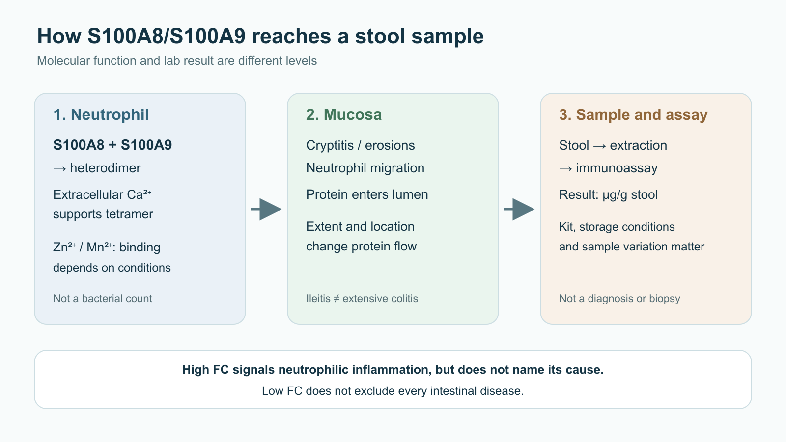
S100A8/S100A9 can act as a DAMP, a damage signal for innate immunity. Experimental activation of TLR4 by the MRP8/MRP14 complex has been shown; interactions with RAGE and downstream cellular responses have been studied in different models. A typical TLR4 axis is MyD88–IRAK–TRAF6–TAK1–IKK, with NF-κB released after IκB degradation; the outcome can be transcription of inflammatory mediators. Describing this cellular pathway does not mean that every elevated FC proves direct TLR4 or RAGE activation in a particular patient. Identifying the ligand, receptor, and causal contribution in human mucosa requires a separate experiment.
S100A8/S100A9 is also associated with phagocyte function and oxidative responses, but superoxide is produced by the assembled NADPH oxidase complex, not by the calprotectin molecule. The correct net electron-transfer reaction for NOX2 is:
This pathway and subsequent oxidant formation belong to activated-neutrophil biology. The measured concentration of stool protein is not a direct measurement of NOX2, hypochlorite, tight-junction damage, or the gut–vascular barrier. Detailed chains such as “FC → a specific VE-cadherin residue → bacteria in the portal vein” are not established by a clinical FC assay and are not used here as proven explanations. Likewise, individual S100A8/S100A9 variants are not a validated genetic test for interpreting FC or predicting pancolitis severity.
In ulcerative colitis, neutrophils enter the crypt epithelium and crypt lumen; cryptitis, crypt abscesses, erosions, and ulcers usually provide a route for protein to enter intestinal contents. In extensive active colitis, FC tends to track endoscopic activity more closely than in a small, limited lesion. Crohn disease can be segmental and transmural. When inflammation is mainly in the small bowel, especially a short segment of ileum, FC may be lower than in comparable colitis. A low result therefore does not exclude isolated small-bowel disease and extensive colitis equally. No universal sensitivity percentage “for ileitis” describes all locations and methods.
Endoscopic indices answer different questions. Mayo and UCEIS assess the mucosal appearance in ulcerative colitis; SES-CD assesses segments, ulcers, and narrowing in Crohn disease. Among patients with similar endoscopic scores, FC can differ because of disease extent, location, and variation in the sample itself. A table claiming “Mayo 0 is always <150, Mayo 3 is always >800 μg/g” would be a false conversion scale. Studies show a statistical association, not a one-to-one mapping.
Histology answers another question: whether neutrophils remain in a biopsy. The Nancy index and Robarts Histopathology Index account for active inflammation at the microscopic level. Low FC increases the likelihood of mucosal healing in some ulcerative-colitis cohorts, but thresholds and accuracy vary by study and by the chosen definition of remission. A patient with low FC may still have residual histological inflammation; biopsy remains the way to establish it when that affects a decision.
Not all chronic diarrhea carries a strong neutrophil signal. In microscopic colitis, endoscopic mucosa often looks normal, and diagnosis is made from biopsies of the right and left colon. Normal or moderately elevated FC does not rule out lymphocytic or collagenous colitis. FC is also heterogeneous in celiac disease, allergic enteropathies, and eosinophilic enteropathies, and does not replace their serological, histological, or clinical criteria.
Neutrophilic inflammation also occurs with intestinal infection, NSAID use, diverticulitis, some neoplastic and other organic lesions. After NSAID exposure, an increase may reflect enteropathy; a clinician decides whether to stop the drug or repeat the test based on the indication for treatment, not a fixed “4–6 weeks” rule for everyone. Proton-pump inhibitors are discussed as a possible contributing factor to modest elevations, but their effect should not be equated with a proven inflammatory diagnosis. Acute infectious gastroenteritis can produce a very high result, and a test repeated after the illness resolves may no longer reflect current activity.
With Clostridioides difficile, FC may accompany mucosal inflammation, but it is not a validated arbiter between asymptomatic carriage and infection. Testing and treatment decisions require clinical symptoms and an accepted toxin/toxigenic-strain algorithm. The statement “>500 plus toxins = 95% specificity” should not be transferred to practice without checking the particular population and diagnostic standard. In patients with an ileal pouch–anal anastomosis, FC can also accompany pouchitis, but symptoms, examination, and pouchoscopy are needed. Thresholds for a normal colon cannot automatically be applied to pouch contents, much less used to prescribe antibiotics or biologic therapy from one number.
Colorectal neoplasia is another reason not to use FC as a “protective” test. It is not a cancer-screening test and a low value does not rule out a tumor. Blood in stool, iron-deficiency anemia, unexplained weight loss, family history, and other alarm features require a separate diagnostic pathway regardless of FC.
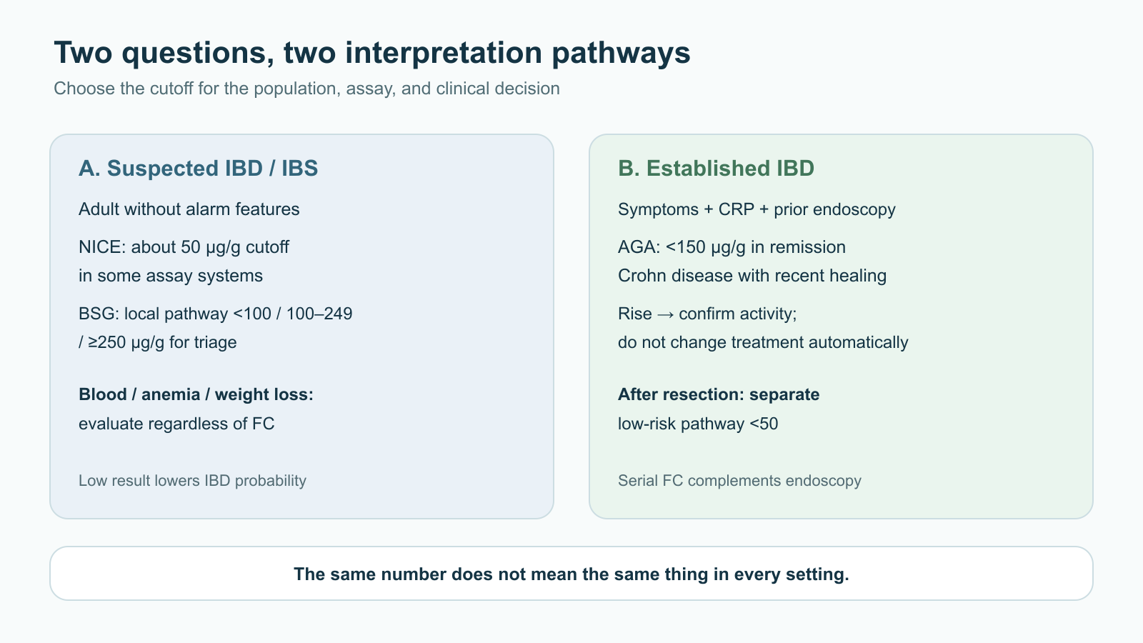
For an adult with chronic pain and altered bowel habits, when clinical assessment has found no alarm features, FC can help select people for further investigation. Sensitivity, specificity, and predictive value depend on the patient spectrum. The prevalence of IBD is substantially higher in a referred endoscopy cohort than in primary care, so negative predictive value will differ even if test characteristics remain the same. It is not accurate to say that FC below 50 μg/g “guarantees no organic disease” or that its NPV is always 98–99%.
Published pathways use different cutoffs. In NICE diagnostic-test assessments, a threshold around 50 μg/g often matched the manufacturer’s instructions and served as a starting point for comparing assay systems. British guidance for an adult with probable IBS and no alarm features describes a local pathway with a normal zone below 100, repeat testing at 100–249, and referral at ≥250 μg/g. These are not conflicting “biological norms”: developers balanced sensitivity, repeat testing, and colonoscopy differently for particular care settings. The laboratory and patient should use one validated local pathway; a <50 / 50–250 / >250 split should not be presented as a mandatory international rule.
Even a precise analytical cutoff does not define constant clinical accuracy. Sensitivity is the fraction of people with the target disease who test positive; specificity is the fraction without it who test negative. Positive and negative predictive values also depend on the proportion of IBD among those tested. Formally, if is sensitivity, is specificity, and is pretest probability, then:
This explains why a high FC produces more false suspicion of IBD in primary care than the same result in a referred high-risk group, and why a low FC is less reassuring when alarm features are pronounced. For a conditional calculation—not the performance of a particular assay—if , , and , then among 1,000 patients there would be about 45 true positives, 5 false negatives, 190 false positives, and 760 true negatives. The modeled arises from the low prevalence of IBD, not from an ability of the test to “rule out all organic disease.”
| Context and pathway source | What the example value means | The next step depends on |
|---|---|---|
| Adult with suspected IBD versus IBS; laboratory assay with a 50 μg/g cutoff | A low result reduces the probability of neutrophilic inflammation; it does not exclude cancer, microscopic colitis, or every form of Crohn disease | Symptoms, alarm features, pretest probability, and assay instructions |
| Adult in a British local pathway for probable IBS | <100 μg/g is a low zone; 100–249 calls for repeat testing and review of transient factors; ≥250 prompts further IBD assessment | The adopted regional pathway, laboratory, and symptom duration |
| Child assessed by a specialist for suspected IBD | FC may be a sensitive triage tool, but an adult scale cannot be transferred to an infant | Age, growth, anemia, bleeding, family history, and pediatric assay method |
| Established IBD, activity assessment | A threshold near 150 μg/g appears in specific AGA recommendations with a defined history of endoscopic monitoring | Current symptoms, CRP, recent endoscopy, treatment, and IBD type |
Repeating a result in the gray zone may help when symptoms are stable, alarm features are absent, and transient causes are plausible—for example, recent gastroenteritis, NSAIDs, or differences between samples. Record the medication, infection date, assay method, and result before repeating. No fixed “exactly 4–6 weeks” interval suits everyone: timing depends on the local pathway and clinical urgency. Persistent elevation or worsening symptoms calls for in-person assessment and selection of endoscopy, small-bowel imaging, or other tests. Bleeding, anemia, weight loss, or marked deterioration should not wait for a repeat FC test.
NICE assessed pediatric secondary-care evidence separately. Across the studies it considered, at 50 μg/g sensitivity of about 99% and specificity of 74% were reported for IBD versus other causes; at 100 μg/g, about 94% and 82%, respectively. These are properties of those studies, age ranges, and analyzers. Raising the cutoff reduces some false positives at the risk of missing some cases; it does not make 100 μg/g a universal “pediatric normal.” These results cannot be directly compared with adult primary-care studies, where IBD prevalence and referral criteria differ.
FC and CRP complement each other: one is measured in stool, while the other reflects a systemic acute-phase response. Normal CRP does not exclude localized mucosal inflammation, and elevated CRP is not specific to the intestine. Fecal lactoferrin is another neutrophil marker and should not be arbitrarily combined with FC into an “activity index.” Eosinophil-derived neurotoxin reflects a different type of inflammatory response and is not an accepted substitute for FC in the basic IBD-versus-IBS pathway.
After diagnosis, the question changes. In Crohn disease or ulcerative colitis, a patient may report less pain and diarrhea while endoscopic inflammation persists; the reverse can also occur, with symptoms continuing despite controlled inflammation. STRIDE-II treats biomarkers as intermediate targets on the way to longer-term mucosal healing. It describes FC around 100–250 μg/g as a target range that depends on disease and assay, not a universal diagnostic cutoff or proof of histological remission. FC is one coordinate alongside symptoms, CRP, and objective assessment of the bowel.
AGA guidance for established Crohn disease specifies a particular scenario: in a patient in symptomatic remission on stable therapy with recently confirmed endoscopic remission, FC <150 μg/g together with normal CRP can help avoid routine endoscopy. Without recent confirmation, low FC alone does not replace endoscopy. If biomarkers are elevated during asymptomatic remission, confirm activity endoscopically rather than automatically intensifying medication. In patients with mild symptoms, neither a normal nor an elevated value always removes the need for examination. AGA guidance for ulcerative colitis also links about 150 μg/g to defined activity-assessment scenarios, but decisions depend on symptom severity and combination with CRP; the number does not imply an identical probability of lesions in colitis and Crohn disease.
CALM tested tight control in Crohn disease: decisions to escalate adalimumab used a composite of CDAI, CRP, FC, and prednisone use, compared with clinical management. At week 48, endoscopic improvement was more frequent with the strategy (45.9% versus 30.3% for the specified criterion CDEIS <4 without deep ulcers). This is an outcome of a decision program, not proof that lowering FC or one home sample alone improves outcomes. An early biomarker shift during induction may support response assessment, but neither “normalization required by week 8” nor a universal rate of FC decline is validated across all medicines.
Serial FC measurements may precede relapse in established disease. Cohort studies found that rising values can be informative, but the relationship depends on diagnosis, treatment, assay, relapse definition, and follow-up time. A single high result during stool variability, infection, or NSAID use needs contextual review. Serial comparison is more informative when the same analytical method and a similar collection approach are used. Decisions to switch a biologic should also consider disease severity, objective activity, drug and antibody concentrations when therapeutic drug monitoring is indicated, and clinical safety. FC does not directly measure drug clearance.
After ileocecal resection for Crohn disease, early endoscopic recurrence using the modified Rutgeerts index becomes clinically important. FC may help select patients for endoscopy but does not replace it in every risk group. The AGA low threshold of <50 μg/g during the first 12 months applies to a defined postoperative scenario involving lower-risk patients or prophylaxis, not to every adult with suspected IBD. Pouchitis monitoring after IPAA also requires separate evidence and a locally validated method.
FC is usually reported as micrograms of protein per gram of stool, μg/g. The equivalent notation is mg/kg because . This is a mass concentration in a heterogeneous biological matrix, not a serum concentration or a fraction of neutrophils. The laboratory takes a defined mass of stool, extracts protein into buffer at a specified mass-to-volume ratio, and calibrates the signal against a standard. In simplified form:
where accounts for dilutions and calibration for the particular assay; this expression does not replace the manufacturer’s instructions. Incomplete homogenization, liquid stool, and uneven sampling from a heterogeneous portion add variation. Two samples cannot be treated as fully independent of collection method.
Methods include laboratory enzyme immunoassays, automated immunoturbidimetric and chemiluminescent systems, and quantitative or semiquantitative point-of-care tests. Antibodies recognize different epitopes, extraction devices differ, and cross-manufacturer standardization is incomplete. Comparisons among assays have found inter-method discrepancies. Thus, “180 μg/g” on one instrument is not necessarily equivalent to “180 μg/g” on another; for monitoring over time, it is useful to keep the laboratory, method, and units consistent. When the method changes, record a new baseline and do not call a small difference a definite biological response without analytical context.
Calprotectin is relatively stable in stool, which makes testing practical, but “stable for 7 days at room temperature” is not universal. The acceptable interval, temperature, container, extraction buffer, and whether freezing is allowed are determined by the validated instructions of that laboratory. Collect stool in a clean, dry container and avoid toilet water; collection date and storage conditions should be known. Visible blood, menstrual contamination, acute infection, or current NSAID use should be discussed in context; do not apply an arbitrary correction factor. Within-day and within-stool differences are especially apparent in active IBD. Repeat samples may be appropriate when the result conflicts with the clinical picture, but averaging should not delay urgent evaluation.
Home kits and smartphone readers may support monitoring if validated against a laboratory method and incorporated into an agreed clinical pathway. Lighting, camera, strip, read time, and extraction procedure affect quality. A value entered by a patient into an app does not establish endoscopic remission and is not an automatic instruction to increase a dose. The source text cited specific reductions in hospitalization and time to treatment change from telemonitoring; without a reliable corresponding trial, these should not be treated as expected effects of home FC testing.
| Source of uncertainty | What may change | How to keep interpretation grounded |
|---|---|---|
| Site and extent of disease | Extensive colitis often contributes more neutrophil protein to a sample than limited ileitis | Compare with symptoms, history, and examination of the relevant bowel segment |
| Liquid or heterogeneous stool | Concentration per unit mass and the sampled portion may differ | Follow laboratory instructions; consider repeat sampling if results conflict |
| Infection, NSAIDs, and other causes | FC may rise without IBD | Ask about transient factors and investigate according to the clinical picture |
| Different assays and calibrations | Absolute values may not be equivalent | Compare trends within one system and record the platform |
| Age and population | Baseline concentrations and IBD prevalence differ | Use an age- and context-specific pathway, not a universal adult cutoff |
| Low pretest probability | Positive predictive value changes even if sensitivity is unchanged | Interpret against pretest probability and alarm features |
In the first months of life, FC concentrations in healthy infants are higher and more variable than in adults; observational studies have also found effects of age and feeding conditions. Therefore, adult 50 μg/g is not a universal infant reference value. In an older child, possible IBD is assessed in light of growth, weight, delayed puberty, blood in stool, anemia, inflammatory markers, and family history. NICE considered pediatric evidence in secondary care separately from adult primary care: even high sensitivity in that sample does not authorize ruling out disease in an individual child without assessment.
For an older person with chronic watery diarrhea, medications and microscopic colitis matter; biopsies may be needed even when the mucosa looks normal. Suspected celiac disease requires appropriate serology and, in the relevant pathway, biopsy; FC neither confirms nor rules it out. Blood in stool and iron-deficiency anemia call for a separate evaluation of bleeding and neoplastic sources. If infection is suspected, use microbiological tests rather than diagnosing from one inflammatory protein.
When anemia is present, hemoglobin and ferritin require their own iron-status assessment; even those results do not turn FC into a cancer-screening test. In a person without signs of inflammation, discussion of fiber and prebiotics concerns symptom and nutrition management, not diagnostic interpretation of FC.
Nutritional interventions can affect disease and therefore FC trends, but they should not be selected by a “personal FC threshold” without a diagnosis. Exclusive enteral nutrition is an established induction option for pediatric Crohn disease in an appropriate clinical context; FC may be one marker of response. The source text assigned specific percentages of achieving FC <150 and a molecular pathway through CXCL8 that cannot be generalized across children and formulas. Exclusion diets for Crohn disease are studied in defined protocols, but a decrease in FC does not establish that one emulsifier or gluten caused IBD. For a person with probable IBS, an elevated FC first needs diagnostic interpretation, not self-directed food restriction.
A false-positive signal during diagnostic triage can cause worry, repeat sampling, and unnecessary colonoscopy. A false negative can delay IBD diagnosis if the result is treated as final despite progressive symptoms. More dangerous is substituting FC for investigation when cancer is suspected: the test was not designed for that negative-predictive task. These risks differ, so an algorithm does not choose a cutoff only by maximizing area under the ROC curve. It also weighs the cost of missed disease, endoscopy access, local IBD prevalence, and accompanying clinical signals.
In established IBD, a different kind of error is possible: automatic escalation after one high FC creates adverse-effect and treatment-cost risks without confirmed inflammation, while reassurance from one low number can miss limited ileitis or symptoms from another cause. If the biomarker and how someone feels do not agree, clarify which bowel segment needs assessment, whether healing was objectively demonstrated recently, and whether the laboratory method changed. CRP, a complete blood count, and iron tests help describe systemic response and consequences of disease but do not localize inflammation. Endoscopy with biopsy shows mucosa and histology; suspected small-bowel disease may require imaging of the relevant segment. Each test resolves a different part of the uncertainty.
FC can reduce costs when used to select patients with an appropriate pretest probability and when a defined pathway follows the result. An economic evaluation in the source bibliography modeled fewer unnecessary endoscopies under specific assumptions about costs and accuracy. This is not a universal claim that FC reduces spending in every healthcare system: test and repeat costs, false referrals, and colonoscopy access vary locally. For home monitoring, correlation between a strip and a laboratory assay is likewise insufficient; the decision pathway tied to the measurement must improve meaningful outcomes without causing overtreatment.
First state the question: initial differentiation between probable IBS and IBD, assessment of established IBD, postoperative monitoring, or investigation of a mismatch between symptoms and objective findings. Then record age, sample date and conditions, method and its cutoff, medications, recent infection, and alarm features. Only then compare the number with the appropriate pathway. A low FC may reduce the need for immediate colonoscopy in an adult without alarm features and with a low pretest probability of IBD. It does not replace investigation for blood, anemia, or suspected cancer; it does not rule out microscopic colitis or every small-bowel lesion. A high FC signals an objective inflammatory process or another cause of tissue injury and needs investigation before IBD treatment is started or intensified.
The value of FC lies in matching three things: the biological source of the protein, a validated measurement, and the right clinical question. Laboratory cutoffs, cellular alarmin effects, and observations in established IBD answer different questions. Keeping those distinctions clear allows the test to guide reasonable investigation and monitoring without turning a marker into a diagnosis.
It reflects the amount of the S100A8/S100A9 protein complex in a stool sample and serves as a marker of mucosal neutrophilic inflammation, but does not identify the cause by itself.
Low FC reduces the likelihood of marked neutrophilic inflammation, especially colitis, but does not rule out limited small-bowel disease or other conditions. The decision depends on symptoms and evaluation.
They are used in different laboratory systems, populations, and clinical goals: initial triage for suspected IBD, assessment of established IBD, or postoperative monitoring.
Yes. Infection, NSAIDs, and other causes of intestinal injury can also raise the value, so a high result is not the same as an IBD diagnosis.
No. Blood in stool, anemia, weight loss, and other alarm features require separate evaluation regardless of FC.
Sometimes, if the condition is stable and there are no alarm features. Timing and next steps depend on the local pathway, medicines, recent infection, and assay.
Usually transient causes are considered and disease activity is confirmed with symptoms, CRP, endoscopy, or other investigations first; the clinician makes the treatment decision.
No. Healthy infants have higher and more variable concentrations; results are interpreted by age and pediatric context.
Different antibodies, extraction devices, and calibrations can produce discrepancies. A method change can look like a change in disease.