
Dietary lectins: glycan binding, the intestinal barrier, and real-world exposure
How WGA and PHA bind glycans, what permeability models show, why undercooked beans can cause acute poisoning, and why clinical effects of cooked wheat remain uncertain.
A lectin recognizes a carbohydrate pattern on a cell surface, but binding alone does not mean that the mucosa is damaged. In an experiment with purified wheat germ agglutinin (WGA), concentration and contact time can be selected so that epithelial cells change. People eat grain after milling, mixing, heating, and digestion, together with starch, proteins, and other constituents. The dietary link is much clearer for bean phytohaemagglutinin (PHA): undercooked beans can cause acute poisoning, while adequate moist heat inactivates the hazardous activity. The distinction among molecule, prepared food, and clinical outcome guides this review.
1. What dietary lectins share—and what separates them
Lectins are proteins with carbohydrate-recognition sites. They bind certain glycans noncovalently and, when several sites are accessible, can cross-link cells or glycoconjugates. They do not “cut” sugar chains as glycosidases do; their action depends on oligosaccharide shape, epitope density, and protein geometry. Erythrocyte haemagglutination is a useful laboratory activity assay for some lectins, but it does not directly measure the toxicity of a serving: food matrix, erythrocyte type, and preparation all affect the result.
WGA is concentrated in the wheat germ. Its small, cysteine-rich hevein-like domains form a scaffold stabilized by disulfide bonds; several carbohydrate-binding sites on the protein create avidity when it encounters a dense cellular glycocalyx. Structural experiments show WGA binding N-acetylglucosamine (GlcNAc), chitobiose/chitotriose, and some sialylated glycans. Affinity of one site for a soluble sugar must be distinguished from the strength of multivalent adhesion to a surface: a Kd measured in one system is not a dose at which bread is harmful. Structural work by Wright, Harata, and Schwefel refines the locations of binding sites, dimer state, and glycan accessibility; in intestinal contents, however, those same sites compete with mucins and other molecules.
PHA from beans of the genus Phaseolus is not a form of WGA. It is a family of bean haemagglutinins with its own glycan specificity and marked toxicity in raw or undercooked seeds. The literature distinguishes isoforms that preferentially bind erythrocytes or leukocytes; the result depends on cultivar, processing, and assay. SBA, soy agglutinin, has been studied chiefly in raw soy and inadequately processed feed models; findings on growth and intestinal morphology in piglets and rodents do not automatically translate into risk from ordinary cooked soy foods. ConA, concanavalin A from Canavalia, is widely used as a laboratory mitogen and probe for α-mannosyl/α-glucosyl motifs, but it is not a typical molecule in the daily diet. Grouping all four under “lectins” without specifying species, food, and activity erases the most important differences.
| Lectin and typical matrix | Carbohydrate target in the laboratory | Reliable conclusion about dietary exposure |
|---|---|---|
| WGA, wheat germ | GlcNAc-containing and some sialylated glycans | Can bind mucosal structures in experiments; active WGA dose in a prepared meal and its clinical effect require separate measurements |
| PHA, raw beans | Glycans on intestinal cells and erythrocytes | Acute risk from inadequate cooking is documented in people |
| SBA, soy | Specific glycans in agglutination tests | Effects of raw material in animal models do not describe all cooked soy foods |
| ConA, Canavalia | Mannosyl/glucosyl motifs | Useful experimental mitogen; not a universal dietary risk |
1.1. Specificity is spatial chemistry, not a list of “harmful sugars”
The energy of a single noncovalent lectin–monosaccharide contact reflects hydrogen bonds, van der Waals interactions, and the position of water in the binding pocket. Replacing one residue in an oligosaccharide, changing its orientation, or adding sialic acid can alter affinity. On a cell, the spacing between epitopes and the possibility of simultaneous binding at several sites matter: multivalent avidity can sometimes retain a protein even when the affinity of each individual site is relatively weak. Conversely, an abundance of a suitable sugar in solution can compete for one site without necessarily disrupting an already formed multivalent complex. Thus, “WGA likes GlcNAc” begins a description of its substrate; it does not adequately predict an intestinal outcome.
The label “antinutrient” also needs context. In a model feed containing a high proportion of raw seed, active lectin may affect nutrient absorption and animal growth; in people, boiling, fermentation, seed maturity, and serving size change the initial amount and activity. Comparing “milligrams of WGA per kilogram of grain” with “micrograms of WGA per millilitre of culture” is impossible without accounting for extraction, heat losses, intestinal contents, and glycan competition. Haemagglutination units from different laboratories are not always comparable because activity depends on erythrocytes and incubation conditions. For a dietary conclusion, protein quantification should be combined with a functional binding assay after cooking and a digestion model.
1.2. Different raw-material forms pose different causal questions
Wheat germ and bran are not the same as purified flour, and adding germ to a smoothie is not the same as eating baked bread. Even if an analytical method detects a WGA fragment, whether its carbohydrate-binding sites remain functional is an open question. Beans are different: raw or insufficiently heated seeds can retain substantial PHA activity, and risk is primarily associated with a specific cooking failure. Soy foods also differ: raw flour, heat-treated tofu, and fermented products do not belong in one exposure category. Finally, ConA often appears in diagrams as a convenient research tool, but its experimental doses cannot be added to a typical human diet.
2. How a lectin encounters the intestinal wall
2.1. Glycocalyx, mucins, and multivalent adhesion
The enterocyte brush border is coated by a glycocalyx: transmembrane glycoproteins, glycolipids, and membrane-associated mucins display different sugar chains. The secreted MUC2 mucin layer between microbes and epithelium is especially important in the colon; the composition and thickness of the mucus layer differ in the small intestine. Therefore, the statement “WGA bound GlcNAc” should prompt the question: where—on free mucin, an apical protein, a bacterial surface, or the cell culture used? Binding to one substrate can reduce access to the others.
Multivalency allows WGA to cross-link neighbouring glycoproteins and alter their mobility; in an experiment this may accompany changes in membrane microdomains, endocytosis, or cytoskeletal responses. Yet glycans on healthy epithelium are not an “unprotected target”: mucus, dietary sugars, proteolysis, and rapid epithelial turnover alter actual contact. Physiological relevance requires the concentration of active lectin at the surface after a meal, not just protein content in dry raw material.
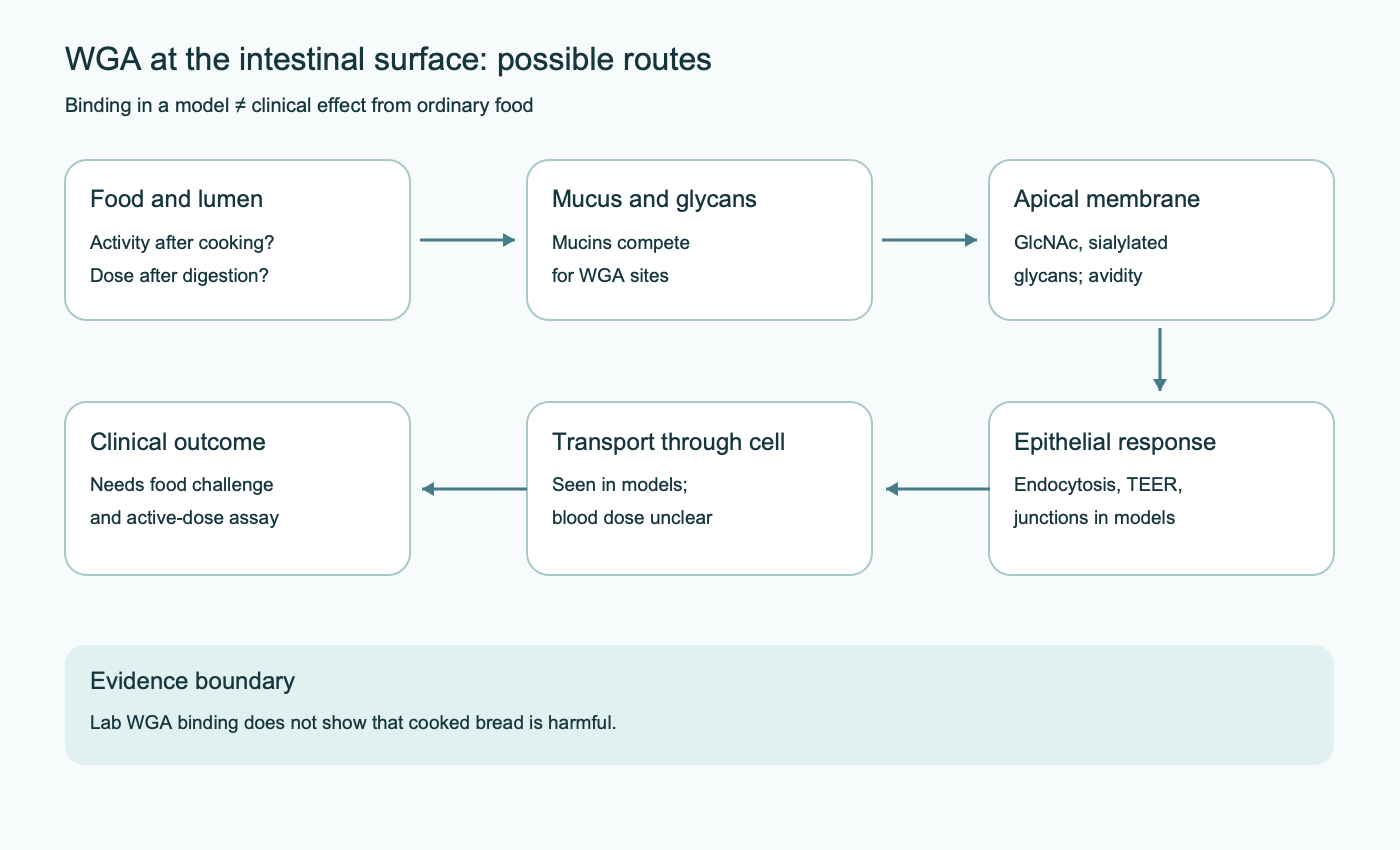
Microbial and dietary glycans add another interpretive boundary. If labelled WGA is located in mucus, that does not necessarily mean it bound an enterocyte. If mucus was washed away before an experiment, or a cell monolayer without a normal MUC2 layer was used, access to apical glycans is easier than in tissue. Inflammation can change mucin composition and epithelial glycosylation; this matters to a susceptibility hypothesis but does not allow inflammation in a person to be retrospectively attributed to lectin exposure.
2.2. Microvilli, tight junctions, and permeability measurements
Microvilli increase absorptive surface area; actin bundles connect to the membrane through proteins such as ezrin and villin. High-dose experiments with purified lectins describe shortened microvilli, changes in brush-border enzymes, and passage of large molecules. Studies by Sjölander in rat mucosa and Dalla Pellegrina in model human epithelium/immune cells support the possibility of a local response. But a cell line, isolated tissue, or administration of a purified substance bypasses part of food processing and mucosal protection; the time and concentration in such an experiment cannot silently be called an ordinary exposure after porridge or bread.
Tight junctions are not a single “lock” but a dynamic system of claudins, occludin, ZO proteins, and the actomyosin ring. The MLCK → myosin light-chain phosphorylation → perijunctional ring contraction pathway can increase paracellular permeability in response to inflammatory signalling. A direct or indirect role for this pathway after high lectin doses in a model must be demonstrated separately from an observed fall in transepithelial electrical resistance (TEER). TEER depends on ion transport and the number of viable cells; fluorescent-dextran flux, junction morphology, and viability help distinguish “opening” of a tight junction from cell death. In people, an increased urinary lactulose-to-mannitol excretion ratio indicates barrier status under a specified protocol, but does not identify lectin as the cause.
At least three fundamentally different processes can lower TEER in a model: remodelling of the paracellular route without cell loss, suppression of electrogenic ion transport, and cell injury or detachment. Tracers of different sizes, localization of ZO-1 and claudins, cell-death staining, and recovery over time help separate them. The paracellular “pore” route, regulated by particular claudins, is not equivalent to the larger “leak” route. One permeability value therefore cannot show that lectins “opened all tight junctions.” Even a confirmed change in an epithelial culture must still be compared with the active dose after a meal and with symptoms.
2.3. Endocytosis and transcytosis do not equal systemic toxicity
Binding to apical glycans may promote uptake of labelled WGA by enterocytes or M cells. The molecule may remain in an endosome, be degraded, return to the surface, or pass through the cell. Transport observed in culture or in a rat establishes that a route is possible under those conditions, not the amount of active WGA in human plasma after an ordinary meal. An older human study detected WGA in intestinal contents; this indicates persistence of recognizable protein in the lumen, not “90–95% survival,” systemic absorption, or entry into the brain. Drug-delivery technologies use WGA as a ligand to attach a carrier to mucosa; a purpose-built conjugate cannot be equated with grain that has been eaten.
| Link in the chain | What is measured | What the result alone does not establish |
|---|---|---|
| Binding | Crystal structure, microcalorimetry, sugar competition | Tissue damage |
| Activity in food | Haemagglutination/glycan assay after processing | Concentration at the epithelium after digestion |
| Epithelial response | TEER, tracer flux, microscopy, cytokines | A human symptom or disease |
| Systemic exposure | Specific detection of intact protein in blood with sensitivity controls | Action at a receptor in brain, joint, or adipose tissue |
| Clinical causality | Controlled food challenge with a specified food and dose | Universal harm from all lectins |
2.4. Dose, time, and model species define the limit of an inference
Experimental models can be arranged as a sequence of questions. Protein structure shows which glycans it can recognize. Epithelial culture asks what can happen with direct contact with purified protein. Isolated intestine or an animal model adds mucus, blood flow, and immune cells, but often uses an unusual route of administration or a diet high in raw lectin. A human food study adds preparation, digestion, behaviour, and symptoms, but has more difficulty identifying one molecule among dozens of food constituents. Nor does the absence of a human effect in a small or poorly measured study prove absolute safety for every product; the exposure and outcome must be specified precisely.
Concentration requires a denominator and a compartment. “10 μg/mL in culture” describes the medium above cells, not an equivalent concentration of active WGA in fluid above the human brush border. Dietary protein is diluted by contents, adsorbed to the matrix, and exposed to mucins and proteases; local contact may also be uneven. One cannot mechanically multiply a culture concentration by intestinal volume and declare a safe serving, or assume that every microgram detected in the lumen is biologically active. Bridging the models requires the same functional assay of residual activity.
3. Immune and endocrine signals: where the evidence lies
PHA and ConA are widely used to stimulate lymphocytes in vitro. This lets researchers cross-link glycosylated surface proteins at a specified concentration; it does not show that an ordinary serving of cooked beans causes polyclonal T-cell activation or a systemic “cytokine storm.” WGA can alter inflammatory signals in epithelial and immune cell cultures, but experiments need controls for preparation purity, endotoxin contamination, cell viability, and the concentration that could actually occur at mucosa. Mast-cell or basophil degranulation after direct laboratory exposure does not establish a clinical “WGA pseudo-allergy” syndrome from eating bread. Wheat allergy is assessed from clinical history and validated allergy methods.
Studies by Cuatrecasas, Livingston, and Shechter showed that purified lectins can interact with the glycosylated insulin receptor or alter glucose use in isolated cells. This is useful for understanding glycan-dependent receptor cross-linking and the difference between an agonist and a physiological hormone. A claim that “dietary WGA causes insulin resistance,” however, would require evidence that intact protein reaches the blood, its concentration near adipocytes, duration of action, and a controlled metabolic outcome. Listing the IR–IRS–PI3K–Akt–GLUT4 pathway does not fill this gap. Similar claims that lectins block the leptin receptor, stimulate EGFR, or cause hyperphagia have not been confirmed in clinical food trials and should not be presented as established disease.
Labelled WGA has long been used as a neuroanatomical tracer by injecting it into animal tissue or blood; carrier development also uses its ability to bind endothelial cells. The route of administration is crucial: intravenous or local injection bypasses the stomach, cooking, and intestinal barrier. Movement of an injected tracer along a neuron cannot show that whole-grain food delivers WGA across the blood–brain barrier or causes “brain fog,” multiple sclerosis, or neuroinflammation. For autoimmune diseases, thyroiditis, and arthritis, the original manuscript constructs plausible glycan analogies but does not provide a causal human demonstration of dietary exposure.
Microbiota and bile-acid effects also depend on the system. A lectin may agglutinate bacteria with suitable surface sugars, but the relative abundance of Akkermansia, Bifidobacterium, or E. coli is shaped by diet, mucus, and competition. The hypothesis that dietary WGA blocks the ASBT transporter and causes bile-acid diarrhoea is not established merely because ASBT is glycosylated. It would require direct binding, measurement of bile-acid transport at an attainable dose, and clinical replication. Microbial and dietary effects on mucosa are discussed separately in the article on dietary fibre and prebiotics.
3.1. Where the boundary lies between receptor biochemistry and disease
Binding of WGA to glycoproteins can affect cell signalling in experimental systems. A glycoprotein receptor can bind lectin in a test tube because accessible sugars are present; the insulin receptor need not respond as it does to insulin. Signal direction depends on conformation, internalization, and cell composition. Early work with adipocytes and purified proteins studied receptor physiology, not the consequences of eating bread. Delivery route is even more important for leptin, EGFR, and other pathways: distribution after injecting labelled WGA directly into a vessel, brain, or culture says nothing about the fraction that survives cooking and gastrointestinal digestion.
Claims of “systemic inflammation” need the same distinction. Cytokine secretion by a cell after direct protein exposure demonstrates that a signal is possible. Changes in autoantibodies, clinical exacerbation of autoimmune disease, and long-term symptom improvement on a diet are different outcomes. Between them lie exposure, immune tolerance, genetics, microbiota, and the effect of eliminating entire food groups. None of those transitions is established simply because a lectin is multivalent.
An inflammatory marker or an antibody may change without a clinical disease. Conversely, symptoms can occur for reasons unrelated to a lectin, including allergy, celiac disease, fermentable carbohydrates, infection, or other components of a meal. A mechanistic model is useful when it identifies a testable causal chain; it should not be presented as a diagnosis or a treatment indication.
4. Clinical boundary: beans, wheat, and diagnosis
4.1. PHA in raw or undercooked beans is an established acute hazard
Foodborne outbreaks after red kidney beans have been linked to nausea, vomiting, abdominal pain, and diarrhoea beginning rapidly when beans were raw or inadequately heated. Symptoms typically start within 1–3 hours, and a small number of raw or undercooked beans may be enough. This is not chronic “hidden poisoning” from all legumes; it is a recognizable foodborne illness with a specific condition: heat treatment failed to reach a sufficient temperature throughout the batch. Reports by Noah and Rodhouse describe human cases; EFSA’s 2026 risk assessment found enough evidence to characterize dietary risk quantitatively only for PHA among plant lectins. Its conclusion is that undercooked beans are hazardous, whereas adequate soaking and boiling deactivate lectins and remove this risk during ordinary consumption.
Mechanistically, PHA binds mucosal glycans and at high doses may disrupt enterocyte function. Epidemiological poisoning reports do not establish a precise chain of “PHA → complete loss of SGLT1/NHE3 → paralysis of peristalsis,” or obligatory endoscopic atrophy in every patient. Food-poisoning assessment considers dehydration and other causes of gastroenteritis; severe or prolonged vomiting, marked diarrhoea, or signs of dehydration need medical attention. Here, cooking prevention is more concrete than any nutraceutical “lectin binder.”
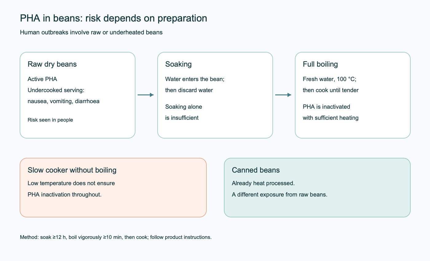
The meal context and clinical picture can guide action without a speculative “lectin-load index.” If several people develop rapid vomiting and diarrhoea after a shared meal containing undercooked beans, a likely foodborne cause should be assessed as acute poisoning. Symptoms are nonspecific, however, and microbial pathogens and other toxins remain possible causes. Official preparation advice concerns dry beans; it does not require people without a reaction to avoid all legumes. Beans with adequately inactivated PHA remain a source of protein and dietary fibre.
The causal interpretation of an outbreak still requires attention to timing, the batch and cooking method, and other possible causes. Symptoms are nonspecific: microbial pathogens and other toxins remain in the differential. Official preparation recommendations concern dry beans; they do not require all people without a reaction to avoid every legume. Beans in which PHA has been adequately inactivated remain sources of protein and dietary fibre.
4.2. WGA, celiac disease, allergy, and wheat sensitivity
Celiac disease is an immune-mediated condition triggered by gluten in a susceptible person; diagnosis relies on serology while gluten is being consumed and, when indicated, biopsy. Wheat allergy is a different reaction assessed clinically and by an allergy specialist, and may cause rapid symptoms. Non-celiac wheat/gluten sensitivity (NCWS/NCGS) describes symptoms after wheat once celiac disease and allergy have been excluded; potential wheat components include gluten, fructans/FODMAPs, amylase/trypsin inhibitors, and other substances. Carroccio’s double-blind food-challenge work and the Salerno criteria emphasize reproducible, controlled symptom assessment, but do not establish WGA as the sole or leading cause of the syndrome.
Confusing dietary exposure with experiments on purified WGA is especially problematic when interpreting wheat: grain, bran, germ, flour, sourdough, and bread have different protein concentrations and heat histories. The original manuscript gave exact amounts of “active WGA” in bread and near-complete retention after baking without comparable analysis of specific products; these values cannot be used to calculate personal risk. A valid comparison needs activity after processing, serving size, bioavailability, and control of other wheat components. Symptoms associated with food choices may depend on fibre or FODMAPs rather than WGA. General information about dietary glycaemic and fibre composition does not replace celiac evaluation.
| Condition | Important diagnostic route | What is not sufficient evidence |
|---|---|---|
| Celiac disease | Serology and clinical/histological assessment while gluten intake continues | “WGA antibodies” or a self-directed gluten-free trial before evaluation |
| Wheat allergy | Match symptoms and timing with specific IgE/skin tests and, when indicated, a controlled challenge | A single positive or negative laboratory test without clinical context |
| NCWS/NCGS | Exclude celiac disease/allergy, then use a reproducible food challenge that assesses placebo and other components | Automatically attributing symptoms to WGA |
| PHA poisoning | Recent raw/undercooked beans and acute onset of gastroenteritis | A diagnosis of chronic “lectin intolerance” |
When assessing recurrent symptoms after wheat, timing, reproducibility, meal composition, and symptoms outside the gut matter. Rash, wheezing, or a rapid systemic reaction calls for an allergy pathway. Persistent diarrhoea, anaemia, weight loss, or a family history of celiac disease calls for evaluation before gluten is removed: after withdrawal, serology may become falsely reassuring. For NCWS, food challenge should, where possible, separate expectation effects from other wheat components. Comparing wholegrain with refined products changes fibre, fructans, and many phytochemicals at once; it is not a clean WGA experiment.
4.3. Why antibodies and “barrier panels” do not establish WGA disease
Immune exposure to a food antigen can produce antibodies without disease. IgG or IgA to WGA does not by itself establish intestinal damage, lectin entry into a joint, food allergy, or a need to exclude grains. Lactulose/mannitol tests describe overall permeability under a specified protocol, not its molecular cause; the sugars reflect different transport routes, dose, urine-collection timing, and intestinal surface area. Commercial “zonulin” assays deserve particular caution because some kits have specificity problems. LBP, sCD14, calprotectin, I-FABP, and citrulline reflect different inflammatory or tissue processes; none is a validated “WGA intoxication” marker. Numerical thresholds for these tests from the original text were not carried over as a diagnostic algorithm for lectin injury.
FUT2 secretor status affects some mucosal glycans and microbial interactions, making it scientifically interesting. But a FUT2 variant does not establish individual sensitivity to WGA or PHA and does not justify genetic testing before ordinary cooked food. Demonstrating effect modification would require comparison of people with different genotypes under standardized servings and blinded challenge, measuring active dose and clinical outcome.
5. Preparation determines the active dose
Protein lectins can lose structure and carbohydrate-recognition ability during moist heating. Soaking helps beans cook evenly and removes some soluble constituents; by itself it does not guarantee PHA inactivation. A practical measure for dry beans is to soak them first, discard the water, then boil in fresh water at a full boil: Hong Kong’s Centre for Food Safety gives, as an example, soaking for at least 12 hours and vigorous boiling for at least 10 minutes, followed by cooking until tender. In a low-temperature slow cooker, the centre of the beans may never reach boiling; studies have found PHA activity even after an hour at 85 °C. Canned beans have already undergone industrial heat processing. Cooking at altitude, large batches, and uneven heating mean that actual temperature matters, not just the timer setting.
A pressure cooker reaches high temperatures and can be effective, but the original text’s single rule of “35–45 minutes at 121–125 °C for any red or black bean” is not a universal requirement for every cooker and batch size. A separate table assigning residual-lectin percentages to quinoa, buckwheat, tomatoes, eggplant, and peppers is also unsupported by comparable measurements and could unjustifiably extend caution from raw beans to ordinary vegetables. Dietary risk should be evaluated for the specific lectin and preparation method. Properly cooked legumes do not require “sugar traps” or avoidance of entire vegetable groups.
During grain fermentation, microbes and enzymes can break down some proteins and change dough structure. Studies of gluten peptides do not automatically confirm 90–99% WGA inactivation in every sourdough loaf. Soaking and germination change seed enzymes and soluble compounds but do not provide a universal constant for residual lectin activity. Activity can be measured by haemagglutination, a specific glycan-binding assay, and, when needed, mass spectrometry, while recording the product, temperature, and serving. Protein may remain detectable after processing while losing binding function; conversely, a small amount of active protein may signal in a sensitive assay without proving clinical harm.
Experimental blockade of WGA by soluble GlcNAc or chitin oligosaccharides shows competitive binding in a defined system. It does not establish a “sugar therapy” for people: a safe delivery route, competitor concentration in the same luminal compartment, lack of unwanted effects, and clinical-outcome trials would be needed. Prescribing N-acetylglucosamine, glutamine, zinc carnosine, butyrate, or probiotics at doses proposed in the original text for “WGA rehabilitation” is not supported by a diagnosis and controlled testing of that regimen. For symptoms after wheat or legumes, clinical assessment and safe preparation matter more than an unproven supplement protocol.
5.1. Processing conditions and the analytical trap
Thermal denaturation depends on time at the temperature actually reached, moisture, product thickness, initial activity, and matrix. Dry heat and moist boiling cannot be treated as the same process at the same appliance setting. Water promotes protein unfolding; soaking lets water enter the seed, but without subsequent boiling it does not neutralize raw PHA. Reheating canned beans usually does not raise an active-PHA concern because industrial processing has already occurred. The appliance name cannot resolve the question: slow cookers, pressure cookers, and pots produce different temperature profiles that must be compared with instructions and actual heating.
Measuring only immunoreactive protein may detect denatured remnants. Haemagglutination, in contrast, shows functional ability to cross-link the selected erythrocytes, but assay sensitivity varies with protocol. Results should be confirmed with a glycan-binding test and a stated detection limit; when two products are compared, samples should undergo the same digestion protocol. Otherwise, a percentage “reduction in lectins” can imply false precision. Even an accurately measured reduction in bread activity does not show that the initial activity caused disease or that lowering it treats symptoms.
5.2. Nutritional balance and uncertainty
A restrictive “anti-lectin” diet removes many legumes and whole grains, although the established acute PHA hazard concerns undercooked beans, not ordinary consumption of properly cooked foods. Any symptom improvement after elimination could reflect changes in FODMAPs, serving size, fibre, or expectation. If a trial diet is medically indicated, it should be tied to a specific diagnosis, symptoms, and a plan for reintroduction while maintaining nutritional adequacy. There is no basis for promising “tight-junction recovery in 6–8 weeks” based on one marker or for prescribing supplements instead of investigating the cause of symptoms.
6. Synthesis for practice and research
Lectins have real biochemistry: WGA binds GlcNAc- and sialylated structures multivalently; active PHA in raw beans can cause acute illness. The strength of these conclusions differs. Structural, cellular, and animal data show possible ways WGA can contact the intestinal wall, but do not establish that ordinary exposure to prepared wheat disrupts the barrier, causes NCWS, or produces systemic autoimmune or neurological disease. For PHA, poisoning reports and food-risk assessment support a simple action: soak dry beans carefully and boil them thoroughly. Future WGA studies should measure active molecule after cooking and digestion, its local and systemic concentrations, and then test clinical outcomes in a controlled design. Until those links are established, the safety of a prepared food and the experimental effects of purified lectin should not be conflated.
Methodological note
This is a narrative review of the supplied material, separating lectin biochemistry, model exposure to purified protein, and clinical outcomes after food. The references come from primary publications cited in the original manuscript. On 9 October 2026, targeted checks addressed the discrepant PMID and title for the Dalla Pellegrina paper, the meaning of human detection of WGA in intestinal contents, EFSA’s 2026 dietary-risk assessment, and Hong Kong Centre for Food Safety guidance on bean preparation. This was not a systematic search of all 50 entries, a GRADE assessment, or formal peer review. The remaining publications were not independently audited line by line; available evidence does not establish disease from ordinary dietary WGA exposure.
FAQ
Are lectins destroyed by cooking?
Activity depends on the protein, moisture, temperature, and time. For PHA in dry beans, adequate soaking and boiling are critical; residual WGA activity in a particular wheat food cannot be inferred from the process name alone.
Is canned beans dangerous because of PHA?
Canned beans undergo industrial heat processing. The documented acute risk chiefly concerns raw or insufficiently heated dry beans.
Do WGA experiments prove that bread causes “leaky gut”?
No. A cell culture or animal model demonstrates a mechanism at a specified dose; this claim would require measurements of active WGA in prepared food and human clinical outcomes.
Can IgG or zonulin diagnose “lectin intolerance”?
WGA antibodies, commercial zonulin, or a permeability test alone do not identify the cause of symptoms. Evaluation depends on the clinical picture and should rule out celiac disease and allergy when indicated.
Should all legumes and grains be eliminated?
A blanket restriction does not follow from PHA evidence. Safe bean preparation and evaluation of specific symptoms are more precise than excluding all lectin-containing foods.
Will GlcNAc or supplements neutralize WGA?
Competitive sugar binding in an experiment does not prove that a supplement prevents symptoms or treats disease in people. No clinically tested WGA-neutralization regimen is established.
Sources
- Wright CS. Structural comparison of the two distinct sugar binding sites in wheat germ agglutinin isolectin II. *J Mol Biol*. 1984;178(1):91-104.
- Wright CS. 2.2 A resolution structure analysis of two refined N-acetylneuraminyl-lactose--wheat germ agglutinin isolectin complexes. *J Mol Biol*. 1990;215(4):635-651.
- Harata K, Nagahora H, Jigami Y. X-ray crystallographic study of wheat germ agglutinin isolectin A and its complex with chitobiose. *Acta Crystallogr D Biol Crystallogr*. 1995;51(Pt 6):1013-1019.
- Schwefel D, Maierhofer C, Beck JG, et al. Structural basis of multivalent carbohydrate recognition by wheat germ agglutinin. *J Am Chem Soc*. 2010;132(25):8704-8719.
- Pusztai A, Greer F, Grant G. Specific uptake of dietary lectins into the systemic circulation of rats. *Biochem Soc Trans*. 1989;17(3):527-528.
- Vasconcelos IM, Oliveira JT. Antinutritional properties of plant lectins. *Toxicon*. 2004;44(4):385-403.
- Dalla Pellegrina C, Perbellini O, Scupoli MT, et al. Effects of wheat germ agglutinin on human gastrointestinal epithelium: insights from an experimental model of immune/epithelial cell interaction. Toxicol Appl Pharmacol. 2009;237(2):146-153.
- Sjölander A, Magnusson KE, Latkovic S. The effect of concanavalin A and wheat germ agglutinin on the ultrastructure and permeability of rat intestinal mucosa. *Int Arch Allergy Appl Immunol*. 1984;75(3):230-236.
- Gabor F, Bogner E, Weissenboeck A, Wirth M. The lectin-cell interaction and its relevance to absorptive cell-mediated drug delivery. *Adv Drug Deliv Rev*. 2004;56(4):459-480.
- Rodewald R. Permeability of the neonatal rat intestinal epithelium to horseradish peroxidase and wheat germ agglutinin. *J Cell Biol*. 1980;85(1):18-32.
- Turner JR. Intestinal mucosal barrier function in health and disease. *Nat Rev Immunol*. 2009;9(11):799-809.
- Shen L, Weber CR, Raleigh DR, Turner JR. Tight junction pore and leak pathways: a dynamic duplex. *Annu Rev Physiol*. 2011;73:283-309.
- Cuatrecasas P, Tell GP. Insulin-like activity of concanavalin A and wheat germ agglutinin--direct interactions with insulin receptors. *Proc Natl Acad Sci U S A*. 1973;70(2):485-489.
- Livingston JN, Purvis BJ. Effects of wheat germ agglutinin on insulin binding and insulin action in isolated adipocytes. *Am J Physiol*. 1980;238(3):E267-E275.
- Shechter Y. Bound lectins that mimic insulin produce persistent stimulation of hexose transport. *Endocrinology*. 1983;113(5):1921-1926.
- Sapone A, Bai JC, Ciacci C, et al. Spectrum of gluten-related disorders: consensus on new nomenclature and classification. *BMC Med*. 2012;10:13.
- Catassi C, Elli L, Bonaz B, et al. Diagnosis of Non-Celiac Gluten Sensitivity (NCGS): The Salerno Experts' Criteria. *Nutrients*. 2015;7(6):4966-4977.
- Carroccio A, Mansueto P, Iacono G, et al. Non-celiac wheat sensitivity investigated by double-blind placebo-controlled challenge. *Am J Gastroenterol*. 2012;107(12):1898-1906.
- Junker Y, Zeissig S, Kim SJ, et al. Wheat amylase trypsin inhibitors drive intestinal inflammation via activation of toll-like receptor 4. *J Exp Med*. 2012;209(13):2395-2408.
- Noah ND, Bender AE, Reaidi GB, Gilbert RJ. Food poisoning from raw red kidney beans. *Br Med J*. 1980;281(6234):236-237.
- Rodhouse JC, Haugh CA, Roberts D, Gilbert RJ. Red kidney bean poisoning in the UK: an analysis of 50 suspected incidents between 1976 and 1989. *Epidemiol Infect*. 1990;105(3):485-491.
- Grant G, More LJ, McKenzie NH, Stewart JC, Pusztai A. A survey of the nutritional value for rats of commercially available soya products in Britain. *Br J Nutr*. 1988;60(2):369-379.
- Pusztai A, Ewen SW, Grant G, et al. The relationship between tissue growth and protein catabolism in the gut of rats fed Phaseolus vulgaris lectin. *Br J Nutr*. 1990;64(2):543-555.
- Bischoff SC, Barbara G, Buurman W, et al. Intestinal permeability in health and disease: an overview. *BMC Gastroenterol*. 2014;14:189.
- Sharon N, Lis H. History of lectins: from hemagglutinins to biological recognition molecules. *Glycobiology*. 2004;14(11):53R-62R.
- Gabor F, Stangl M, Wirth M. Lectin-mediated bioadhesion: binding characteristics of plant lectins to intestinal enterocytes. *Int J Pharm*. 1997;158(1):55-64.
- EFSA CONTAM Panel. Risks for human health related to the presence of plant lectins in food. EFSA Journal. 2026;24:e9850.
- Centre for Food Safety, Hong Kong. Food Safety Focus: Natural Toxins in Red Kidney Beans.
- Identification of the dietary lectin, wheat germ agglutinin, in human intestinal contents. Gastroenterology. 1978.