Ubiquinol or ubiquinone: Q cycle, bioavailability and trial evidence — NutriFit
Ubiquinol and ubiquinone: the CoQ10 redox cycle, absorption and clinical limits
An in-depth comparison of coenzyme Q10 forms: the Q cycle, reductase network, micelles and chylomicrons, plasma pharmacokinetics and clinical-outcome trials, without claiming universal superiority of either form.
The question “which form is better?” has several meanings. Ubiquinone and ubiquinol continuously interconvert in membranes; a preparation must first release its hydrophobic molecule from its matrix, pass through the intestine and enter lipoproteins, while a change in plasma abundance does not establish its concentration in heart muscle or the outcome of disease. Redox form and manufacturing technology are therefore inseparable in a pharmacokinetic experiment, but clinical effectiveness needs separate study. This comparison starts with chemistry and ends with the question each trial actually addressed.
1. The quinone molecule and its three states
1.1. From isolation to biological function
Crane and colleagues, working with heart mitochondria, identified a lipophilic carrier reduced by electrons from respiratory substrates; Morton established the name “ubiquinone,” and structural studies by Folkers’ group determined the quinone head and long isoprenoid chain. This discovery does not make one pharmaceutical redox form more physiological: the active system cycles through both forms and the semiquinone intermediate. The number of isoprene units differs between species; CoQ₁₀ predominates in humans, whereas CoQ₉ makes up a substantial proportion in mice and rats. Quantitative rodent tissue findings therefore require consideration of Q-pool composition before extrapolation to humans.
1.2. The quinone ring, decaprenyl tail and phase environment
Oxidized CoQ₁₀ has the formula C59H90O4; the two carbonyl groups of 1,4-benzoquinone accept electrons and protons, while methoxy groups and a methyl substituent modify the ring’s electronic and steric properties. The tail of ten isoprene units keeps the compound in the lipid phase, allowing its quinone head to interact with protein pockets in respiratory complexes and the membrane interface. Chain length, configuration and local lipid packing influence diffusion. A “firm anchor” does not mean immobility: lateral movement and exchange between membrane microdomains are necessary for carrier function.
High lipophilicity has two opposing consequences. It retains the mitochondrial Q pool near electron donors and complex III, but creates a dissolution problem in the aqueous intestinal environment. The position of the quinone head in the bilayer changes with redox state, membrane composition and protein surroundings. A single immersion depth, lateral diffusion rate or redox potential for every tissue would incorrectly substitute a universal value for experimental conditions.
1.3. Ubiquinone, semiquinone and ubiquinol
Two-electron reduction is written as
CoQ10+2e−+2H+⇌CoQ10H2.
Between these endpoints appears semiquinone, a protonated or anionic radical whose proportion depends on pH and stabilization at a particular protein site. It is a short-lived participant in useful electron transfer, rather than an automatically toxic product. Ubiquinone accepts electrons from enzymes and does not directly donate hydrogen to a lipid peroxyl radical; ubiquinol can interrupt these chains, itself entering a radical or oxidized state. Both endpoint states belong to one cycle, so “only the reduced form is active” is an incomplete description of respiration.
The redox potential of a half-reaction depends on pH, concentrations, phase environment and protein binding. Values for a free molecule cannot automatically be inserted into the mechanism of complex III: Qo and Qi stabilize the intermediate differently. The plasma ubiquinol fraction is also sensitive to oxidation in the collection tube, lipoprotein composition and analytical protocol. Age or disease may alter the ratio, but no fixed “young: 95%, elderly: 50%” table applies to all tissues and laboratories.
2. The mitochondrial Q pool and the complex III Q cycle
2.1. Several donors converge on one pool
In the inner mitochondrial membrane, CoQ receives reducing equivalents from complex I through FMN and Fe–S centers and from complex II through FAD and iron–sulfur centers; the former pumps protons, the latter does not. Other inputs include electron-transferring flavoprotein:ubiquinone oxidoreductase ETFDH during fatty-acid and some amino-acid oxidation, mitochondrial glycerol-3-phosphate dehydrogenase GPD2, dihydroorotate dehydrogenase DHODH in pyrimidine synthesis, and sulfide:quinone oxidoreductase SQOR. Their fluxes vary with substrate, tissue and respiratory state. Thus “CoQ content” alone cannot replace measurements of reduced-to-oxidized proportions, coupling to oxygen and ADP, and local distribution among protein complexes.
2.2. Two sites in the bc1 complex
Complex III, or ubiquinol:cytochrome-c oxidoreductase, couples ubiquinol oxidation at the intermembrane-space-facing Qo site to ubiquinone reduction at the matrix-facing Qi site. Structures of bc1, including 1BGY, show catalytic subunits: cytochrome b with bL and bH hemes, cytochrome c1, and the mobile Rieske protein with its [2Fe−2S] cluster. A structural snapshot does not establish one fixed tunneling rate or Rieske-head movement angle for every state; its changing position is part of reaction coupling, not a pharmacokinetic argument for a capsule form.
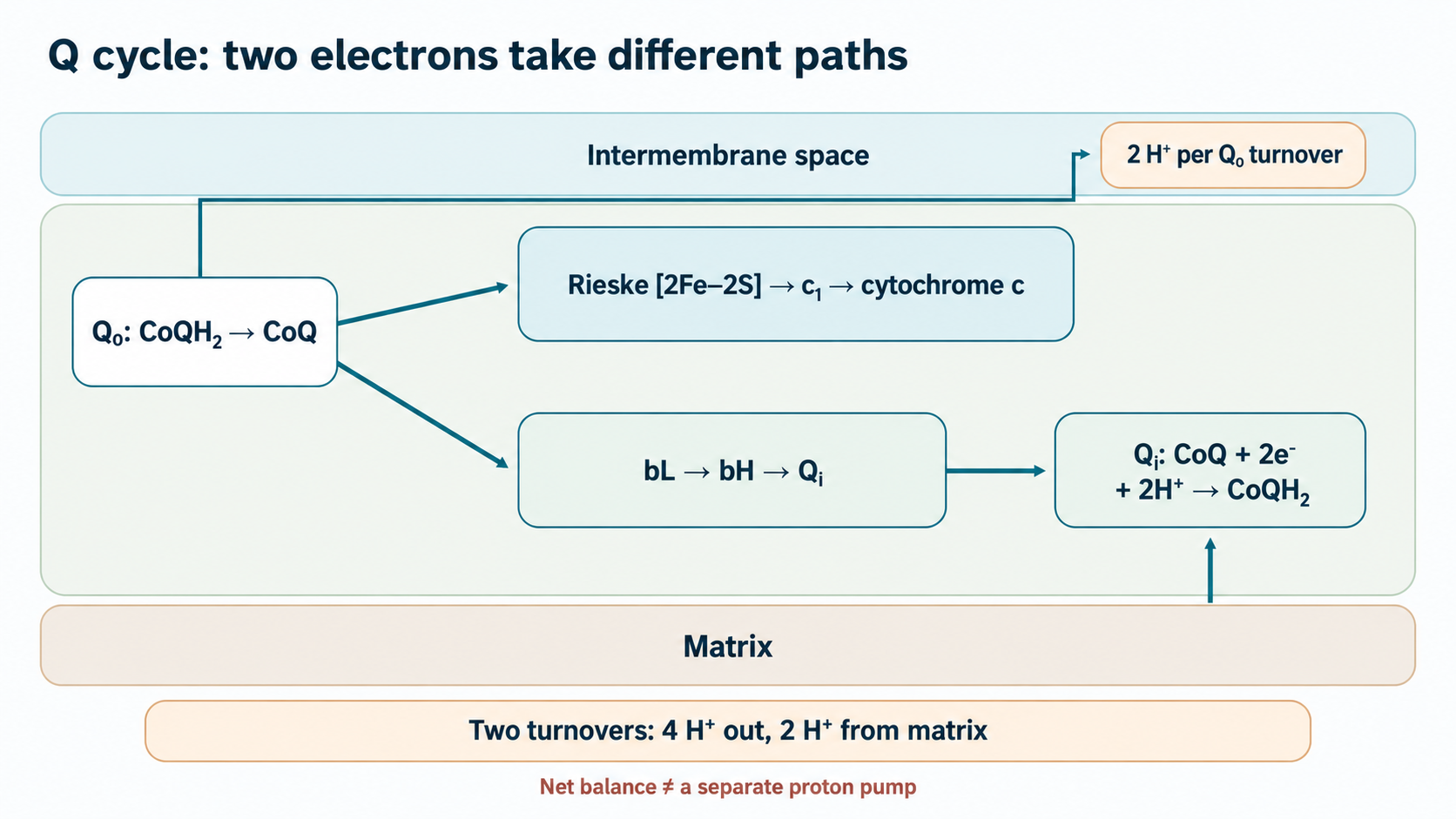
At Qo, ubiquinol’s first electron travels through the Rieske center to heme c1 and then soluble cytochrome c; the second passes through bL and bH to Qi. Oxidation of each molecule at Qo releases two protons on the outer side of the inner membrane. After two turnovers, Qi receives two electrons and two matrix protons, reducing one CoQ molecule. The overall scheme is
CoQH2+2cytc3++2Hmatrix+⟶CoQ+2cytc2++4HIMS+.
This is formal net stoichiometry for two electrons reaching cytochrome c, not independent mechanical pumping of four protons by protein b. Proton release at Qo and uptake at Qi contribute to the proton-motive force used by ATP synthase; the sign and magnitude of membrane potential depend on convention and metabolic state. Electron movement between hemes depends on distance, reorganization energy and the protein environment. Tunneling models explain kinetics, but a transfer time below 10 µs is not a universal constant.
2.3. Semiquinone, superoxide and membrane protection
If the second-electron route from Qo is obstructed, the reaction intermediate may persist longer and react with oxygen to form superoxide. Antimycin A, which blocks Qi, and Qo inhibitors such as myxothiazol and stigmatellin help distinguish these routes. Their effects depend on binding site and pool state; experimental respiratory blockade cannot quantify radicals during ordinary CoQ₁₀ intake. Reverse electron transport through complex I is another reactive-oxygen source when the Q pool is strongly reduced and membrane potential permits it.
Reduced CoQ can donate a reducing equivalent to lipid peroxyl radicals and help regenerate tocopherol; enzymatic systems subsequently renew its own redox state. The inner membrane is rich in cardiolipin, making control of lipid peroxidation relevant to protein-complex integrity and the membrane barrier. Yet “oral ubiquinol → less cardiolipin damage → mPTP closure → prevention of patient apoptosis” comprises several separately testable steps, rather than an established clinical mechanism. Membrane-chain physics provides a useful comparison with other mitochondrial redox interventions, but does not demonstrate synergy.
3. How cells return quinone to the reduced pool
3.1. NQO1: two-electron chemistry and its limits
NAD(P)H:quinone oxidoreductase NQO1 is an FAD-dependent homodimer using NADH or NADPH to reduce quinones. In the first half-reaction, reducing equivalents move from nicotinamide to bound FAD; after the oxidized cofactor leaves, reduced flavin transfers equivalents to the quinone substrate in the second half-reaction. For CoQ, the overall reaction is NAD(P)H+H++CoQ→NAD(P)++CoQH2. Two-electron transfer reduces free semiquinone formation compared with sequential one-electron reduction, but does not make NQO1 the sole enzyme maintaining membrane ubiquinol.
NQO1 is studied mainly in the cytosol; its contribution to the plasma-membrane redox system and relationship with membrane CoQ require specification of cell type and electron-flow direction. Michaelis constants and turnover numbers obtained with one quinone cannot be transferred to CoQ₁₀ with different lipophilicity, preparation and delivery to the active site. Within individual compartments, NAD(P)H, FAD and quinone availability matter alongside enzyme composition. The dependence of the nicotinamide pool on precursors is discussed in the vitamin B3 review; increased blood NAD⁺ does not prove stronger NQO1 flux in a particular tissue.
3.2. Other reductases and the misleading “sole activator” concept
Thioredoxin reductase 1 contains FAD and a C-terminal selenocysteine motif; it receives electrons from NADPH and can reduce CoQ in experimental systems. Cytochrome-b5 reductase CYB5R3 accepts NADH, while membrane partners determine subsequent electron destinations. Lipoamide dehydrogenase DLD is the FAD-dependent E3 subunit of pyruvate, α-ketoglutarate and branched-chain ketoacid dehydrogenase complexes; its secondary quinone-reductase reactions have been described biochemically. Activity of a purified protein alone does not establish any of these reactions as a guaranteed mitochondrial backup during human ischemia.
Compartment matters fundamentally: matrix DLD is separated from the outer surface of the inner membrane hosting other redox-network components; NADH donors and local CoQ availability differ. CoQ is also reduced through the usual respiratory-chain electron donors. Partial NQO1 loss therefore does not equal complete inability to convert ubiquinone into ubiquinol, and greater plasma exposure to one formulation does not show that it bypassed a single obligatory enzymatic step.
3.3. NQO1 Pro187Ser, Nrf2 and age: findings at different levels
NQO1 c.609C>T, rs1800566, replaces Pro187 with Ser. Structural and cellular studies associate it with lower stability, impaired FAD binding and accelerated proteasomal protein turnover; enzyme activity can differ substantially between genotypes. This is a meaningful pharmacogenetic hypothesis for CoQ metabolism research, but the supplied literature contains no clinical trial establishing that T/T carriers cannot absorb oral ubiquinone and must receive only ubiquinol. Genotype is not an indication for a commercial form without direct genotype-specific comparisons of exposure and outcomes.
NQO1 expression responds to electrophilic stress through Keap1–Nrf2–ARE: basal Keap1-CUL3 directs Nrf2 to degradation; modification of Keap1 sensor cysteines reduces turnover, allowing nuclear Nrf2 to interact with small Maf proteins and regulate targets including NQO1. This is regulated cellular adaptation, not a simple universal age-dependent switch. Age, disease, diet, medication and blood sampling can change the CoQ-form ratio; fixed age percentages establish neither tissue deficiency nor ubiquinone failure in every older patient.
4. Intestinal absorption and lipoprotein transport
4.1. From solid phase to mixed micelle
CoQ₁₀ is large and exceptionally lipophilic; crystalline material disperses poorly in aqueous chyme. Its melting point may exceed body temperature, but that does not mean intestinal absorption always requires “melting the crystal”: dissolution, oil dispersion, particle size, emulsification and formulation alter availability to micelles. One physical characteristic cannot establish 1–3% absorption for all crystalline ubiquinone. The oxidized form is also supplied in oil and dispersed matrices, so a fair comparison controls both chemical redox form and formulation.
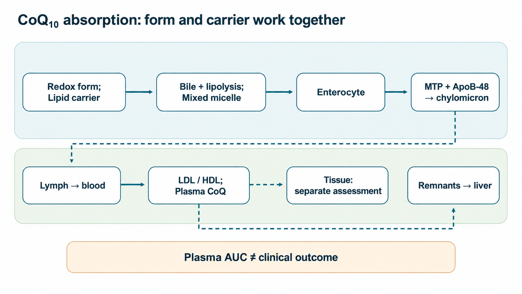
After meals, bile secretion and pancreatic enzymes create an aqueous–lipid mixture. Bile salts, phosphatidylcholine, free fatty acids and 2-monoacylglycerols form mixed structures incorporating hydrophobic CoQ and carrying it across the unstirred water layer to the brush border. Micelle size, critical concentration and diffusion rate depend on chyme composition and measurement method. Bile deficiency, impaired fat digestion or an altered mucosal surface can modify exposure to either form, including a pre-solubilized one.
4.2. Enterocytes, ApoB-48 and lymph
SR-BI/SCARB1 and NPC1L1 are discussed as contributors to apical transfer of lipophilic material alongside passive membrane partitioning. Their contribution depends on the model; a “hydrophobic SR-BI channel” or obligatory 30–50% CoQ blockade by ezetimibe is not established for all products and people. Inside enterocytes, CoQ partitions among intracellular membranes and lipid particles. MTP helps assemble triglyceride particles containing ApoB-48, released into villous lacteals and reaching systemic blood through the thoracic duct. This explains a delayed postprandial peak, but not permanent “liver bypass”: chylomicron remnants are subsequently taken up by the liver, with further distribution and lipoprotein exchange.
CoQ is later found in VLDL, LDL and HDL. Each fraction depends on particle concentration and composition; serum concentration without lipid adjustment may reflect carrier numbers rather than cell supply. Entry into cardiomyocytes, skeletal muscle and brain involves separate transport and distribution questions. Plasma AUC is neither quantitatively nor causally equivalent to myocardial mitochondrial concentration. CoQ interacts with tocopherol and other redox systems in lipoprotein antioxidant protection, but these observations do not establish fewer atherosclerotic events.
5. Comparing ubiquinol and ubiquinone pharmacokinetics
5.1. Preparation source and interconversion
Ubiquinol can oxidize during storage and manufacturing, making oxygen protection and capsule-content control important. Oxidized CoQ can be reduced in the body; administered ubiquinol also undergoes oxidation and reduction again. A raw-material name, including Kaneka QH™, is not a separate physiological mechanism: carrier oil, dispersion, dose, food and form stability can influence results. Inevitable enterocyte NQO1 saturation and P-glycoprotein-mediated CoQ efflux are not established as a general pathway and do not justify calling oxidized CoQ “energetically expensive” or ineffective.
5.2. Cmax, Tmax, AUC and repeated dosing
After a single dose, Cmax is the observed maximum, Tmax its timing, and AUC0−t=∫0tC(τ)dτ the measured plasma exposure through the last sampling point. During repeated dosing, steady state depends on duration, dosing interval, food context and lipoprotein turnover. Long-chain CoQ requires adjustment for endogenous background, while blood redox form must be preserved during sampling and processing. Total CoQ measurement alone establishes neither tissue reduction nor better mitochondrial function.
Langsjoen’s small sequential study in 12 healthy volunteers compared four weeks of 200 mg/day ubiquinone with four weeks of 200 mg/day ubiquinol after washout, using identical capsule excipients. Total plasma CoQ rose from approximately 0.9 to 2.5 µg/mL with ubiquinone and to 4.3 µg/mL with ubiquinol; cholesterol ratios also differed. The design supports an advantage of this product pair for this surrogate in this group, but sequential order, small sample and absent clinical outcomes preclude a universal “3–8 times better.” In López-Lluch’s crossover study of 14 healthy participants, the two most absorbed of seven formulations were one ubiquinone and one ubiquinol capsule. This emphasizes carrier and solubilization effects: a direct redox-form conclusion requires equal dose, comparable matrix and controlled food. Hosoe’s work primarily studied ubiquinol safety and exposure rather than superiority over every ubiquinone product.
Measurement
Supported interpretation
What it does not establish by itself
Single-dose Cmax and Tmax
Height and timing of plasma peak under these conditions
Mitochondrial CoQ content or duration of benefit
AUC0−t
Integrated exposure in sampled plasma
Whether redox form or formulation technology caused the difference
CoQ/cholesterol and ubiquinol fraction
Blood lipoprotein and redox-pool characteristics in a preserved sample
A universal diagnostic threshold or clinical response
Symptoms, events and function
Clinical outcome in the studied population
Superiority of a form not directly compared
The moment area AUMC0−t=∫0tτC(τ)dτ and ratio MRT=AUMC/AUC are useful descriptively, but endogenous background, lymphatic entry and recirculation complicate their interpretation for exogenous CoQ. Longer plasma MRT does not show “deep saturation of tissue stores.” Nor can one therapeutic serum CoQ threshold be assigned to every form, disease and laboratory.
5.3. Older age, malabsorption and medication
Heart failure, intestinal and hepatobiliary disease, dietary changes and altered lipoprotein profiles may modify CoQ entry. These factors make formulation important but do not prove that ubiquinol automatically overcomes bile deficiency, ezetimibe or any NQO1 variant. Each subgroup needs direct study with comparable formulation technology and pharmacokinetic and clinical endpoints. Comparative findings support selecting an evaluated product for the research question, rather than treating molecular redox state as a prescription on its own.
6. Clinical evidence: CoQ₁₀ effects are not synonymous with ubiquinol effects
6.1. Heart failure and Q-SYMBIO
Q-SYMBIO randomized patients with symptomatic heart failure to CoQ₁₀ 100 mg three times daily or placebo in addition to contemporary standard therapy, with roughly two years of follow-up. Among 420 participants, the major cardiovascular composite occurred in about 15% versus 26%; differences in cardiovascular and all-cause mortality were also reported. This deserves attention because the outcome comprised events, not merely laboratory concentration. However, ubiquinol and ubiquinone were not compared, so superiority of one redox form for survival is not established. Formulation, background treatment and treatment era limit application to every current patient; CoQ does not replace proven heart-failure medication.
The original manuscript linked this result to purported increases in stroke volume and cardiac index and obligatory mPTP closure. The trial did not establish that causal chain. Mitochondrial membrane permeability depends on calcium load, reactive oxygen species and regulators such as cyclophilin D, but molecular explanation of clinical outcomes requires intermediary measurements in the relevant tissue, not biochemical plausibility alone.
6.2. KiSel-10: a combination and prolonged follow-up
KiSel-10 gave 443 older Swedish residents selenium and CoQ₁₀ together or placebo; fewer cardiovascular deaths and differences in NT-proBNP and echocardiographic measures were reported. Longer follow-up of the originally randomized groups found a persisting mortality difference after supplementation ended. The intervention tested a combination in a particular northern European population, not separate selenium or CoQ effects or ubiquinol superiority. Baseline selenium status, food context, treatment changes over the years and multiple secondary analyses matter for generalizability.
Selenium is required for selenoproteins, including thioredoxin reductases and GPx4, creating a biochemical possibility of interaction with the CoQ redox network. But a two-component intervention cannot quantify “synergy” without separate groups for each substance. Later biomarker analyses and follow-up are not a second independent randomization and do not validate multi-ingredient “stacks.”
6.3. Statins and muscle symptoms
HMG-CoA reductase inhibition reduces mevalonate-pathway flux; isoprenoid intermediates are needed for cholesterol, the CoQ side chain and protein prenylation. Falling plasma CoQ during statin use may partly reflect fewer LDL carriers. Muscle symptoms have heterogeneous causes and do not establish myocyte CoQ deficiency: complaints, creatine kinase, objective muscle pathology and responses to withdrawal or rechallenge need distinction. General biochemistry cannot establish CoQ prevention of fiber necrosis through cyclophilin D.
Qu and colleagues pooled 12 small randomized trials with 575 patients and reported improvement in some subjective muscle symptoms. An earlier analysis in the same literature found no convincing effect; differences in design, SAMS criteria, doses, duration and pain scales weaken confidence in one universal effect. “Normalizes creatine kinase and restores statin adherence” does not follow from these findings. Even if CoQ helps some patients tolerate treatment, symptoms need separate assessment and statin decisions depend on cardiovascular risk. These trials do not establish a redox-form comparison for clinical SAMS relief.
6.4. Endothelium, metabolic markers and reproduction
In models, oxidation of tetrahydrobiopterin BH₄ can uncouple eNOS, promoting superoxide rather than NO. CoQ’s antioxidant role and lipoprotein interactions make an effect plausible, but “ubiquinol restores BH₄ and heals endothelium” requires causal testing. A meta-analysis of small randomized trials described changes in brachial-artery flow-mediated dilation; FMD depends on method and vascular state and remains a surrogate rather than proof of fewer infarctions.
Oocyte and sperm mitochondria depend on electron transfer, providing a rationale for CoQ research in reproductive medicine. Ben-Meir’s work on oocyte function and fertility, however, mainly concerns an aging-mouse model and cannot establish increased pregnancy rates with ubiquinol in women undergoing IVF. Seminal-plasma CoQ measurements do not demonstrate a particular increase in motility, concentration or decrease in DNA fragmentation after supplementation. Clinical sperm-parameter findings must be distinguished from live birth and direct form comparisons.
Context
Evidence actually measured
Unresolved question for form selection
Q-SYMBIO in heart failure
Randomized CoQ–placebo comparison of clinical events
Ubiquinol and ubiquinone were not compared for those events
KiSel-10 in older adults
CoQ plus selenium versus placebo and subsequent follow-up
Components cannot be separated or synergy inferred
SAMS
Small heterogeneous trials and conflicting symptom meta-analyses
No established form advantage for pain or maintaining statins
FMD and reproductive measures
Surrogates and individual preclinical or early clinical studies
Tissue exposure, long-term outcomes and form effects need trials
7. Practical interpretation, combinations and safety
7.1. Dose and food depend on the question, not the form’s name
For a lipophilic compound, dietary fat, bile and capsule composition can substantially alter absorption; taking it with food reasonably approximates conditions in many pharmacokinetic studies. However, obligatory 10–15 g of fat, a ban on evening use, universal split dosing attributed to saturable SR-BI, and a “therapeutic” plasma threshold above 3.5 µg/mL are not established general clinical standards. A dose studied in a particular randomized trial should be discussed together with its population, formulation, duration, background treatment and endpoint. High plasma concentration is not a clinical response.
7.2. Combinations: biochemical compatibility is not a factorial trial
Selenium supports active sites of some redox enzymes, while cell and animal experiments link PQQ to CREB/PGC-1α and mitochondrial biogenesis. Astaxanthin partitions into membranes; shilajit contains heterogeneous organic components. None of these schemes proves that adding ingredients to CoQ increases human ATP by a specified percentage, stabilizes semiquinone specifically in complex III or prevents disease. Using KiSel-10 to validate a package of selenium, PQQ, shilajit and other ingredients is especially inappropriate. Each added component increases uncertainty about composition and interactions, and potential adverse effects. The dietary polyphenol review likewise distinguishes biomarker activity from clinical outcomes.
7.3. Safety and medication monitoring
CoQ has generally been tolerated satisfactorily in studied oral regimens, but this does not establish safety for every dose, form or multi-ingredient product. Dyspepsia, individual reactions, product quality and stability, pregnancy, severe comorbidity and concomitant medication require consideration. Possible reduction of warfarin’s anticoagulant effect is discussed because of vitamin-K structural similarity and clinical reports; combined use requires INR monitoring according to clinical practice. Statins, heart-failure therapy or anticoagulants should not be stopped for CoQ without a physician. A preclinical NOAEL cannot be mechanically converted into a recommended human dose; claims of absent feedback on endogenous synthesis or no withdrawal “rebound” have limited experimental support.
8. Review method and practical decision
This is a narrative analysis of the available literature with targeted primary-publication checks of key conflicts on October 4, 2026; no systematic search, formal risk-of-bias assessment or GRADE evaluation was performed. Sources span structural and cellular biochemistry, pharmacokinetics and selected human trials, so causal confidence varies. In particular, healthy-volunteer AUC comparisons establish neither mortality, pain nor fertility differences; clinical CoQ trials do not isolate ubiquinol’s independent effect. Some older studies predate changes in background treatment standards.
Practical selection starts with the goal: confirmed primary CoQ deficiency, a possible adjunct in a studied population, or simply increasing a plasma marker are different situations. The specific formulation, tolerability, comorbidities and medication are then considered. Among quality products, ubiquinol may provide higher exposure under certain conditions, but the best form for an individual is not determined by redox-state name alone. The next needed study is a direct, adequately powered comparison controlling carrier and food, assessing tissue biology where ethically feasible and selecting a clinical endpoint in advance.
FAQ
Is ubiquinol always absorbed better than ubiquinone?
No. Some comparisons show higher plasma exposure to a particular ubiquinol product, but carrier lipids and solubilization may be equally important; universal form superiority and clinical benefit are not established.
Does NQO1 Pro187Ser make ubiquinone useless?
The variant reduces NQO1 activity, but other CoQ reduction pathways exist. Direct clinical evidence for genotype-based form selection is insufficient.
Do Q-SYMBIO and KiSel-10 prove ubiquinol superiority?
No. Q-SYMBIO compared CoQ10 with placebo and KiSel-10 compared CoQ10 plus selenium with placebo. Neither directly compared redox forms for clinical events.
Can CoQ10 replace heart-failure medication or a statin?
No. CoQ10 was studied as an adjunct; baseline treatment decisions depend on clinical risk and proven benefit.
Does everyone need plasma CoQ10 above 3.5 µg/mL?
No universal target is validated. Plasma levels depend on lipoproteins and do not automatically indicate tissue concentration or functional effect.